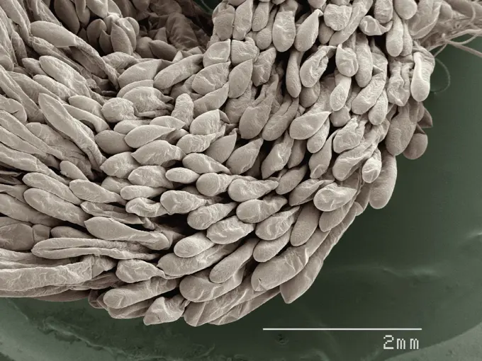
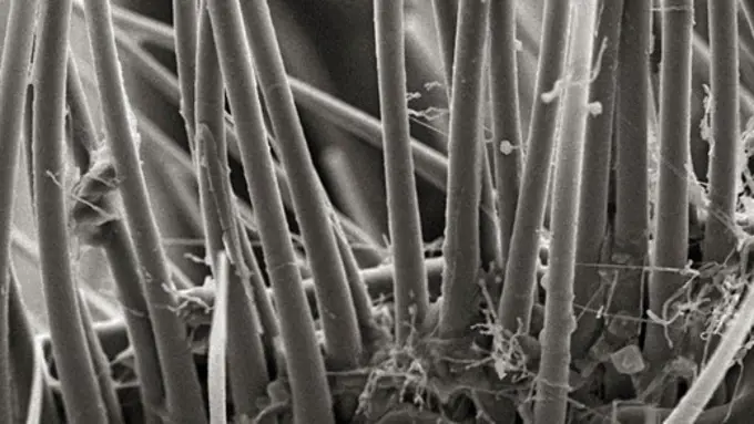
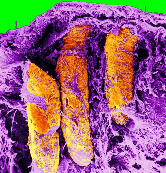
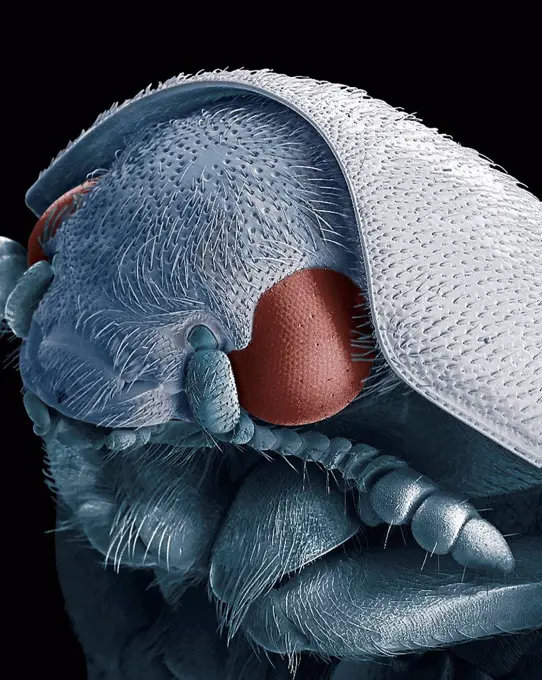
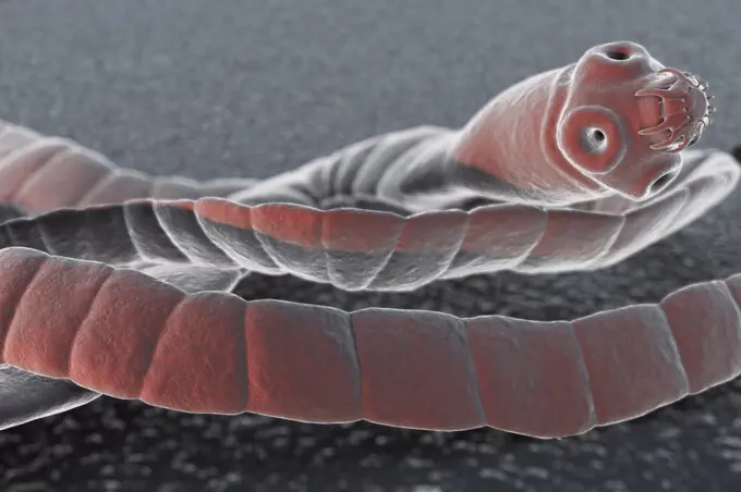
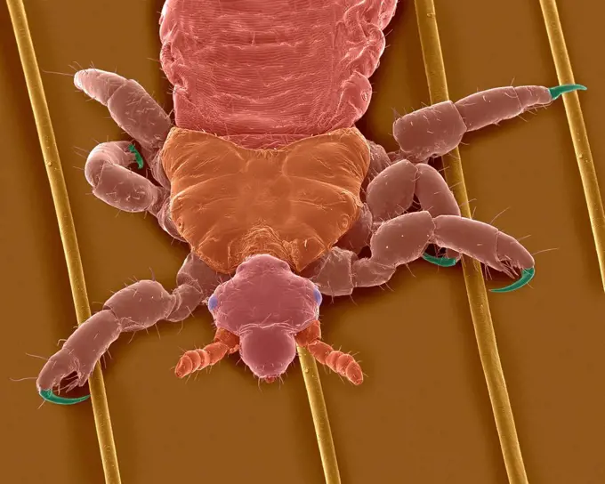
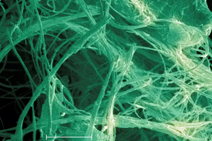
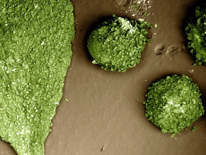
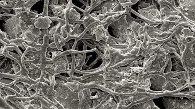
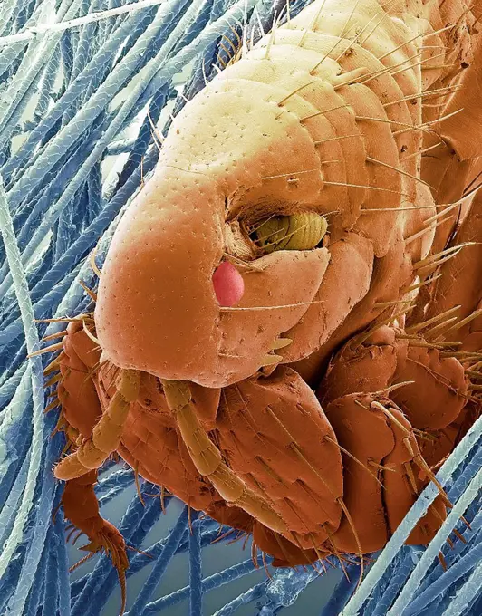
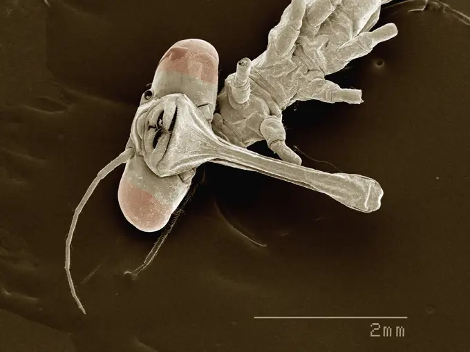
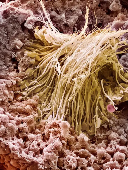
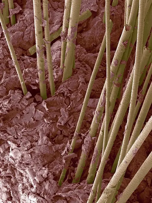
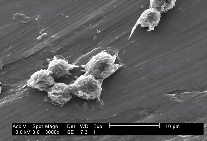
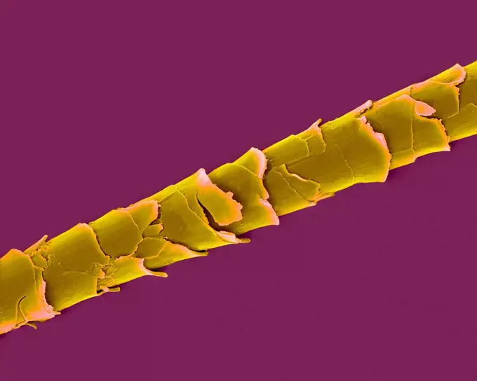
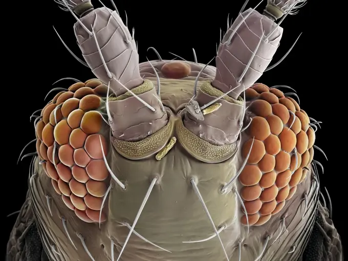
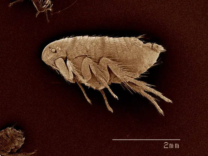
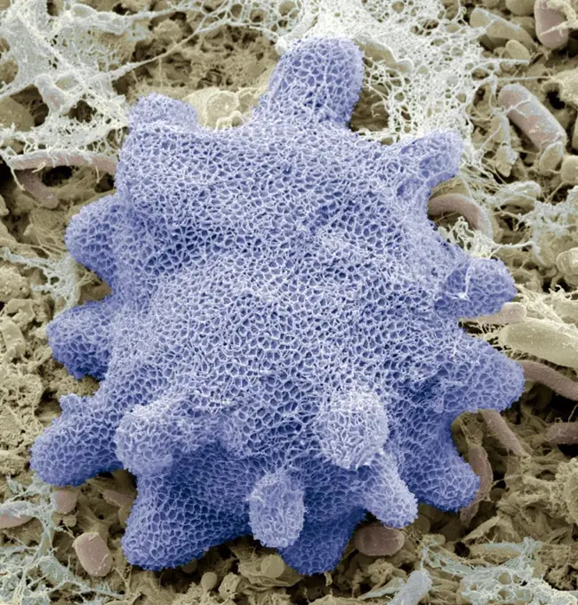
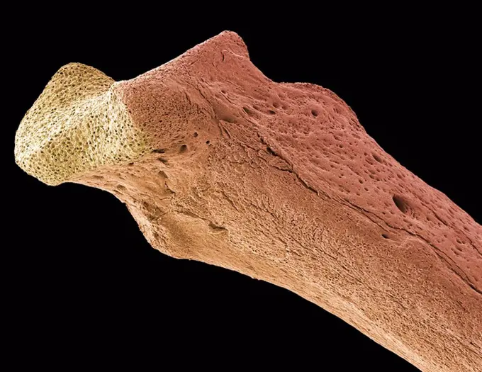
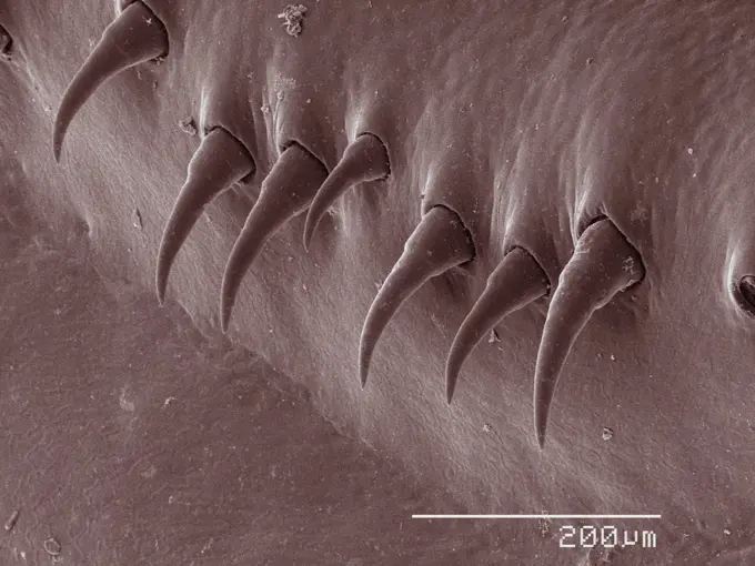
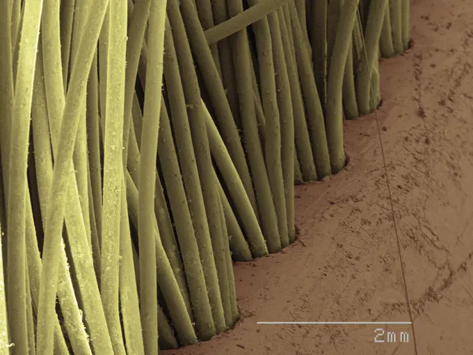
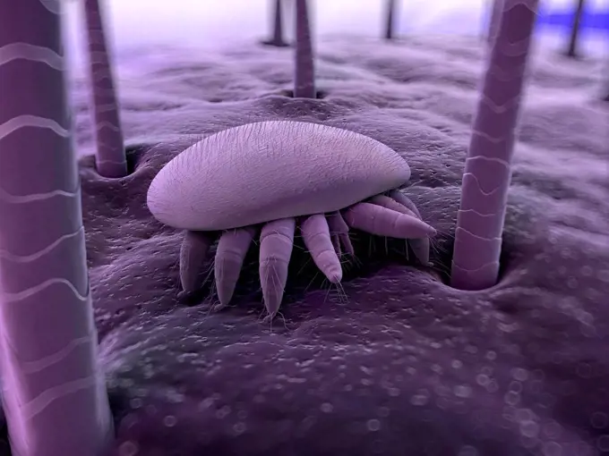
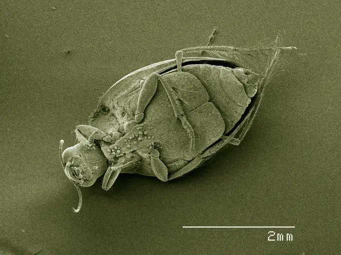
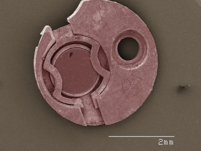
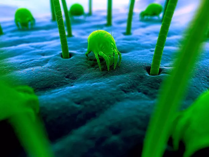
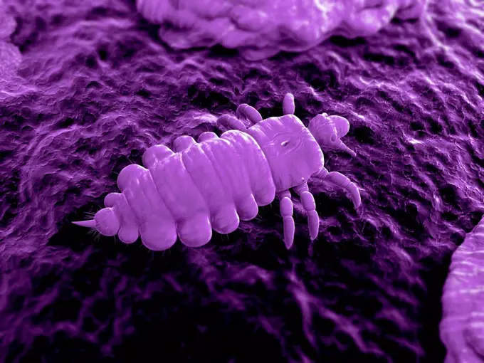
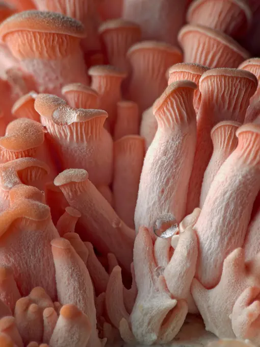
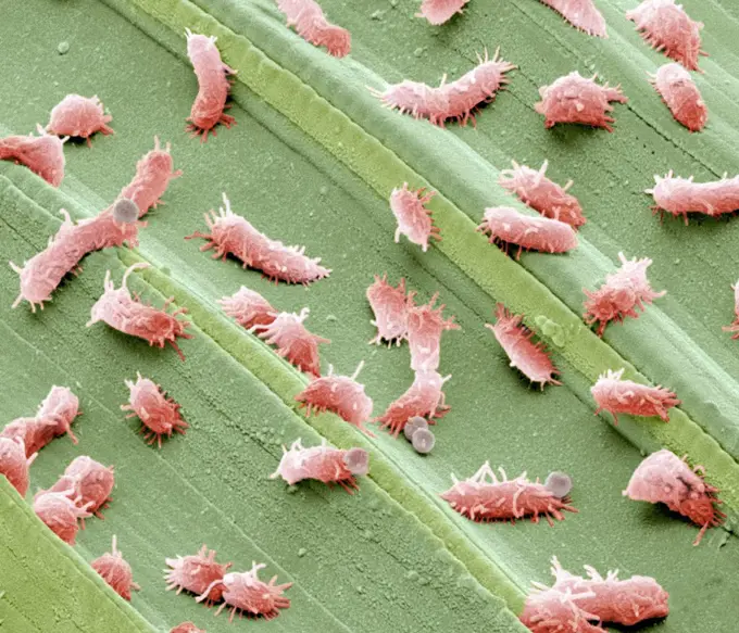
Microscopic Insect Views

Extreme close-ups of insects and arachnids captured under a microscope, revealing intricate details and textures.

Closeup view of textured organic patterns under magnification.
Scanning Electron Micrograph of a Beetle's Exoskeletal SurfaceTransversal section of the colon. SEM x 150.Dermestid beetle. Scanning electron micrograph of a dermestid beetle. Dermestidae are a family of Coleoptera that are commonly referred to as skin bee...Scale insects. Coloured scanning electron micrograph (SEM) of scale insects (superfamily Coccoidea) on a leaf. This pest feeds on the plant's sap. It ...A cestode, a parasitic tapeworm.Coloured scanning electron micrograph (SEM) of Human head louse on human hair (Pediculus humanus capitis). The head louse (Pediculus humanus capitis) ...Scanning Electron Micrograph of Brown RecluseCloseup view of a cellular structureScanning Electron micrograph of  asbestos, 5000xSEM image of budsExoskeleton of Unidentified InsectVein, semIntricate details of a microscopic world a closeup view under the electron microscope.Stomach lining with gastric pits, SEMTextured Surface of Honeydew MelonCat flea, SEMA closeup look into the micro world An intricate ant viewed through a scanning electron microscope.Sperm production site, SEMCloseup of an insect's compound eyes and textured antennaeHuman hair, SEMMicroscopic DebrisMagnified 3000X, this 2002 scanning electron microscope (SEM) image revealed part of the ultrastructure exhibited by a number of Hartmannella vermiformis amoeba cysts. The amoeba assumes the cystic stage of its life cycle under harsh conditions such as drought, or when there is an accumulation of toxins in its environment, which can reduce its metabolic needs. Under these circumstances, the protozoan produces a protective coat and goes dormant to wait for better conditionsIntricate natural structure in stunning closeupExtreme closeup of an ant under the microscopeColoured scanning electron micrograph (SEM) Dog hair (shih tzu) with damaged cuticle. Dogs have two types of hair in their coats. The first type is sh...False-coloured scanning electron micrograph of a Thrip's (Thysanoptera) head, the bulging compound eyes can be seen on either side of the head, with sensory hairs sprouting between the individual units (ommatidia) that make up each compound eye. This thrip's head is 0.15 millimetres across in real life. Derbyshire, UK. July. Image x650 magnification when printed at 10cm wide.Closeup view of a precise metal coil under a microscopeUpclose view of a flea under microscopic imagingKorotnevella. Coloured scanning electron micrograph (SEM) of the scales covering a 'naked' amoeba of the genus Korotnevella. an Amoeba with finger-lik...Scanning electron micrograph of filamentous Ebola virus particles budding from an infected VERO E6 cell (35,000x magnification). Image captured at the NIAID Integrated Research Facility in Ft. Detrick, Maryland.Credit: NIAID.Osteoporotic bone, SEMMicroscopic Detail of Spiderwort Flower PetalUpclose look at an insect under a microscopeCloseup of a bizarre microorganism under the microscopeCloseup view of cat tongue texture under a microscopeCloseup view of green matchsticks arranged in an orderly patternHoneybee mite (Varroa destructor), computer artwork.Scorpion fly foot, SEMCloseup of a dust mite under the microscopeCloseup of a microscopic mechanical deviceLipid layer with molecules lipid layer with molecules copyright: xzoonar.com/dr.xnorbertxlangeex 14177740Dust mites. Computer artwork of house dust mites Dermatophagoides pteronyssinus on human skin.Head louse Pediculus humanus capitis, computer artwork.Tiny pink oyster mushrooms. A macro studio photo of small pink oyster mushrooms in their early stages of growth. Copyright: xZoonar.com/GregoryxJohnstonx 21693899Upclose view of a green microstructure resembling a coil or spring.Stagnant water bacteria, SEMDiatom, SEM