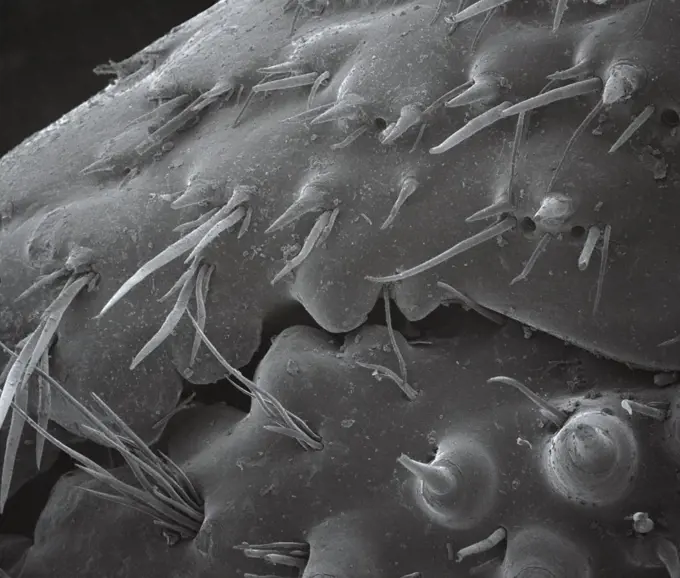
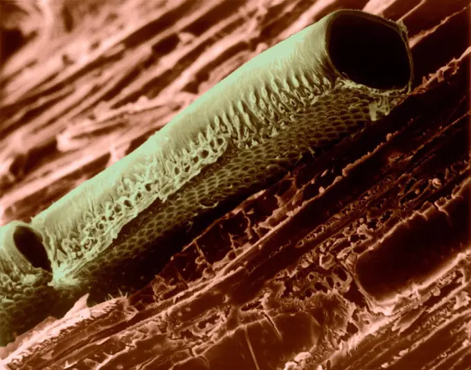
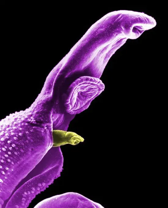
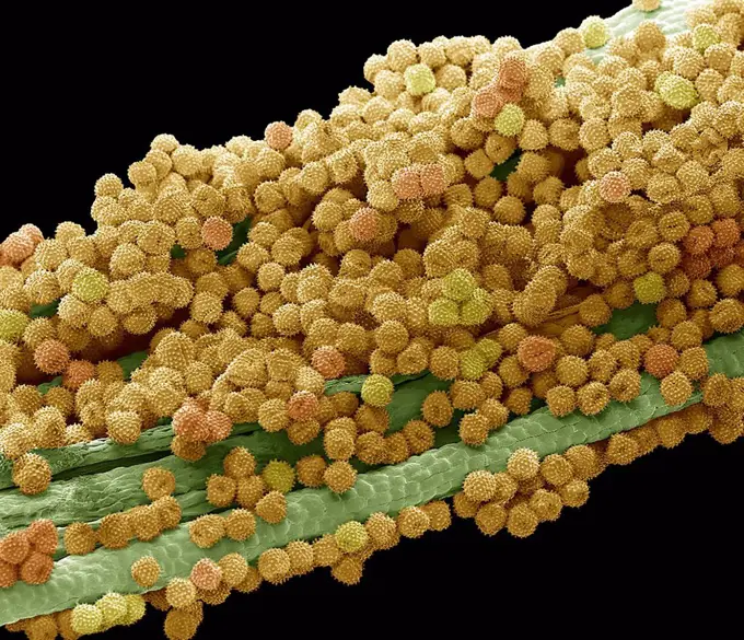
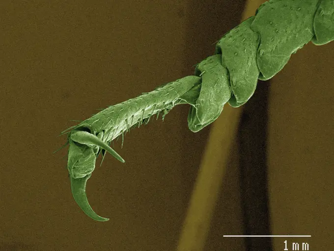
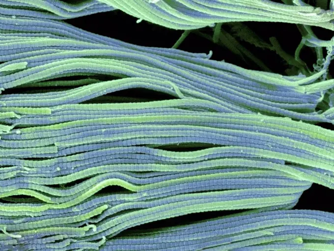
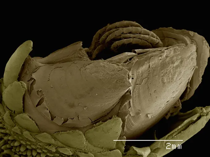
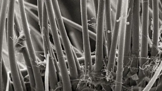
Fine ArtVintageFootageSubscriptionBoardsFree ResearchPricing
  • Fine Art
  • Vintage
  • Footage
  • Subscription
  • Boards
  • Free Research
  • Pricing

Microscopic Insect Views

Extreme close-ups of insects and arachnids captured under a microscope, revealing intricate details and textures.

Closeup view of textured organic patterns under magnification.
148 assets in this story
1439-57940160
Closeup view of an insect under a microscope.
1439-57943375
Microscopic view of a mite on a textured surface
1439-57940156
Extreme closeup of a spider's body under a microscope showing intricate details
4384-224
Scanning Electron Micrograph of a Spider's Prey
1439-57940104
Extreme close-up view of a housefly head under a scanning electron microscope
4384-239
Scanning Electron Micrograph of Termite's Leg
1439-57940127
Closeup view of textured organic patterns under magnification.
4201-82342
Wolf Spider (Lycosa tarantula) SEM close-up view of chelicerae at 42x magnification
1439-57952637
Closeup of an ant's mandibles in sepia tones
1439-57943357
Upclose exploration of the micro world
1439-57940146
Closeup view of a mosquito under the microscope
1439-57943359
Closeup view of a fracturing green material under a microscope.
1439-57949057
A microscopic landscape of interwoven fibers and textures
1773-209592
Leaf, x3000 magnification
1439-57943294
Extreme closeup of a textured surface revealing intricate details
1439-57940021
Intricate patterns of a microscopic landscape.
1439-57940006
Closeup view of microscopic structures resembling a dense forest
1439-57940126
Extreme closeup of a microscopic structure resembling a heart shape.
1439-57940148
Closeup view of a textured surface revealing intricate fibers
1439-57940084
Intricate microfiber structure in closeup detail
1439-57949080
Microscopic close-up of cellular structures.
1439-57943362
Closeup view of an ant's mandibles under a scanning electron microscope
4201-82343
Assassin Bug (Reduviidae) SEM close-up view of head with mouth part used to suck fluid out of prey at 21x magnification
1439-57940108
Closeup of a flea under the microscope.
4128R-13619936
Used wax dental floss with dental plaque (brown), bacteria (yellow) and cheek cells (purple) on the dental floss fibres (green), coloured scanning ele...
1439-57940002
Closeup of a textured fiber under a microscope
1439-57949070
Microscopic marvel a dust mite in its habitat
4201-21255274
Blue Hound's Tongue (Cynoglossum creticum) hooked seed spines enabling animal dispersal of seeds, native to Mediterranean Basis a noxious pasture weed elsewhere, magnification 97x
1439-57940171
A microscopic creature nestled within a rugged terrain.
1439-57949088
A closeup view of an insect leg under a microscope.
1439-57949028
Closeup of a bedbug hidden within textured fabric under a microscope.
4201-66246
Right-handed Hermit Crab (Paguras sp) SEM close-up view of the claw at 14x magnification
4128R-13622326
Conductive vessel element in mountain mahogany wood (Cercocarpus sp), coloured scanning electron micrograph (SEM). Note the bordered pits in the cellu...
1439-57941088
Intricate microstructures resembling a scifi cityscape
4297-1234
SEM image of the front end of an adult male schistosoma worm parasite
4128-15751145
Marigold pollen, SEM
4384-326
Red Blood Cells Enmeshed in Fibrinous Matrix
1439-57940194
Up close with nature's minute complexity
1439-57952646
Intricate detail of a microscopic organism captured in stunning clarity
4201-66367
Human Louse (Pediculus humanus) SEM close-up view of prehensile leg and human hair at 210x magnification
4384-235
Scanning Electron Micrograph of Mosquito
4128R-3780
Tendon, SEM
1439-57943354
Closeup of a textured surface with intricate patterns and a single protruding fibrous element
1439-57943321
Closeup of microstructures under a scanning electron microscope
4384-304
Giardia Protozoan's Ventral Adhesive Disk and Circular Lesion
1439-57940045
Coloured SEM of ostracod
4384-156
This scanning electron micrograph (SEM) revealed some of the ultrastructural morphology displayed on the ventral surface of a bedbug, Cimex lectularius.  From this view you can see the insects skin-piercing mouthparts used it uses to obtain its blood meal. Although bedbugs have been found naturally-infected with blood-borne pathogens, they are not effective vectors of disease.  The primary medical importance is inflammation associated with their bites (due to allergic reactions to components in
1439-57940011
Closeup of microscopic structures resembling an abstract terrain
4201-66238
SEM close-up of a millipede at 70x magnification showing 2 pair of legs per body section, the attribute distinguishing this group from centipedes which have but a single pair of legs per section
1439-57940074
An intricate closeup view of a microstructure.
4384-202
In this relatively highly magnified scanning electron micrographic (SEM) view, some of the wondrous ultrastructural shapes found on the surface of an unidentified white moth were revealed. At this level of magnification, 1479X, the proximal strut of a single scale could be seen as it inserted into its exoskeletal pore. Imagery such as this brings to life the incredible beauty, which goes unseen, but is abundant all around us. Composed of a substance known as chitin, this molecule is made up of b
1439-57952417
Up close with the fine details of an insect leg
4128R-12700
Fossil debris in chalk. Coloured scanning electron micrograph SEM of a fossilised fragment of a coccolithotrope skeleton. Magnification: x8000 when pr...
4128-V58557947
4128R-3934
Human hair, SEM
4128-111578356
Osteoclast, computer illustration.
4201-66255
Bramble (Rubus sp) SEM close-up view of blackberry spine at 21x magnification
1439-57943363
A striking closeup view under the microscope
1439-57944414
Black and white microscopic view of a spherical object surrounded by textured filaments
4384-427
Microscopic Detail of Insect's Exoskeltal Surface
1439-57943349
Intricate detail of a mosquito's antenna and proboscis under a microscope
4128R-8804
Female copepod, SEM
4201-62787
Mediterranean Mantis (Iris oratoria) detail of spines on front legs at 14x magnification, Spain
4384-217
Scanning Electron Micrograph of Brown Recluse
1439-57940181
Closeup of a microscopic organism
4384-392
Microscopic Detail of Spiderwort Flower Petal
4297-1461
Colorized scanning photomicrograph shows numbers of clustered Gram-negative Salmonella typhimurium bacteria
4384-170
Under a moderate magnification of 2000X, this digitally-colorized scanning electron micrograph (SEM) of an untreated water specimen extracted from a wild stream mainly used to control flooding during inclement weather, revealed the presence of unidentified organisms, which included bacteria, protozoa, and algae. In this particular view, a single copepod-like microorganism was seen occupying the field of view, which seemed to be encased in an outer shell of armour-like plates, or scales. Looking
4128R-13620197
Coloured scanning electron micrograph (SEM) Chicken eggshell cross section (Gallus gallus domesticus). Shown from left to right in the image. hard min...
4128R-2698
Dog hair, coloured scanning electron micrograph SEM.In dogs several hairs emerge from a single follicle.The collagen of the dermis has been exposed by...
1439-57952383
Incredible closeup of an insect under the microscope
1439-57939989
Extreme closeup of a dust mite under the microscope
1439-57940069
Intricate details revealed in a closeup of a microscopic structure
6145-29268311
Hair bundle of Guinea pig cochlea.
4201-21260206
House Pseudoscorpion (Chelifer cancroides), magnified 500 times, Barcelona, Spain
4201-66345
Shrimp Plant (Beloperone sp) SEM close-up view of pollen at 1620x magnifcation
4128R-9299
Asparagus beetle, SEM
4128R-4680
Human hair, SEM
4269-27452
Red cell. Color enhanced scanning electron micrograph (SEM) of human red blood cells. Magnification 11397x.
4128R-12703
Hoverfly head, coloured scanning electron micrograph SEM. Magnification: x30 when printed at 10 centimetres wide.
6145-29294153
Development of the limbs in a mouse embryo. The feet begin as a paddle shapes with the digits being created by the programmed death of tissue in the interdigital regions.
4128R-13192964
Bone tissue. Coloured scanning electron micrograph (SEM) of cancellous (spongy) bone. Bone tissue can be either cortical (compact) or cancellous. Cort...
4128R-2439
Foetal sense organ, SEM
4384-238
Scanning Electron Micrograph of Termite
1439-57952382
A detailed closeup of an insect under the microscope
4201-21247946
Coral Fountain (Russelia equisetiformis) anther, seen under SEM, 4000x magnification
1439-57939982
Microscopic image of a mechanical gear with a scale bar.
4269-6809
Scanning electron micrograph (SEM) of an absorptive epithelial cells lining the surface of the small intestine, showing numerous microvilli. Magnification x20000.
1439-57952628
Extreme closeup of an ant under a microscope showcasing intricate details.
1439-57943310
A closeup view under the microscope revealing the intricate details of a small insect.
1439-57949001
Microscopic closeup of pollen grains clinging to plant stamen.
1439-57940173
Monochrome capture of a closeup view on textured surfaces and organic patterns.
1439-57940136
Closeup of a microchip connector in sepia tone
1439-57940190
Intricate details of an insect leg under a microscope
6145-45247940
This prostate cancer construct was grown during NASA-sponsored bioreactor studies on Earth. Cells are attached to a biodegradable plastic lattice that gives them a head start in growth. Prostate tumor cells are to be grown in a NASA-sponsored Bioreactor experiment aboard the STS-107 Research-1 mission in 2002. Dr. Leland Chung of the University of Virginia is the principal investigator. The NASA Bioreactor provides a low turbulence culture environment which promotes the formation of large, three-dimensional cell clusters. Due to their high level of cellular organization and specialization, samples constructed in the bioreactor more closely resemble the original tumor or tissue found in the body. The Bioreactor is rotated to provide gentle mixing of fresh and spent nutrient without inducing shear forces that would damage the cells. The work is sponsored by NASA's Office of Biological and Physical Research. The bioreactor is managed by the Biotechnology Cell Science Program at NASA's Joh
1439-57944415
SEM image of a spider
4128R-7376
Collagen, SEM
1439-57952408
Closeup of a flea under electron microscopy
1439-57943312
Intricate microstructure maze under electron microscope
4384-213
Scanning Electron Micrograph of a Beetle's Exoskeletal Surface
PREVIOUS
of 2
NEXT
2401 S. Ervay, Suite 206
Dallas, Texas 75215
United States
Browse
CategoriesGalleriesFree Research
For Creators
License Your ContentSuperStock For StudentsFrame of Mind BlogContributor PortalNew Image SpotlightLicense AI Imagery
Resources
About UsLicensingTerms & ConditionsAPI AccessPrivacyPricing
Contact
+1 866 236 0087help@superstock.com Contact form
©2026 SuperStock. All Rights Reserved. -A
This site is protected by reCAPTCHA and Google Privacy Policy and Terms of Service apply.