Fine ArtVintageFootageSubscriptionBoardsFree ResearchPricing
  • Fine Art
  • Vintage
  • Footage
  • Subscription
  • Boards
  • Free Research
  • Pricing

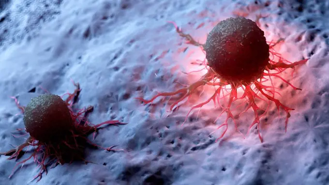
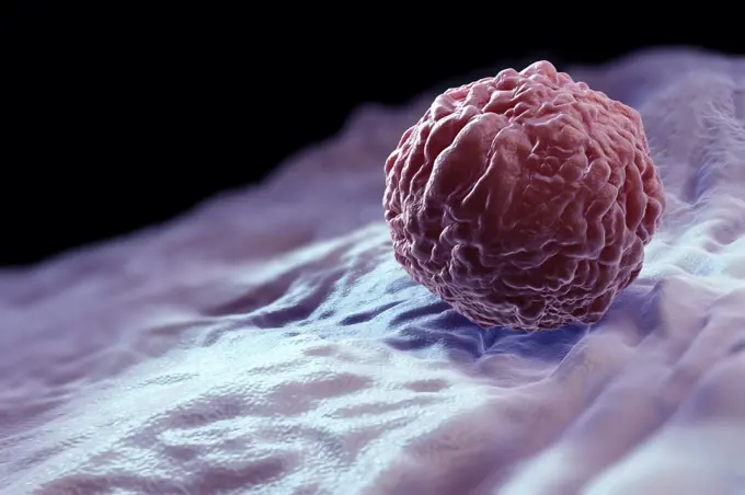
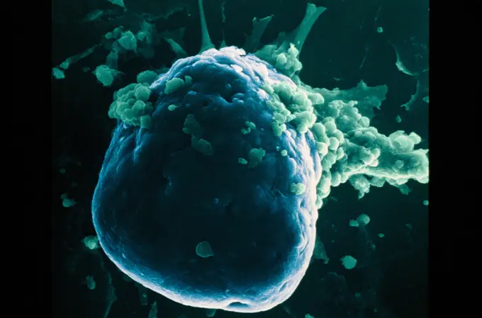
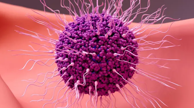
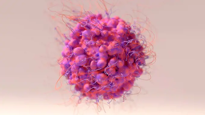
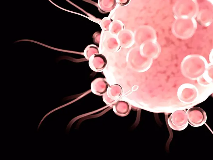
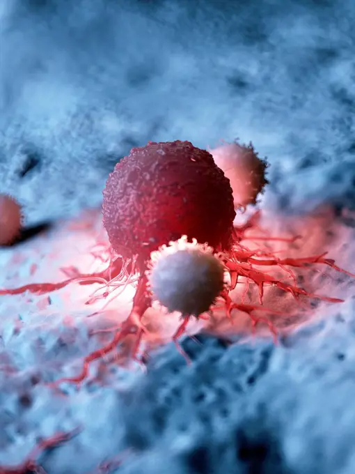
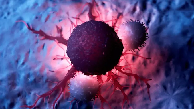
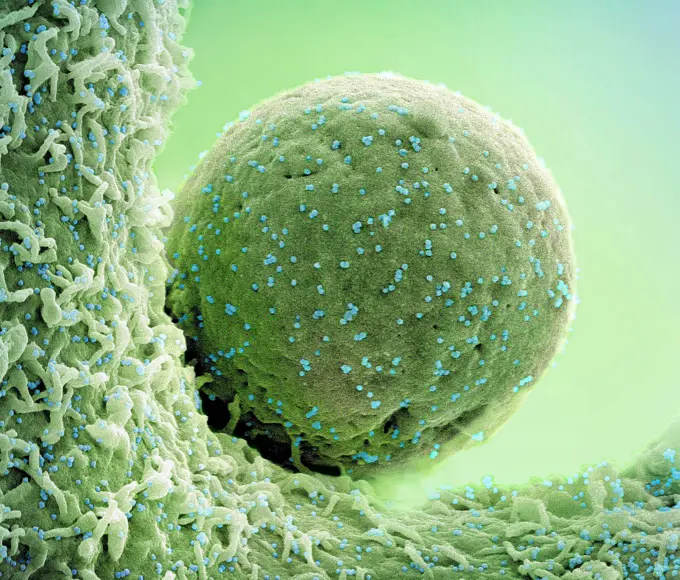
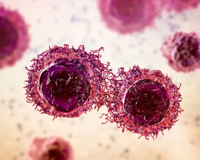
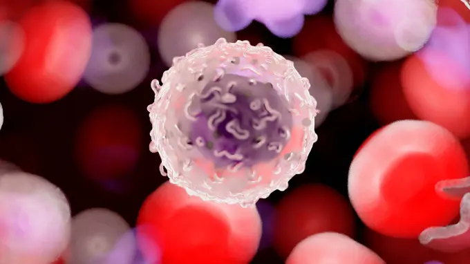
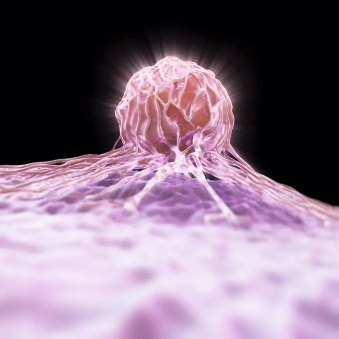
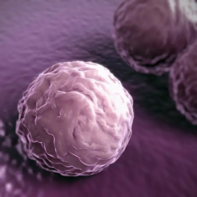
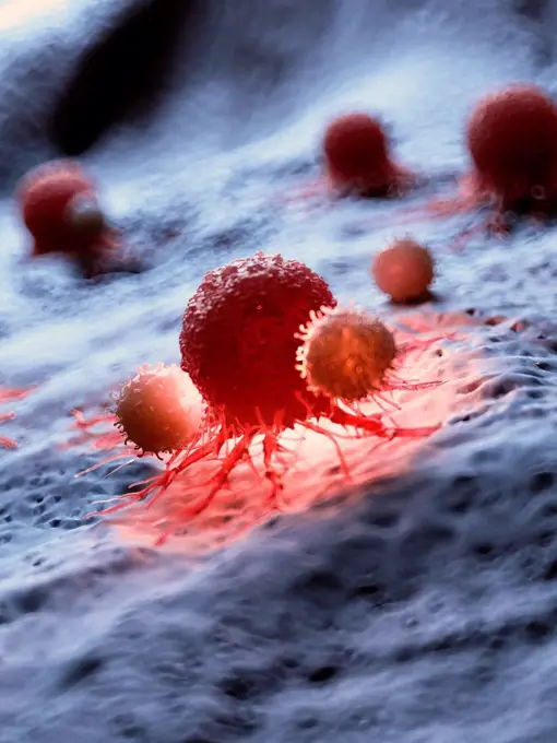
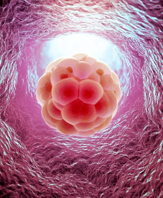
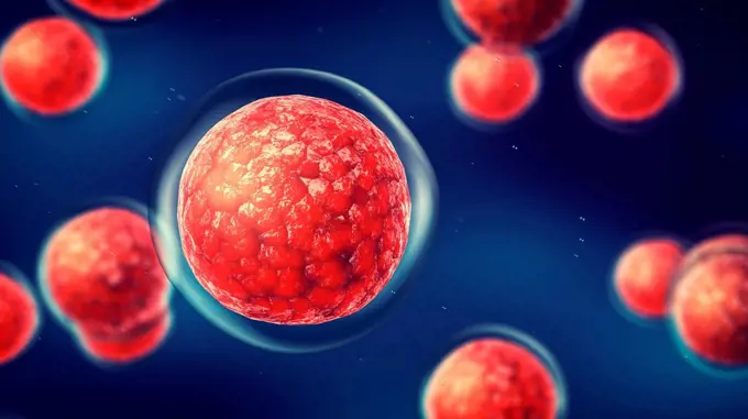
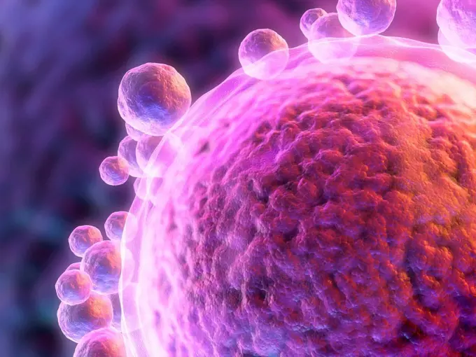
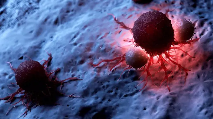
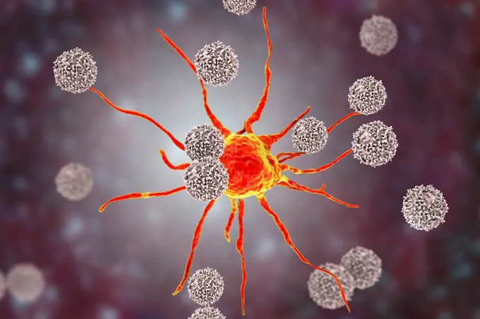
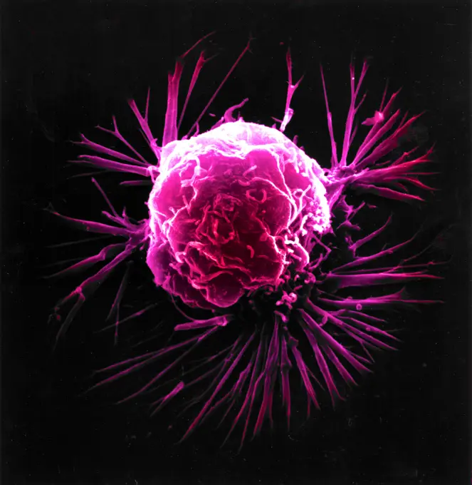
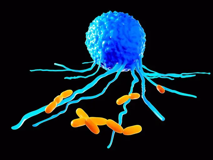
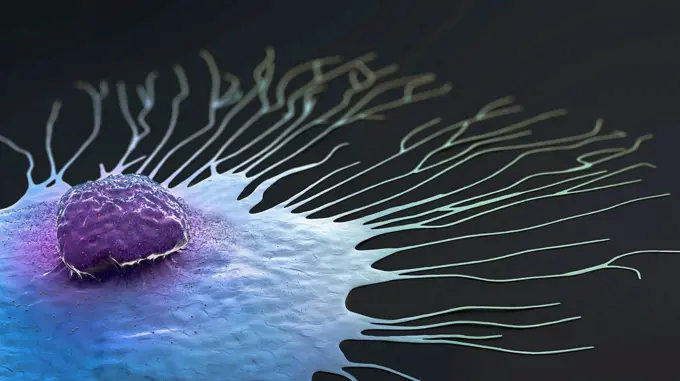
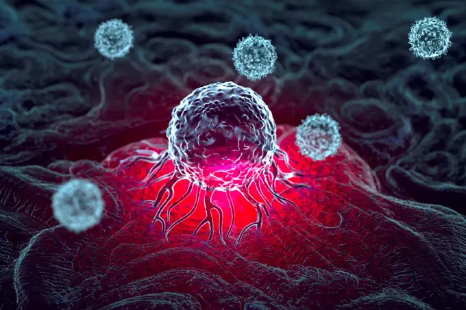
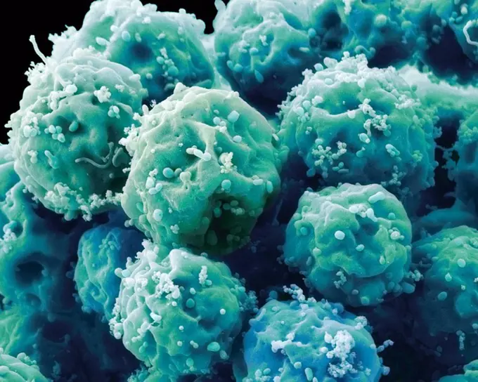
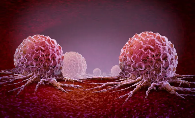
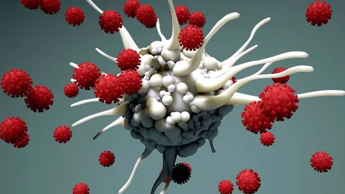
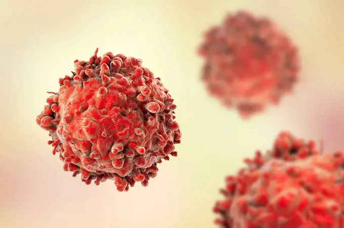
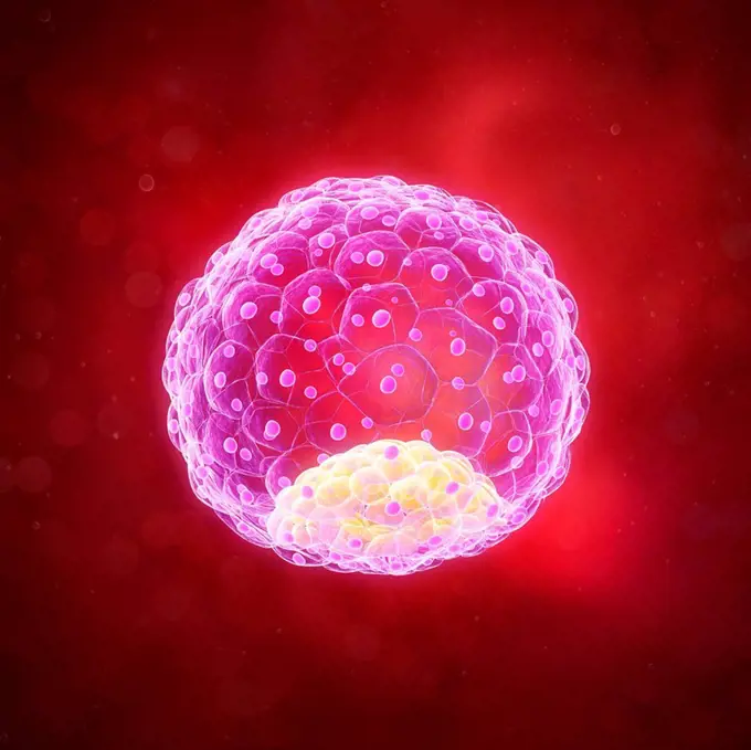
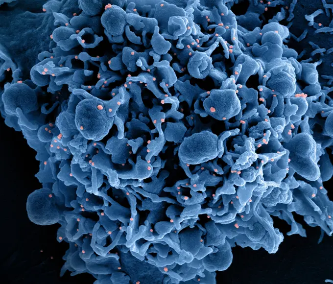
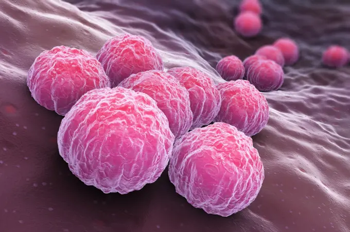
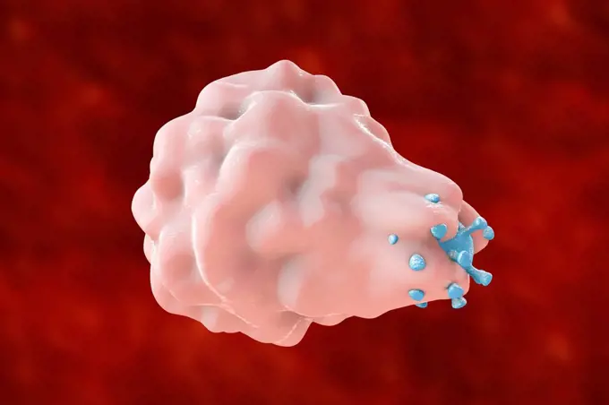
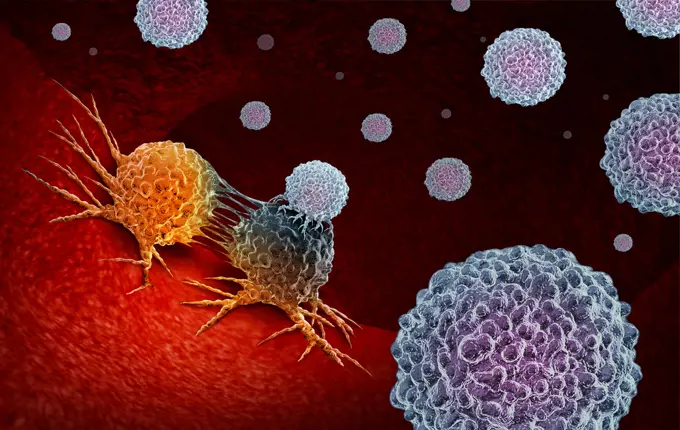
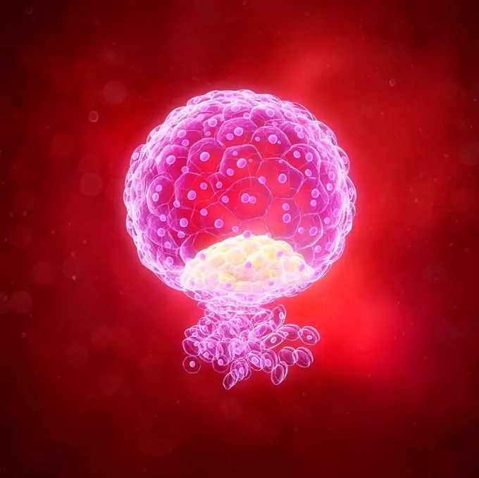
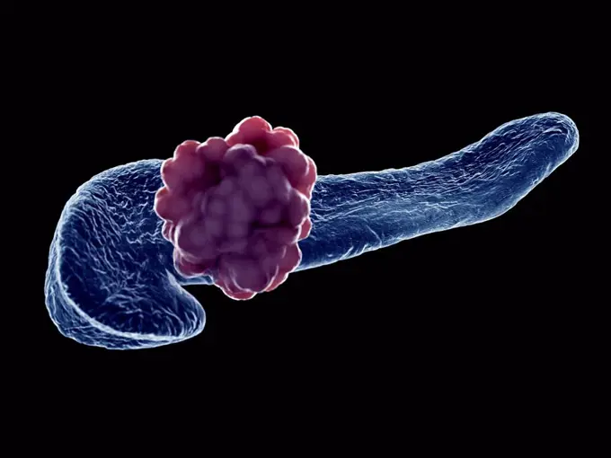
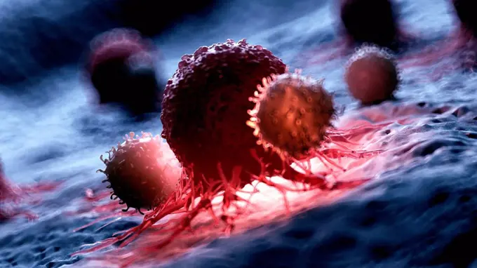
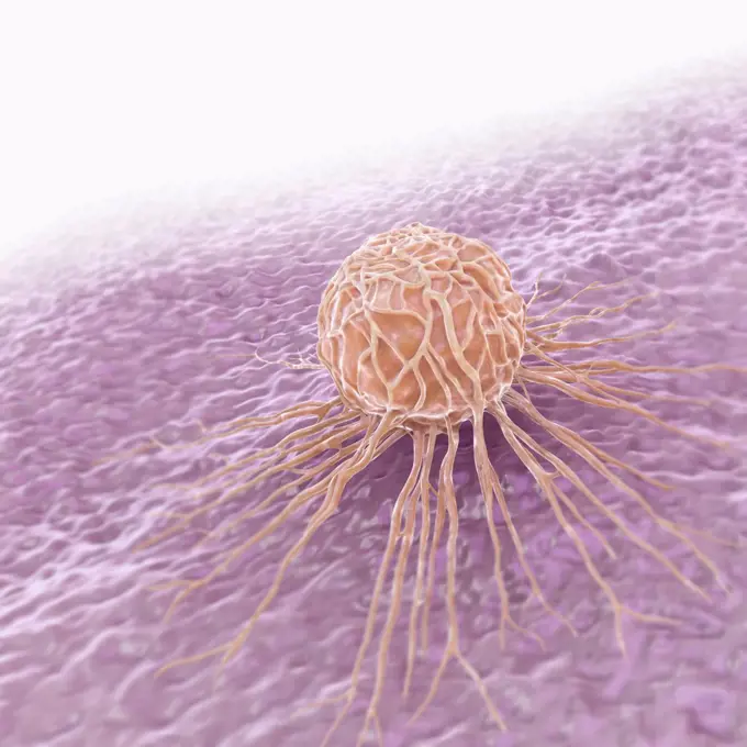
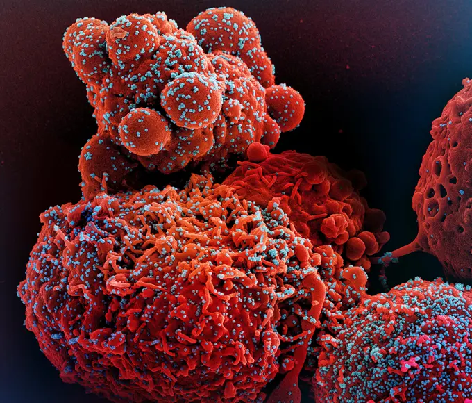
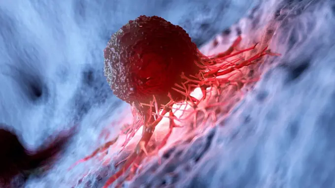
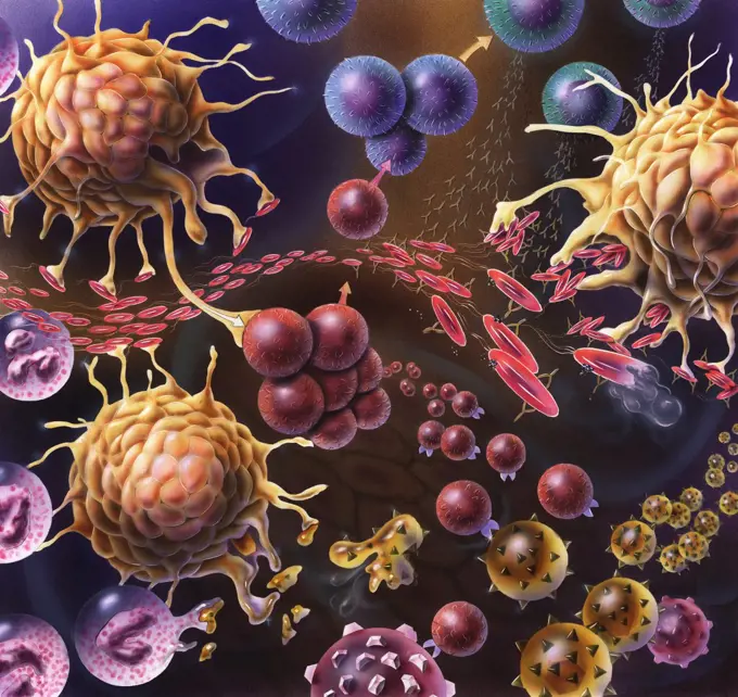
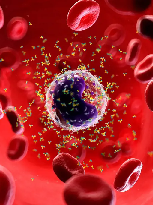
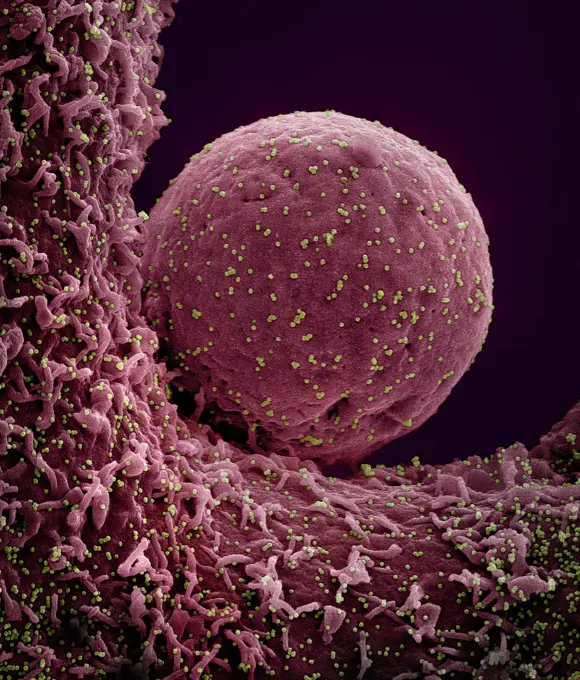
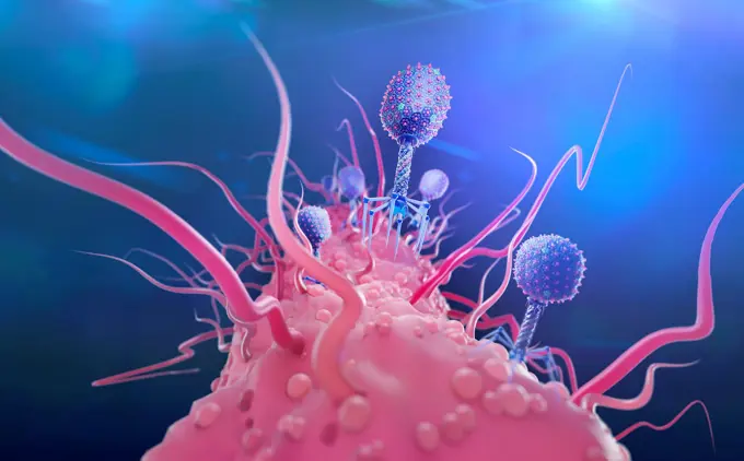
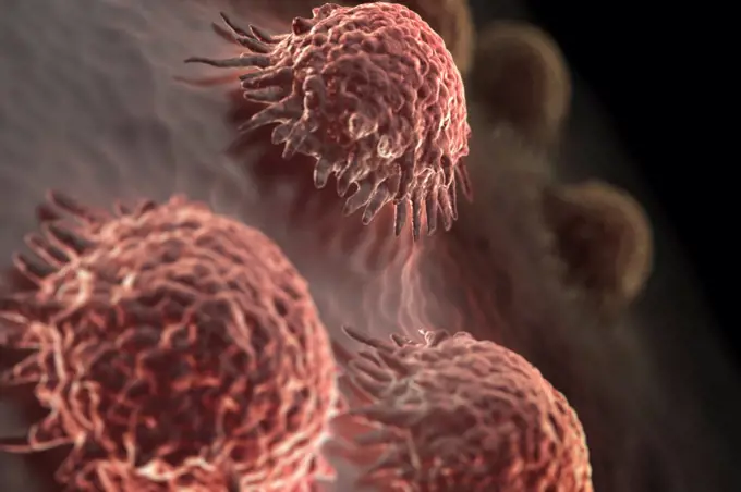
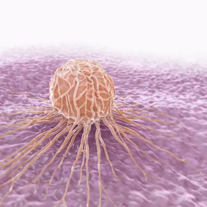
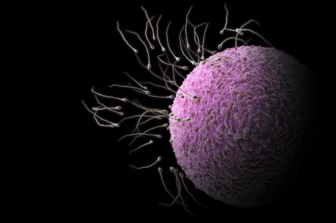
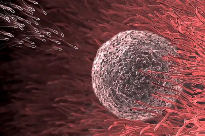
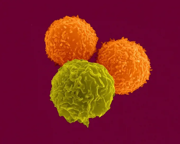
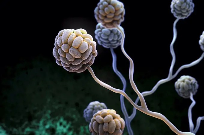
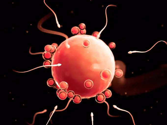
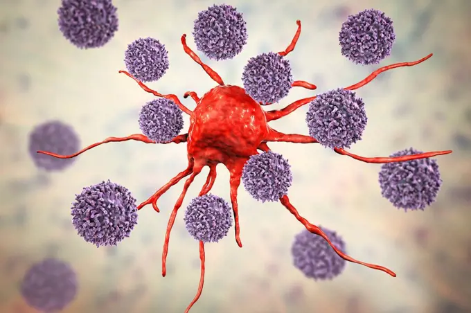
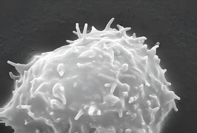
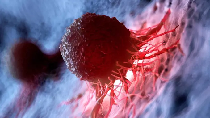
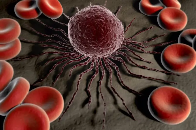
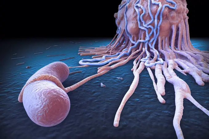
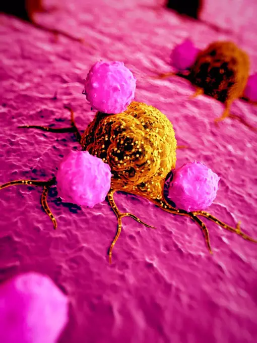
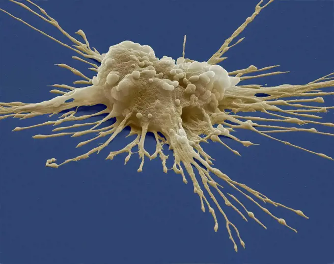
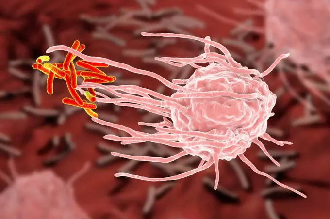
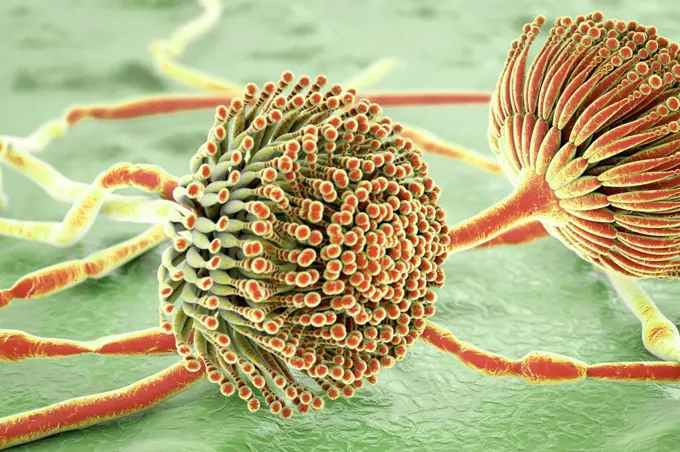
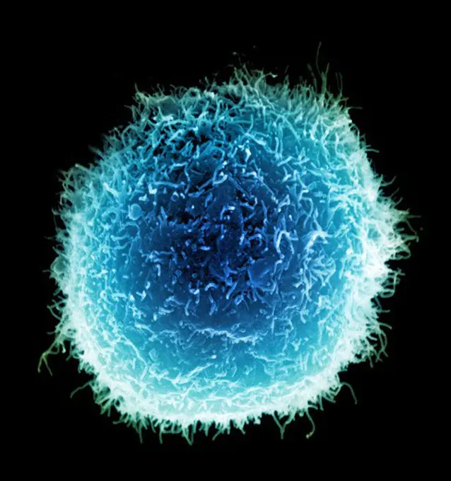
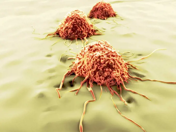
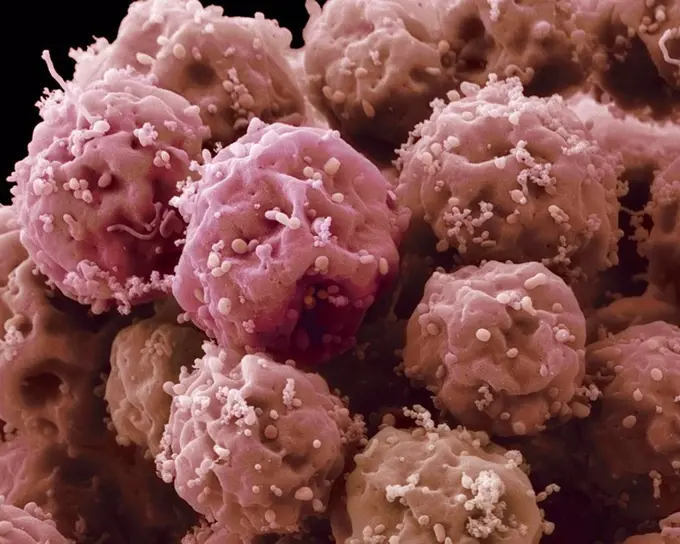
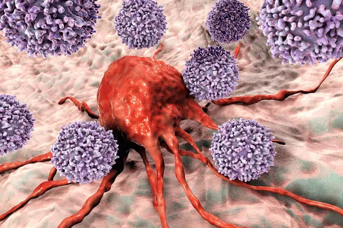
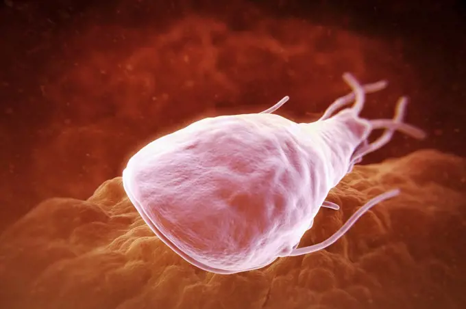
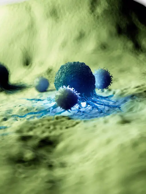
Cellular Structures and Microscopy

High-resolution images of various cells, including stem cells and cancer cells, showcasing intricate details in a scientific context.

Embryonic Stem Cell
Embryonic Stem Cell
194 assets in this story
4269-24762
Langerhan's islets. SEM (Scanning Electron Microscope) of a islet of Langerhan, the endocrine component of the pancreas.
4378-20013769
Embryonic Stem Cell
1525-19757760
cancer cell made in 3d software
4128-20038744
Sperm cells attempting to fertilise egg cell, illustration
4128-18300334
Cancer cells, illustration
4128-15979130
Fertilisation, computer illustration.
4128R-15516275
Illustration of a cancer cell being attacked by white blood
4128R-15516265
Illustration of white blood cells attacking a cancer cell
1899-65662391
Colorized scanning electron micrograph of a cell (green) infected with the Omicron strain of SARS-CoV-2 virus particles (blue), isolated from a patient sample.
824-65830708
Colorized scanning electron micrograph of a cell (green) infected with the Omicron strain of SARS-CoV-2 virus particles (blue), isolated from a patient sample.
4128-18799727
Cancer cells, illustration
4128-30416494
Leukocyte, illustration
4378-3204
Close up view of a single breast cancer cell on a surface.
4378-2817
Group of chlamydia bacteria.
4128R-15516266
Illustration of a cancer cell being attacked by white blood
4128R-31083
Embryo, computer artwork.
4128-19052402
Embryonic stem cells, illustration
4128R-14491
Human egg cell, computer artwork.
4128R-15516262
Illustration of white blood cells attacking a cancer cell
4128R-15405931
T-lymphocytes attacking cancer cell, illustration
4269-27679
Cancer Cell. Scanning electron micrograph (SEM) of a breast cancer cell.
4128R-13587315
Leucocyte, illustration.
4128-15659896
Migrating breast cancer cell, illustration
4128-48286036
Immune response to cancer, illustration
1899-65662352
Colorized scanning electron micrograph of a cell (pink) infected with the Omicron strain of SARS-CoV-2 virus particles (teal), isolated from a patient sample.
4128R-2657
Embryonic stem cells, SEM
824-65831028
Colorized scanning electron micrograph of a cell (red) infected with the Omicron strain of SARS-CoV-2 virus particles (blue), isolated from a patient sample.
1525-56205032
Cancer oncology and Malignant Cancerous Growth and Metastasis anatomy concept as growing tumor cells and Malignancy disease spreading metastasized on an organ inside the human body as a 3D illustration.
4378-5800
Colon Cancer Cells
4128-16171709
Dendritic cell and virus particles, illustration. Dendritic cells are a component of the body's immune system. The cell gets it name from the long mem...
4128R-13682118
Computer illustration of a cancerous white blood cell in leukaemia.
4128R-13682119
Computer illustration of a cancerous white blood cell in leukaemia.
4128R-25365
Blastocyst. Computer artwork of a 100-cell blastocyst embryo.
824-63225870
Colorized scanning electron micrograph of a cell (blue) infected with SARS-CoV-2 virus particles (pink), isolated from a patient sample. Image captured at the NIAID Integrated Research Facility (IRF) in Fort Detrick, Maryland.
4128-V58568584
1525-23580079
Chlamydia trachomatis, an obligate intracellular human pathogen, is one of four bacterial species in the genus Chlamydia. 3D illustration
4128R-13446573
Computer illustration of phagocytosis-like uptake of a virus by a host cell; the first stage of viral infection of the host cell. This type of viral e...
1525-22960577
Cancer Immunotherapy as a human immune system therapy concept as a biomedical or biomedicine oncology treatment using the natural T cell fighting properties of the body as a 3D render.
4128-30418595
Cancer cell, illustration
4128R-25367
Implanted blastocyst. Computer artwork of a blastocyst embryo implanted in the wall of the uterus (womb).
4128-15665326
Pancreas tumour, illustration
4128R-15516255
Illustration of white blood cells attacking a cancer cell
4378-3194
Close up view of a single breast cancer cell on a surface.
1899-65662497
Colorized scanning electron micrograph of a cell (red) infected with the Omicron strain of SARS-CoV-2 virus particles (blue), isolated from a patient sample.
4128R-15516248
Illustration of a human cancer cell
4239-20481110
Artistic representation of the immune system's reaction to bacteria invading the tissues.
4128-111578642
B cell and antibodies, computer illustration.
824-65830658
Colorized scanning electron micrograph of a cell (red) infected with the Omicron strain of SARS-CoV-2 virus particles (yellow-green), isolated from a patient sample.
4128-30419961
T4 bacteriophage infecting E. coli bacterium, illustration
4128-V58572226
4378-1119
Scanning electromicrograph styled osteoblast cells which are related to the reformation of bone matter.
4378-3961
Close up view of a single breast cancer cell on a surface.
4378-3043
Human sperm surrounding an unfertilized ovum prior to conception.
4128-V58562145
4128-V58567495
4128R-14057510
Leishmania parasites, illustration
4128R-13928481
Oesophageal cancer, computer illustration.
4128R-15059
Cancer cell, computer artwork. The uneven surface and cytoplasmic projections are characteristic of cancer cells.
4128-111494704
Bacteriophages infecting bacterium, illustration.
4128R-13376667
Human embryo destruction, illustration
4128R-14057785
Cancer cell and T cell, illustration
4128R-14181662
Cancer cells, illustration
4378-1123
Sperm approaching an ovum before conception.
4128R-13619968
T lymphocytes (pre-T cells) and granulocyte (neutrophil), coloured scanning electron micrograph (SEM). Lymphocytes are involved in the specific immune...
4378-4493
Black Mold Spores
4128R-12578643
Fertilisation of human egg, computer illustration.
4128R-15405938
T-lymphocytes attacking cancer cell, illustration
4128R-11291715
Artwork of a migrating cancer cell.
1525-25739611
Cancer anatomy concept as a growing tumor cell and malignant disease growth on an organ inside the human body as a 3D illustration.. Cancer Anatomy Concept
4128-19056191
Fungi Paracoccidioides braziliensis, illustration
4269-6813
Scanning electron micrograph (SEM) of oocyte with a cluster of cells from the cumulus oophorus.
4128R-15003
Pluripotent stem cell. Coloured scanning electron micrograph SEM of a pluripotent stem cell derived from a macrophage white blood cell. Magnification:...
824-65831029
Colorized scanning electron micrograph of a cell (brown) infected with the Omicron strain of SARS-CoV-2 virus particles (purple), isolated from a patient sample.
824-57659306
This scanning electron microscope (SEM) image revealed some of the ultrastructural features on the surface of a protozoan Acanthamoeba polyphaga. Note the numerous pseudopodia, protruding from the surface of the organisms. These allow the amoeba to move and grasp objects in its environment. CDC/Catherine Armbruster Margaret Williams 2009
4128R-15516246
Illustration of a human cancer cell
4378-999
Microscopic styled view of a stem cell.
4378-3647
A close up of a macrophage cell and bacteria. Macrophages are phagocytes and their function is involved in both innate immunity as well as adaptive immunity.
4128R-13587386
Cancer cells, illustration.
4128R-11543407
Virus, conceptual illustration.
4128R-25665
Fertilisation, computer artwork.
824-63198879
Human coronavirus. Human coronavirus causes respiratory infections and gastroenteritis. Image produced using high-dynamic-range imaging (HDRI) from an image taken with transmission electron microscopy. Viral diameter around 80-160 nm.
4128R-15047074
Transparent cell, illustration
4128R-15004
Pluripotent stem cell. Coloured scanning electron micrograph SEM of a pluripotent stem cell derived from a macrophage white blood cell. Magnification:...
4128R-14324120
Macrophage engulfing tuberculosis bacteria, illustration
824-65831018
Scanning electron micrograph of a cell (purple) infected with the Omicron strain of SARS-CoV-2 virus particles (orange), isolated from a patient sample.
4128R-13587468
Aspergillus fungus, computer illustration. This is a toxic fungus that causes diseases in humans. These include fungal ear, lung and skin infections (...
4297-1596
Scanning electron micrograph of a single macrophage with projectile-looking surface
4378-2761
Close up view of a single breast cancer cell.
4128R-11291719
Artwork of migrating cancer cells.
4128-18800731
Lymphocyte in hairy cell leukaemia, illustration
4128R-15047192
Transparent cells, illustration
824-63225905
Colorized scanning electron micrograph of a cell (blue) infected with a variant strain of SARS-CoV-2 virus particles (UK B.1.1.7 purple), isolated from a patient sample. Image captured at the NIAID Integrated Research Facility (IRF) in Fort Detrick, Maryland.
4128R-13048319
Cancer cell. Computer illustration of a typical cancer cell, showing the numerous blebs (lumps) and microvilli (hair-like) on its surface that are typ...
1815R-14647991
Insemination, 3D Rendering
4128R-1878
Embryonic stem cells, SEM
4239-V53647252
4128R-15405939
T-lymphocytes attacking cancer cell, illustration
4378-3693
Giardia lamblia is a flagellated protozoan parasite. It colonizes and reproduces in the small intestine and causes giardiasis.
4128R-15516270
Illustration of a cancer cell being attacked by white blood
4128R-15516251
Illustration of a human cancer cell
PREVIOUS
of 2
NEXT
2401 S. Ervay, Suite 206
Dallas, Texas 75215
United States
Browse
CategoriesGalleriesFree Research
For Creators
License Your ContentSuperStock For StudentsFrame of Mind BlogContributor PortalNew Image SpotlightLicense AI Imagery
Resources
About UsLicensingTerms & ConditionsAPI AccessPrivacyPricing
Contact
+1 866 236 0087help@superstock.com Contact form
©2026 SuperStock. All Rights Reserved. -B
This site is protected by reCAPTCHA and Google Privacy Policy and Terms of Service apply.