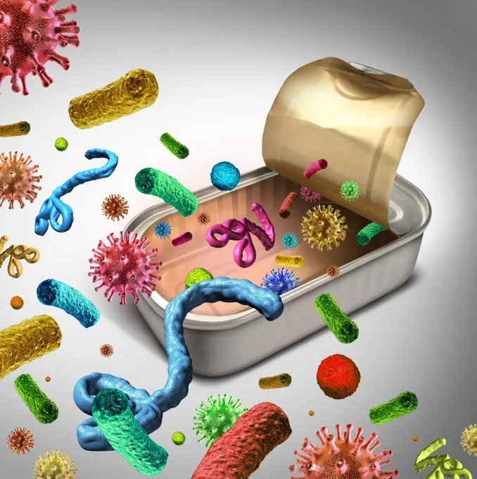
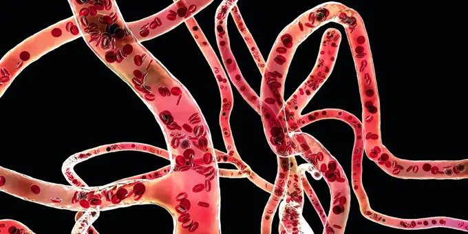
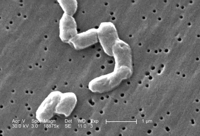
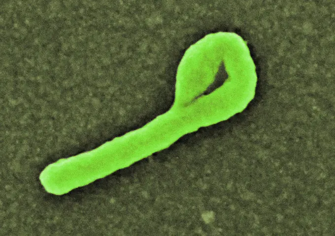
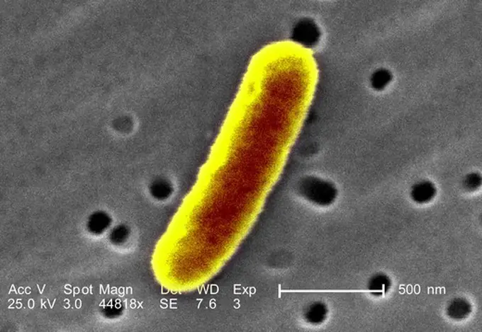
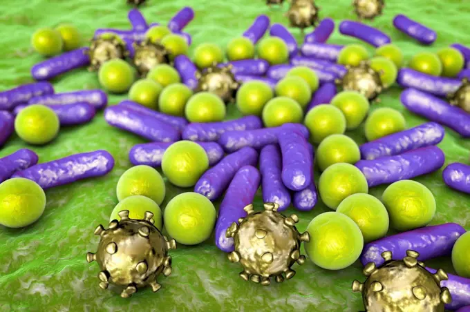
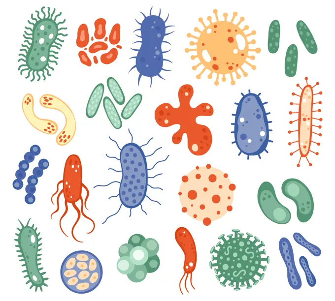
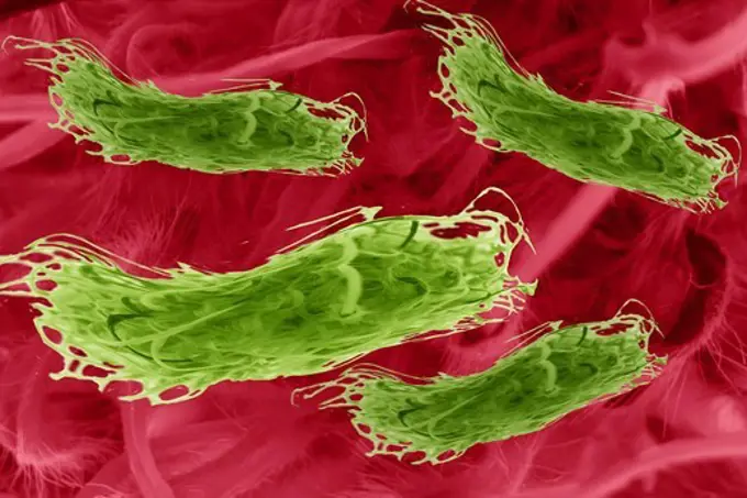
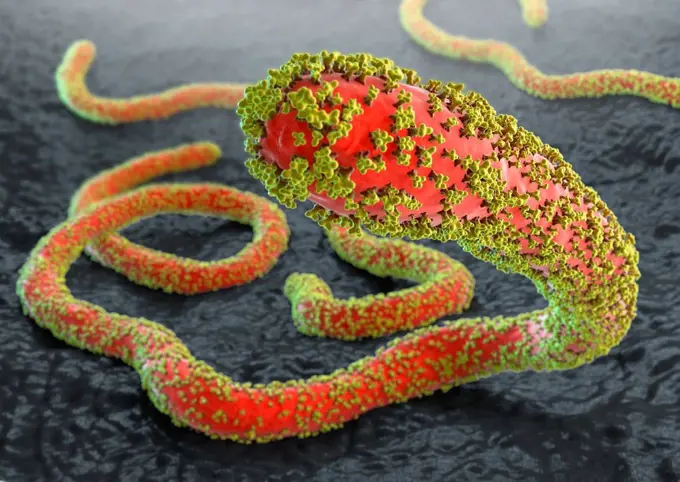
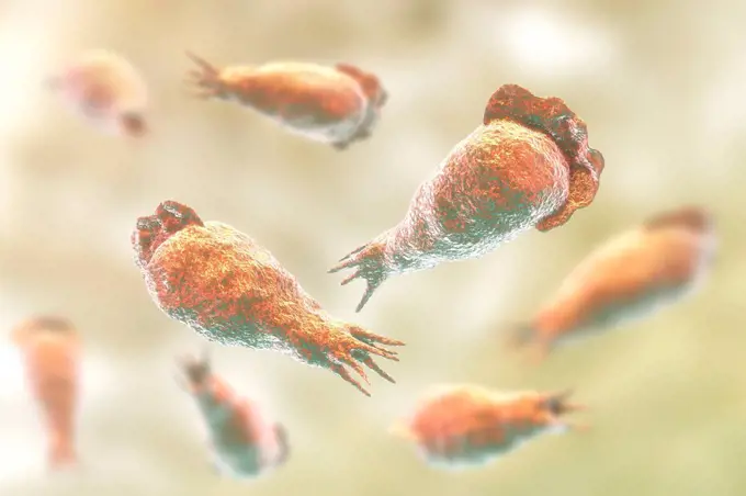
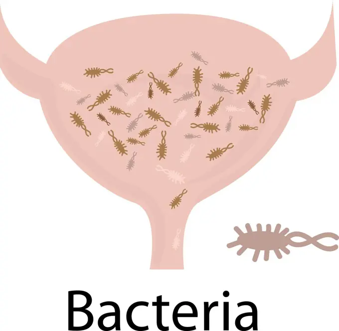
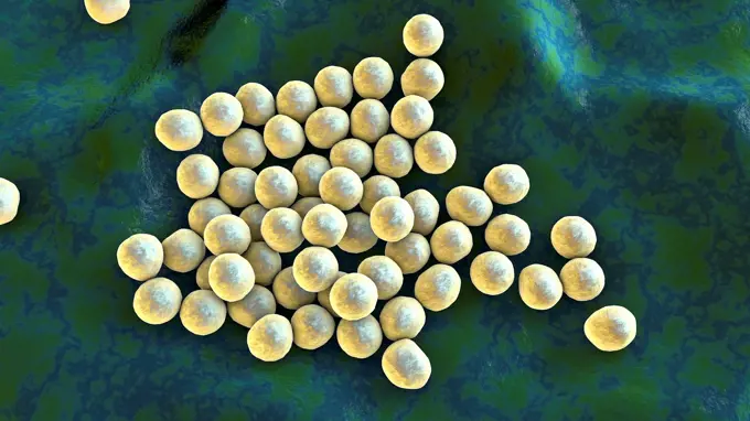
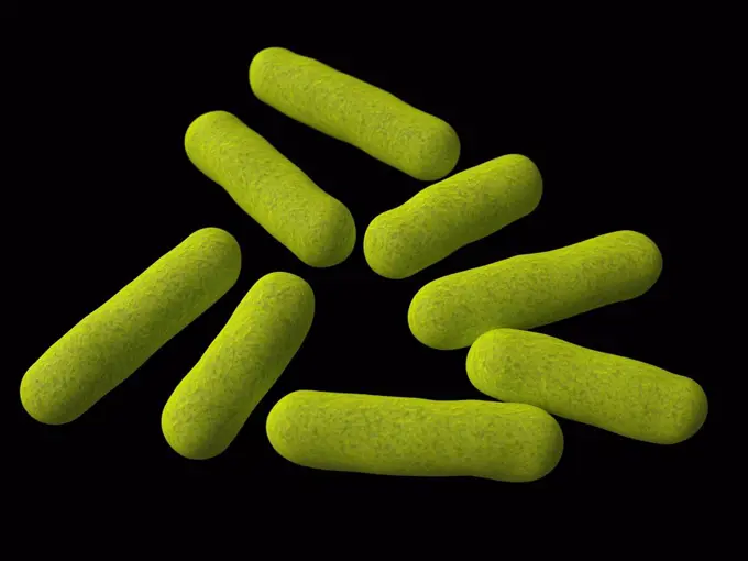
Fine ArtVintageFootageSubscriptionBoardsFree ResearchPricing
  • Fine Art
  • Vintage
  • Footage
  • Subscription
  • Boards
  • Free Research
  • Pricing

Bacterial Illustrations

Illustrations of various types of bacteria including Clostridium and Corynebacterium, depicted in a colorful, scientific style.

Clostridium bacteria, illustration
453 assets in this story
4128-18680804
Y chromosome, illustration
1525-20586608
Contaminated food concept and tainted meal poisoning symbol resulting in illness due to dangerous toxic bacteria parasites and viruse contaminants as salmonella and e coli as an open.
4128-20040218
Blood vessels, illustration
4128-20040408
Fragile X chromosome, illustration
4269-27390
Salmonella infantis. Scanning electron micrograph (SEM) of rod-shaped, motile, Gram-negative Salmonella infantis bacteria. Magnification 18875x. When ingested with spoiled food, the bacteria of the genus Salmonella are responsible for salmonellosis, a small intestine infection (gastroenteritis).
4378-20393190
Bacteria Cross Section
824-63194388
This digitally-colorized scanning electron micrograph (SEM) depicts a single filamentous Ebola virus particle that had been bended from the surface of a VERO cell of the African green monkey kidney epithelial cell line. Ebola hemorrhagic fever (Ebola HF) is one of numerous Viral Hemorrhagic Fevers. It is a severe, often fatal disease in humans and nonhuman primates (such as monkeys, gorillas, and chimpanzees). Ebola HF is caused by infection with a virus of the family Filoviridae, genus Ebolavirus. When infection occurs, symptoms usually begin abruptly. The first Ebolavirus species was discovered in 1976 in what is now the Democratic Republic of the Congo near the Ebola River.
4128R-11286220
Propionibacterium acnes bacteria, computer artwork.
4297-1456
Scanning electron micrograph revealed some of the morphologic details displayed by a single Gram-negative Escherichia coli bacterium
4128R-13587529
Various microbes of different shapes, computer illustration. Mixture of microorganisms including spherical (cocci) and rod-shaped bacteria together wi...
1525-24609233
Biology microorganisms. Biological virus, bacteria, disease microbes, infection germ and infectious agent. Microorganism cells vector symbols. Microscopic bacillus, pandemic influenza. Biology microorganisms. Biological virus, bacteria, disease microbes, infection germ and infectious agent. Microorganism cells vector symbols
4128R-13620082
Coloured scanning electron micrograph (SEM) of Streptococcus pneumoniae, Gram-positive, encapsulated, coccus prokaryote (dividing). Causes (lower resp...
1773R-101713
Illustration of colorful microbes
1558-15246874
Ebola viruses with glycoproteins on gray tissue background
4128-28767217
Mycobacterium ulcerans bacteria, illustration
4128-19052278
Chromosomes, illustration
4128R-14713
Chromosome, computer artwork.
4128R-13446995
Illustration of two chromosomes.
4128R-15466769
XY chromosomes, illustration
4128-V58572055
4128-18680872
Giardia lamblia, illustration
824-63123755
This scanning electron micrograph (SEM) depicts a highly magnified view of a dividing Escherichia coli bacteria, clearly displaying the point at which the bacterias cell wall was dividing; Magnification 21674x. Escherichia coli is a Gram-negative bacter
4378-5200
House Dust Mites
824-63223175
Colorized transmission electron micrograph of a Marburg virus particle (green) harvested from infected VERO E6 cell supernatant. Image captured and color-enhanced at the NIAID Integrated Research Facility in Fort Detrick, Maryland. Credit: NIAID.
4128R-14845727
Naegleria brain-eating amoeba, illustration
6188-67431740
3D Isometrische flache konzeptionelle Illustration der antimikrobiellen Resistenz, medizinisches Lehrdiagrammbr3D Isometrische flache konzeptionelle Illustration der antimikrobiellen Resistenz, medizinisches Lehrdiagramm 3D Isometric Flat Conceptual Illustration of Antimicrobal Resistance, Medical Educational Diagram 3D Isometric Flat Conceptual Illustration of Antimicrobal Resistance, Medical Educational Diagram Copyright: xZoonar.com/AnastasiiaxTorianykx 22948415
4128R-13023030
Helicobacter pylori bacterium, computer illustration. H. pylori is a gram-negative motile bacterium which is associated with formation of gastric and ...
5507-45869191
Urinary tract infection. Bladder full of bacteria infection
4128-28575389
MRSA bacteria, illustration
4128-15659881
Pseudomonas aeruginosa bacteria, illustration
4128R-15559
Computer artwork in wireframe style, depicting two bacteria.
1899-65662361
Colorized electron micrograph of Treponema pallidum, the bacteria that cause syphilis. Several spiral-shaped bacteria have been highlighted in teal.
4128R-11286144
Bacillus bacteria, computer artwork.
4128-111578631
Bacterium, computer illustration.
4128R-13725514
Illustration of human chromosomes.
4378-1246
Hookworms, which are parasites that live in the small intestine.
4128-V58561788
4128R-15290716
Ebola virus particles in blood, illustration
4128-15659889
Pseudomonas aeruginosa bacterium, illustration
1525-27153892
Covid-19 Coronavirus disease design with world map vector illustration
4128R-11479384
Ebola virus particles, computer artwork. This virus is the cause of ebola virus disease in humans and nonhuman primates. It is a severe and often fata...
824-57657620
Human chromosomes (23 pairs of chromosomes - 46 chromosomes, 22 pairs are common to both sexes. The remaining two chromosomes are the sex chromosomes, XY in males and XX in females). They are made up of DNA which carries the genes (about 20,000).
4128R-13818082
Leishmania sp. protozoa, computer illustration. This parasite causes the tropical disease leishmaniasis. This can take several forms, causing open sor...
4239R-8379
Microscopic view of hookworm. The hookworm is a parasitic nematode that lives in the small intestine of its host, which may be a mammal such as a dog, cat, or human.
4128-18680801
Crossing-over of chromatids, illustration
4128R-13818068
Leishmania sp. protozoa, computer illustration. This parasite causes the tropical disease leishmaniasis. This can take several forms, causing open sor...
4128R-11286243
Klebsiella bacteria, computer artwork.
4128R-15545710
E coli, SEM
4128R-13271897
Parasitic nematode worm (Ascaris lumbricoides), computer illustration. A. lumbricoides is a parasite of the human intestine. Female roundworms can lay...
4128R-15465547
Safety of drinking water, conceptual illustration
1525-28211012
Probiotics Bacteria. Biology, Science background Vector illustration. Probiotics Bacteria. Biology, Science background. Vector stock illustration.
4128R-12572046
Bacteria, computer illustration.
4128-28968601
Rhodotorula fungi and beta-carotene molecule, illustration
PREVIOUS
of 5
NEXT
2401 S. Ervay, Suite 206
Dallas, Texas 75215
United States
Browse
CategoriesGalleriesFree Research
For Creators
License Your ContentSuperStock For StudentsFrame of Mind BlogContributor PortalNew Image SpotlightLicense AI Imagery
Resources
About UsLicensingTerms & ConditionsAPI AccessPrivacyPricing
Contact
+1 866 236 0087help@superstock.com Contact form
©2026 SuperStock. All Rights Reserved. -A
This site is protected by reCAPTCHA and Google Privacy Policy and Terms of Service apply.