Fine ArtVintageFootageSubscriptionBoardsFree ResearchPricing
  • Fine Art
  • Vintage
  • Footage
  • Subscription
  • Boards
  • Free Research
  • Pricing

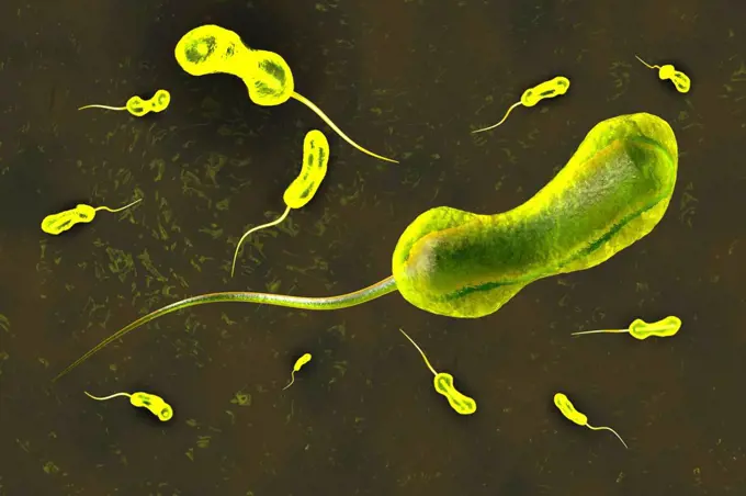
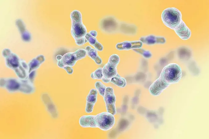
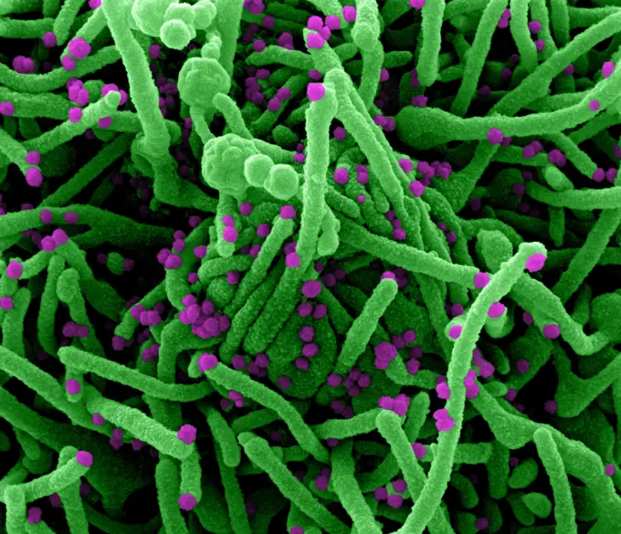
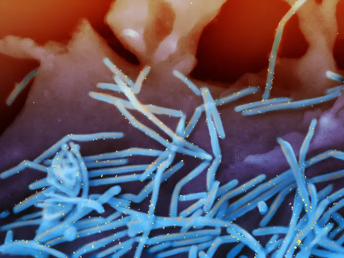
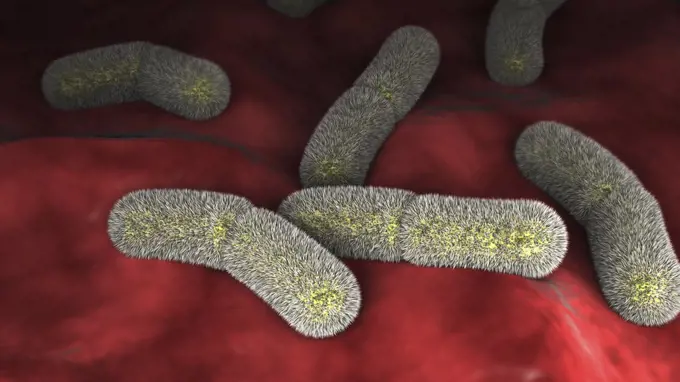
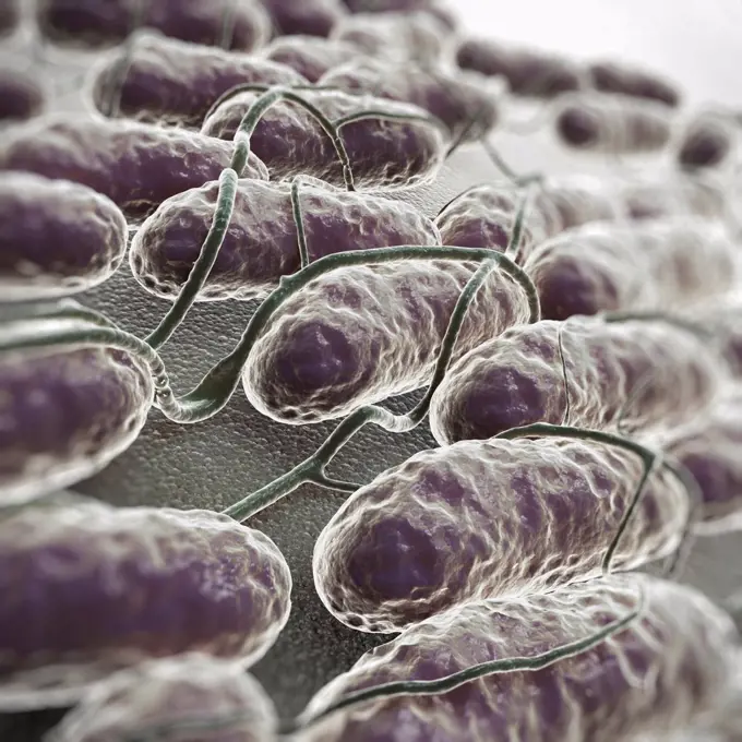
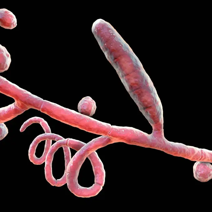
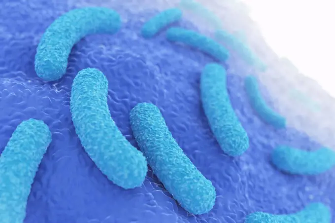
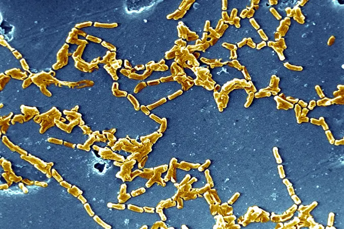
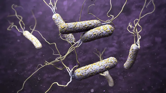
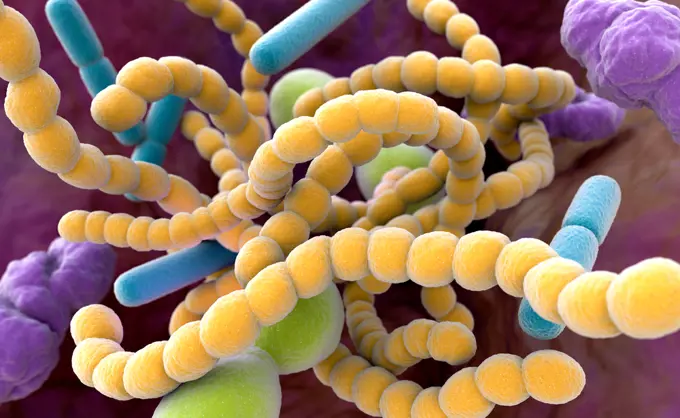
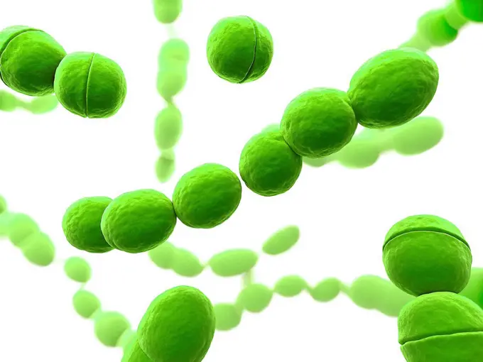
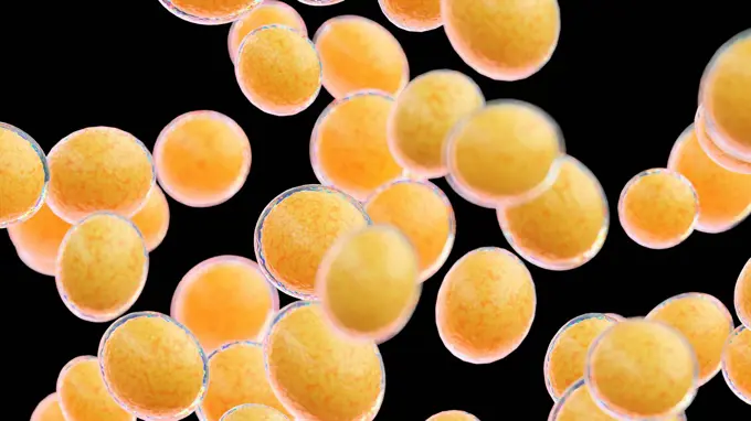
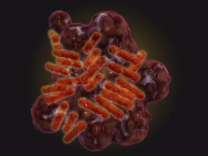
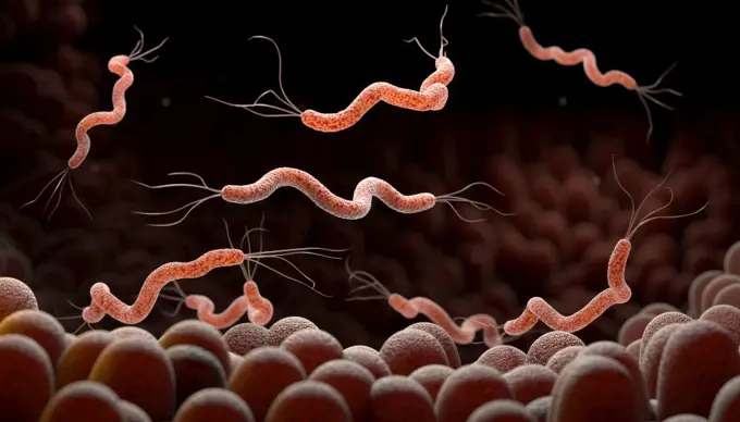
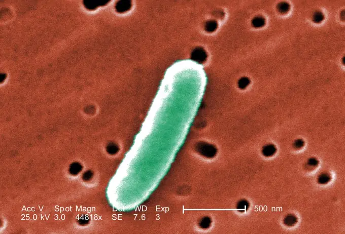
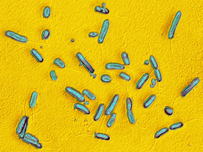
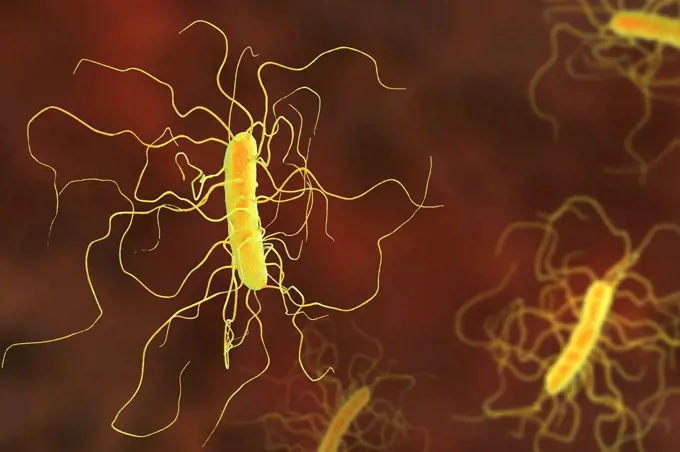
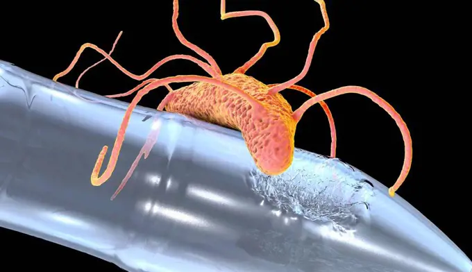
Bacterial Illustrations

Illustrations of various types of bacteria including Clostridium and Corynebacterium, depicted in a colorful, scientific style.

Clostridium bacteria, illustration
453 assets in this story
4128R-14715
Chromosomes, computer artwork.
5514-17938637
Close-up of 3d rendering microscopic blue bacteria.
824-63223244
Colorized scanning electron micrograph of a cell (green) infected with SARS-CoV-2 virus particles (purple), isolated from a patient sample. Image captured at the NIAID Integrated Research Facility (IRF) in Fort Detrick, Maryland. Credit: NIAID.
824-65830892
Scanning electron micrograph of human respiratory syncytial virus (RSV) virions (colorized blue) that are labeled with anti-RSV F protein/gold antibodies (colorized yellow) shedding from the surface of human lung epithelial A549 cells. RSV is a common contagious virus that infects the human respiratory tract.
4128-V58574042
4239-18641893
Microscopic view of Yersinia pestison bacteria on surface.
4378-4505
Salmonella Bacteria
4128R-11286165
Clostridium bacteria, computer artwork.
4128-V58569579
4128-20041261
Athlete's foot fungus, illustration
4128R-14058248
Marburg virus particles, illustration
4128R-13242370
Pseudomonas aeruginosa bacterium, computer illustration. P. aeruginosa is a Gram-negative bacterium which causes multiple antibiotic resistant nosocom...
824-63191190
This illustration depicts a three-dimensional (3D) computer-generated image of a group of extended-spectrum ß-lactamase-producing Enterobacteriaceae (ESBLs) bacteria. The artistic recreation was based upon scanning electron micrographic imagery. This is an excellent visual example of the long, whip-like peritrichous flagellae sprouting from what appear to be random points on the organisms exterior, as well as the numerous shorter, and finer fimbriae, imparting a furry look to the bacteria. Extended-spectrum ß-lactamase is an enzyme that allows bacteria to become resistant to a wide variety of penicillins and cephalosporins. Bacteria that contain this enzyme are known as ESBLs or ESBL-producing bacteria. ESBL-producing Enterobacteriaceae are resistant to strong antibiotics including extended spectrum cephalosporins. An estimated 140,000 healthcare-associated Enterobacteriaceae infections occur in the United States each year. CDC estimates that bloodstream infections caused by ESBL-conta
4128R-14237
Computer artwork depicting a cluster of bacteria.
4128R-13244741
E. coli bacteria, illustration.
4128R-13587522
Various microbes of different shapes, computer illustration. Mixture of microorganisms including spherical (cocci) and rod-shaped bacteria together wi...
4128-V58567294
4128-38524186
Various bacteria, illustration
824-63194465
This colorized negative stained transmission electron micrograph (TEM) showed recreated 1918 influenza virions that were collected from the supernatant of a 1918-infected Madin-Darby Canine Kidney (MDCK) cell culture 18 hours after infection. In order to sequester these virions, the MDCK cells were spun down (centrifugation), and the 1918 virus present in the fluid was immediately fixed for negative staining.
4128R-14742
Streptococcus bacteria, computer artwork.
4128-18497178
Antibiotic resistance mechanisms of bacteria, illustration
1815-18410343
Three dimensional render of green bacteria on human tissue
4269-27412
Marburg virus. Transmission electron micrograph (TEM) of filamentous Marburg virus, magnified 100,000x. This RNA virus of the Filovirus (like Ebola) is known to cause Marburg hemorrhagic fever, which can be fatal.
824-63190261
Salmonella is the bacterium responsible for salmonellosis, a common food-borne infection. Image taken with an optical microscope X 1000.
6019-19982633
Microscopic view of bacteria. 3d illustration
4128R-13587568
Plague bacteria (Yersinia pestis), computer illustration. Y. pestis are gram-negative bacteria from Enterobacteriaceae family, they have ovoid shape w...
4128R-13818038
Clostridium difficile bacterium with peritrichous flagella, computer illustration. C. difficile is Gram-positive, anaerobic, spore forming, rod-shaped...
4128-20244197
Microsporum audouinii fungus, illustration
4128R-15659
Streptococcus bacteria, computer artwork.
1848-53914861
Bacteria, illustration
4378-2711
A number of human chromosomes.
4239-18641839
3D illustration of a cluster of drug resistant, curlicue-shaped, Campylobacter sp. bacteria. This artistic recreation is based upon scanning electron microscopic (SEM) imagery.
6019-19982675
Biofilm of antibiotic resistant bacteria, closeup view.
4128-30420573
Marburg virus, illustration
4128R-13192969
Vaccinia virus particles and bacteria. Coloured scanning electron micrograph (SEM) of vaccinia virus particles (green). Unlike most viruses, vaccinia ...
824-63194689
Under a high magnification of 15549x, this colorized scanning electron micrograph (SEM) depicted some of the ultrastructural details seen in the cell wall configuration of a number of Gram-positive Mycobacterium tuberculosis bacteria. As an obligate aerobic organism M. tuberculosis can only survive in an environment containing oxygen. This bacterium ranges in length between 2 - 4 microns, and a width between 0.2 - 0.5 microns.
4297-1761
Colorized scanning electron micrograph of E.coli O157:H7 at a magnification of 13671x.Escherichia coli bacteria of the strain O157:H7.E.coli O157:H7 is one of hundreds of strains of this bacterium
4128R-13587574
Plague bacteria (Yersinia pestis), computer illustration. Y. pestis are gram-negative bacteria from Enterobacteriaceae family, they have ovoid shape w...
4128-30420566
Marburg viruses, illustration
4128R-13587288
Campylobacter bacteria, illustration.
4378-1122
Scanning electromicrograph styled rod shaped bacteria.
6145-29604697
Collection Dutch Heidematschappij. Damage, plants, mushrooms, split mig. undated
824-63123775
Scanning Electron Micrograph of Mycobacterium chelonae.
4128-V58569651
6188-66486290
3d-Illustration von Cholera-Erregern in dunkel verschmutztem Wasser 3d illustration of cholera pathogens in dark polluted water Copyright: xZoonar.com/ChristophxBurgstedtx 22602851
1525-22561282
Halitosis Bacteria. Close-up of bacteria found in the mouth which can cause halitosis or bad breath. 3D illustration
4128-V58567473
1899-61461226
Salmonella is the bacteria responsible for salmonellosis, a very widespread foodborne illness. Salmonellosis is usually characterized by an acute outbreak of fever, abdominal pain, diarrhea, nausea, severe dehydration, septicemia. Image produced from optical microscopy
1525-22059339
Bacteria virus and germs microorganism cells green inversion seamless pattern vector illustration
4128-V58569121
4378-1048
Microscopic styled visualization of human chromosomes.
4378-4437
Borrelia burgdorferi
4128R-11479426
Ebola virus particles, computer artwork. This virus is the cause of ebola virus disease in humans and nonhuman primates. It is a severe and often fata...
4239-18641825
Under a magnification of 100X, this scanning electron microscopic (SEM) image, reveals some of the ultrastructural morphology exhibited by a grouping of ham mites, Tyrophagus putrescentiae. The ham mite is an agricultural pest, at times, infesting high moisture grain, semi-moist pet foods, cheeses, and dried meats. It is also referred to as the cheese mite, mold mite or copra mite.
4128-111578637
Bacterium, computer illustration.
824-63190221
Cholera bacilli or cholera vibrio (Vibrio cholerae). Cholera is a contagious epidemic enteric poisoning, it is characterized by sudden and very abundant diarrhea. In the absence of treatment, the classic major form is fatal in more than half of the cases. Optical microscopy X 1000.
4128-15659890
Pseudomonas aeruginosa bacterium, illustration
4128-111580085
Legionnaires' disease bacteria. Computer illustration of Legionella pneumophila bacteria, the cause of Legionnaires' disease. These bacilli (rod-shape...
4128R-11310333
Listeria sp. bacteria, computer artwork.
4128R-13192980
Bacteria. Illustration of a colony of bacilli, rod-shaped bacteria. Bacteria are prokaryotic microorganisms with a diversity of shapes ranging from sp...
4378-1012
Scanning electromicrograph styled rod shaped bacteria.
4128-19247845
Bilophila wadsworthia bacteria, computer illustration. Anaerobic bile resistant bacterium found in lower parts of the human gastrointestinal tract, an...
824-63179053
Pneumobacillus Friedlnder (Friedlnder's bacillus) Klebsiella pneumoniae. This bacterium is the cause of certain infections of the respiratory tract, the lungs and more specifically the bronchi. It is also the cause of angina, bile duct infections and surgical wounds. Optical microscopy X 400
824-63190214
Clostridium botulinum bacterium (botulism bacillus). This bacterium secretes a very powerful toxin that inhibits the motor neurons of muscle contraction. This toxin (botox), in localized injection, reduces wrinkles, by paralysis of the muscles responsible for the wrinkle. Optical microscopy X 1000.
4128R-13376697
Leprosy bacteria, illustration
4297-1462
Under a very high magnification of 15000X, this scanning electron micrograph (SEM) revealed the presence of a single gram-negative Salmonella typhimurium bacterium, which was imaged right at the point where it was undergoing the process of cell division
4269-20408848
Mycobacterium tuberculosis bacteria, These Gram-positive rod-shaped bacteria cause the disease tuberculosis, colorized scanning electron micrograph (SEM).
1899-65662481
Scanning electron micrograph of Mycobacterium tuberculosis particles (colorized yellow), the bacterium which causes TB.
4128R-13818042
Clostridium difficile bacterium with peritrichous flagella, computer illustration. C. difficile is Gram-positive, anaerobic, spore forming, rod-shaped...
4128R-13587355
Salmonella, illustration.
4128R-22292
Bacteria, computer artwork.
824-65830612
Colorized scanning electron micrograph of Marburg virus particles (yellow) both budding and attached to the surface of an infected VERO E6 cell (burgundy).
4378-2597
A single human chromosome.
1899-65662480
Scanning electron micrograph of Mycobacterium tuberculosis particles (colorized orange), the bacterium which causes TB.
4128R-15660
Streptococcus bacteria, computer artwork.
4128-30415772
Human fat cells, illustration
4239-18641499
Microscopic view of Rickettsia. Rickettsia is a gram-negative bacteria carried by parasites such as ticks, fleas and lice.
1525-56278033
Spirillum bacteria is the cause of stomach ulcers or bacterial diarrhea , Germ infection , 3d illustration. Spirillum bacteria in the stomach
4138-20003918
Euphyllia ancora, Anchor coral or Crescent-tentacled coral. Showing fluorescent colors when photographed under special blue light and filter. Photographed in aquarium. Portugal
4378-942
Conceptual depiction of a human chromosome.
4128R-11542514
Bacteria, computer illustration.
4128R-2945
Chromosomes and DNA
824-63206954
Clostridium difficile (bacterium responsible for nosocomial diarrhea), visualized by optical microscopy.
4128-28968567
Prevotella bacteria, illustration
824-199
Pneumococcus bacteria (Pneumonia Cells)
6188-65540602
3D-Illustration von transparenten und bunten Protozoen oder einzelligen Organismus 3D illustration of transparent and colorful protozoa or unicellular organism Copyright: xZoonar.com/ChristophxBurgstedtx 22579508
1525-23804092
Vector abstract sphere blue cocci, spirilla bacteria cells concept. Abstract bacteria cells
4048-6173
E. coli, a common cause of bacterial diarrhea. Colorized scanning electron micrograph (SEM). Photo by Janice Haney Carr, 2007.
824-63179058
Pseudomonas aeruginosa or pyocyanin bacillus, pathogenic bacterium, very resistant and often responsible for nosocomial infections. It is one of the most difficult bacteria to eradicate clinically. The pathologies it causes are diverse: eye infections, infections of burns, surgical wounds, urine, lungs, gastrointestinal infections, inoculation meningitis, septicemia. Visualization by optical microscopy.
4128R-13818026
Clostridium difficile bacterium with peritrichous flagella, computer illustration. C. difficile is Gram-positive, anaerobic, spore forming, rod-shaped...
4128-15980131
Computer illustration of Ideonella sakaiensis bacterium degrading a plastic bottle. I. sakaiensis was discovered in 2016 in sediment near a plastic bo...
4128R-15290928
Bacteriophages leaving bacterial cells, illustration
4239-18641502
African trypanosomiasis in the red blood cells. African trypanosomiasis, also known as sleeping sickness, is a parasitic infection caused by protozoa.
6188-65540309
Grün gefärbtes mehrfach antibiotikaresistentes Bakterium Pseudomonas aeruginosa - 3d-Illustration Green colored multiple antibiotic resistant Pseudomonas aeruginosa bacterium - 3d illustration Copyright: xZoonar.com/ChristophxBurgstedtx 22579303
1525-27825332
Digestion bacteria and intestine or gut flora as intestinal bacterium medical anatomy concept as a 3D illustration.
4128-111493709
Ebola virus structure, illustration. Ebola virus particles, computer artwork. This thread-like RNA (ribonucleic acid) virus is the cause of ebola haem...
4128-20244205
Microsporum audouinii fungus, illustration
1899-53511630
Microsporum canis is a species of fungus of the genus Microsporum responsible for the majority of cases of ringworm in dogs and cats, and in humans. X 1000 optical microscopy.
6188-65540498
Einzelnes orangefarbenes mehrfach antibiotikaresistentes Bakterium Pseudomonas aeruginosa - 3d-Illustration Single orange colored multiple antibiotic resistant Pseudomonas aeruginosa bacterium - 3d illustration Copyright: xZoonar.com/ChristophxBurgstedtx 22579422
1815R-14619542
3D rendered Illustration of a anatomically correct convergence to a vibrio cholerae bacterium causing the famous cholera disease
PREVIOUS
of 5
NEXT
2401 S. Ervay, Suite 206
Dallas, Texas 75215
United States
Browse
CategoriesFree ResearchGalleries
For Creators
License Your ContentSuperStock For StudentsFrame of Mind BlogContributor PortalNew Image SpotlightLicense AI Imagery
Resources
About UsLicensingTerms & ConditionsAPI AccessPrivacyPricing
Contact
+1 866 236 0087help@superstock.com Contact form
©2026 SuperStock. All Rights Reserved. -B
This site is protected by reCAPTCHA and Google Privacy Policy and Terms of Service apply.