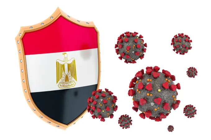
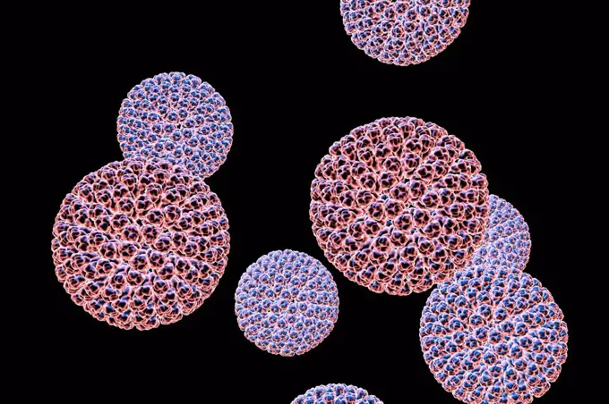
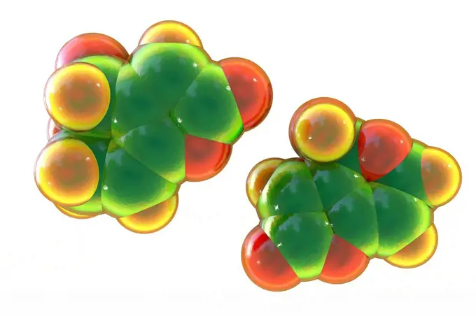
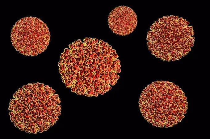
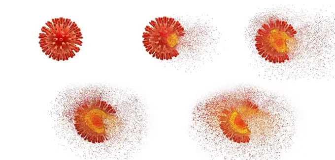
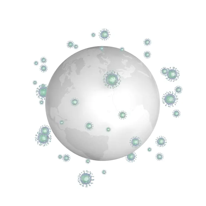
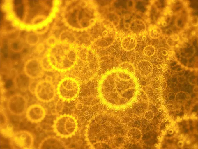
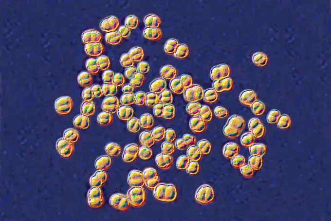
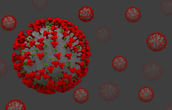
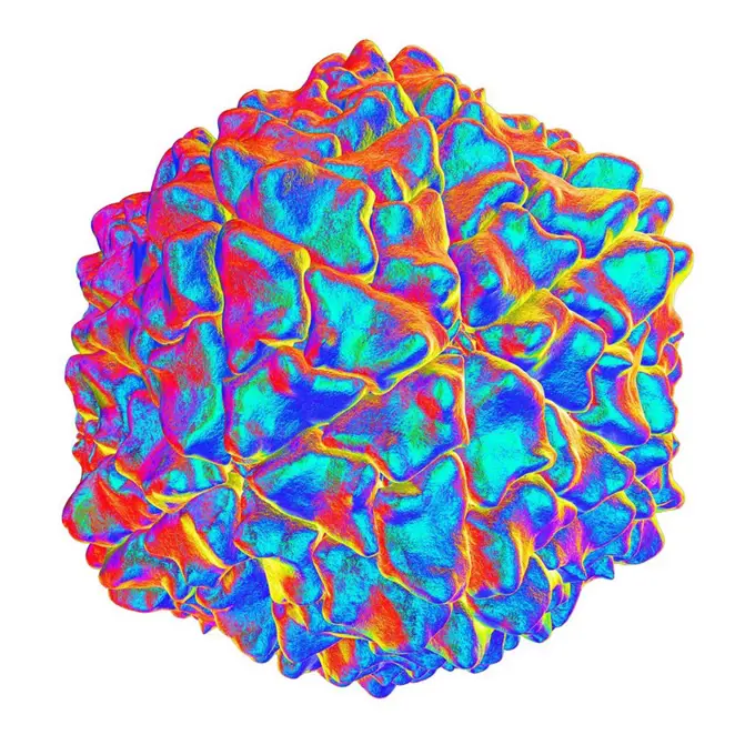
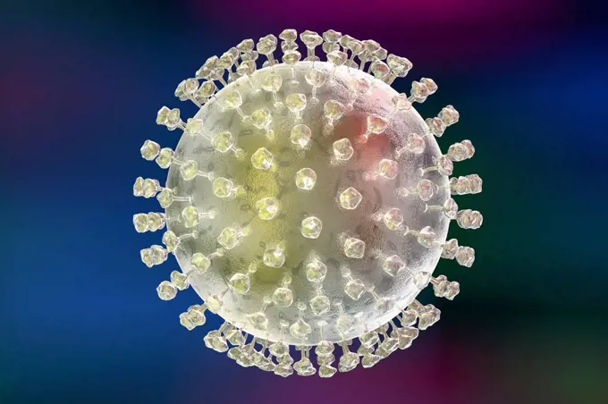
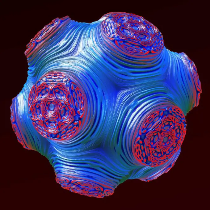
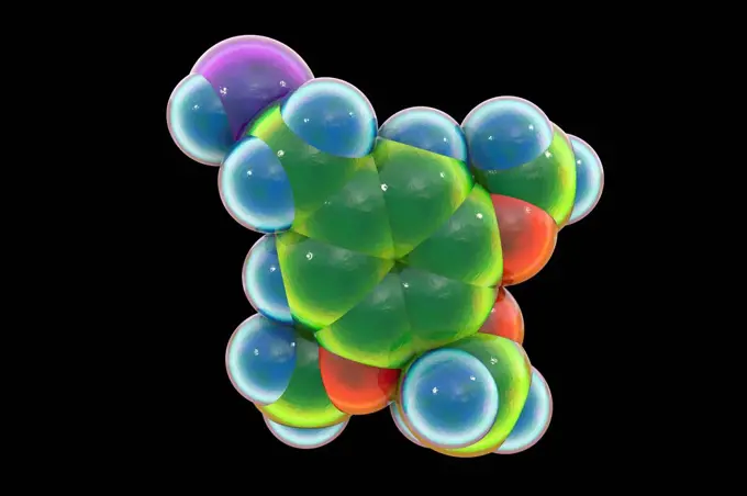
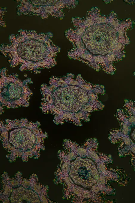
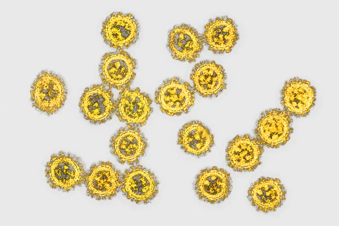
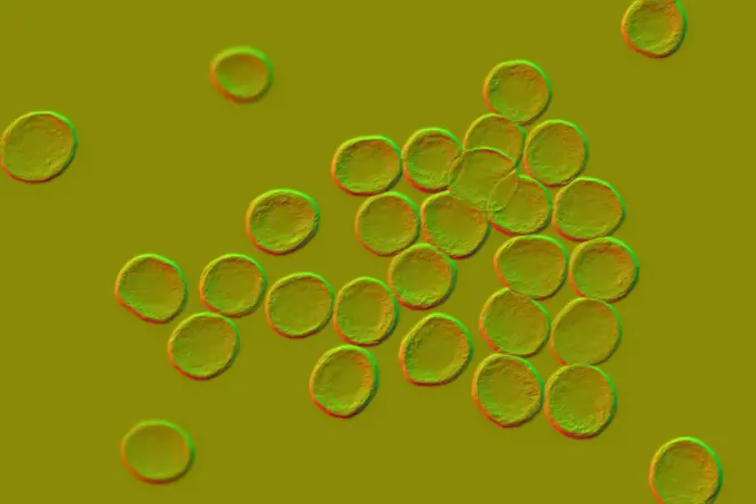
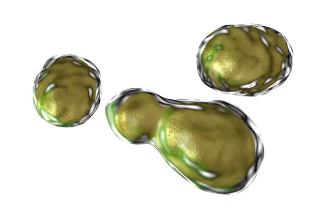
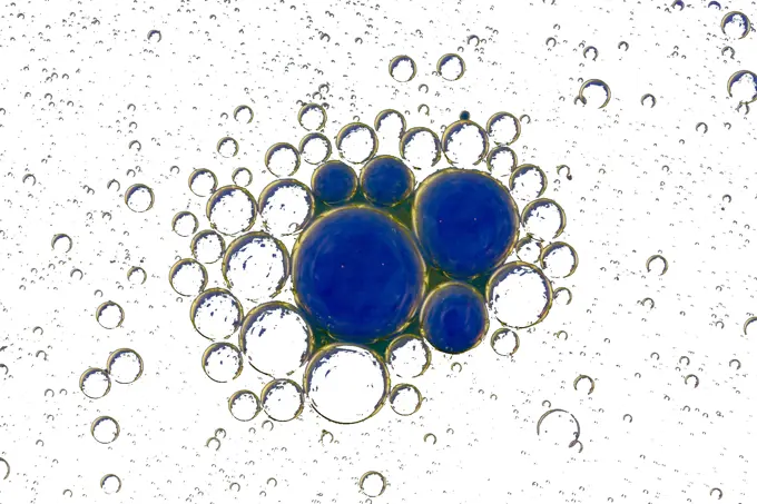
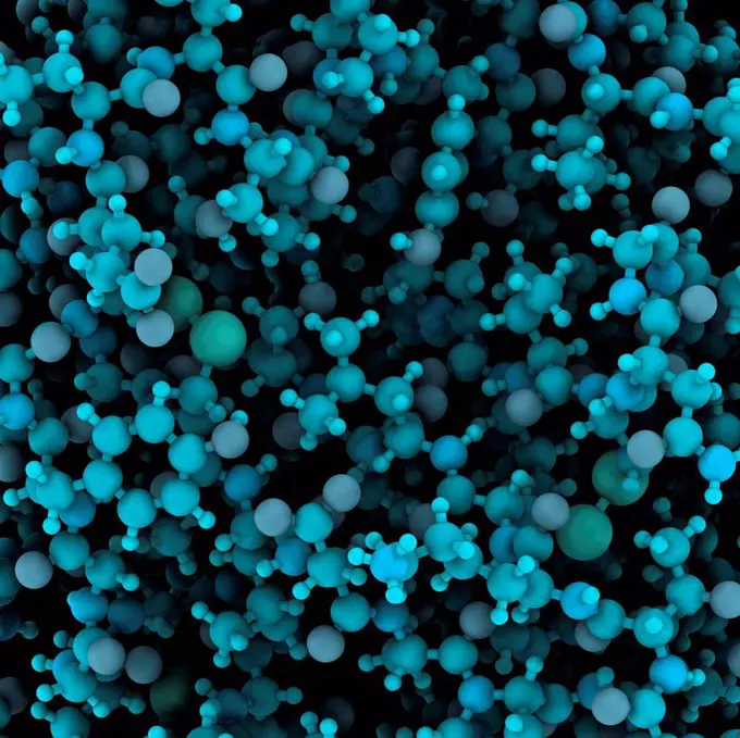
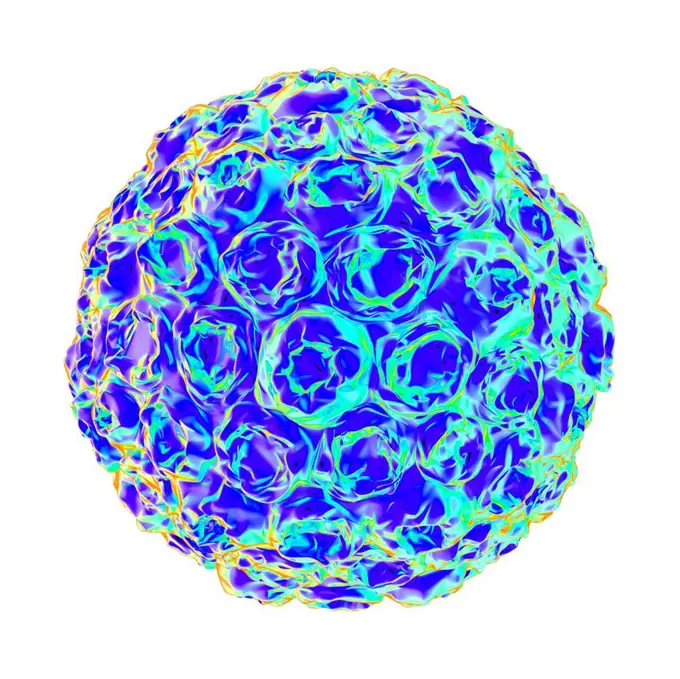
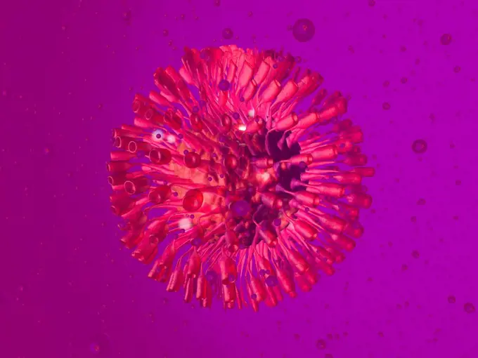
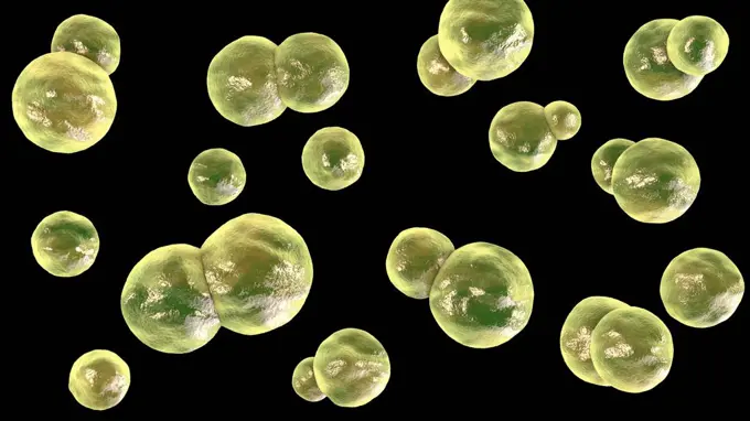
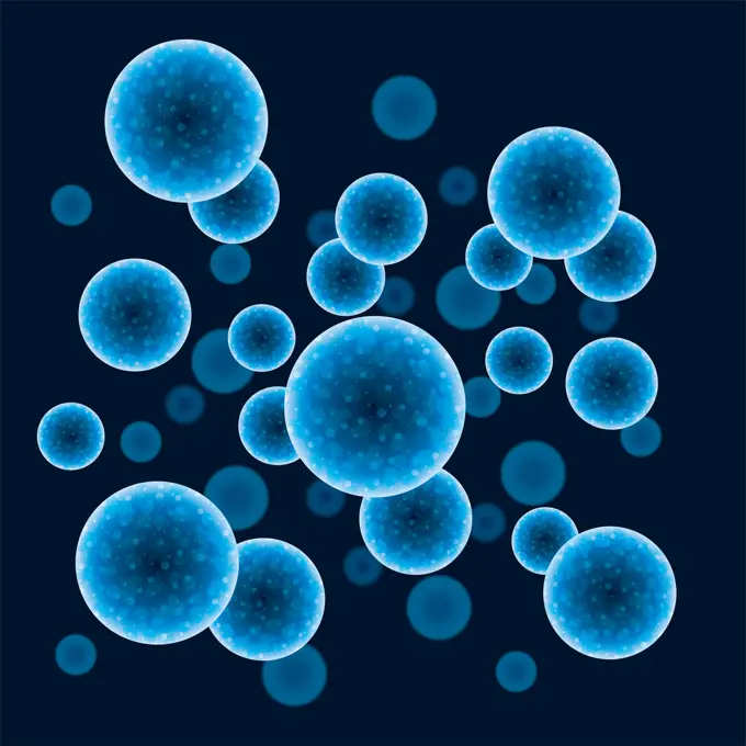
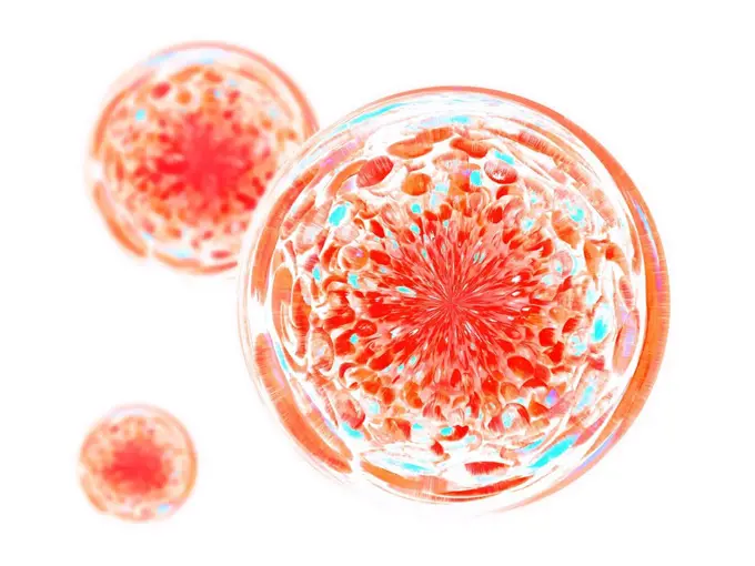
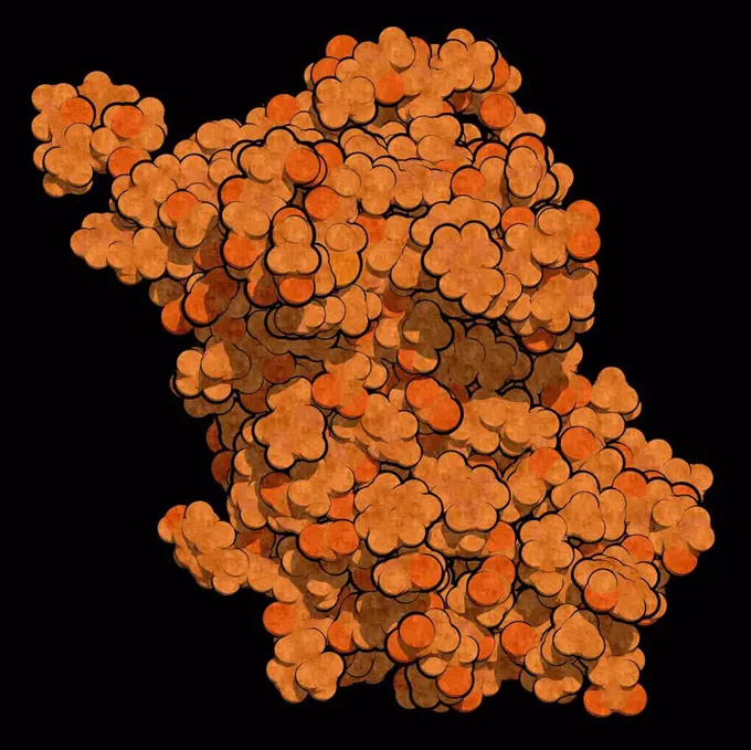
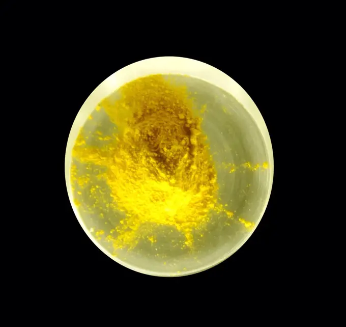
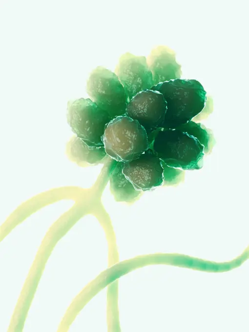
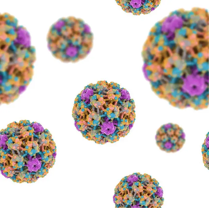
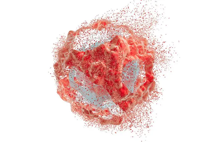
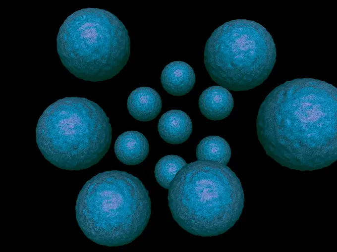
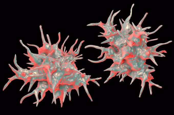
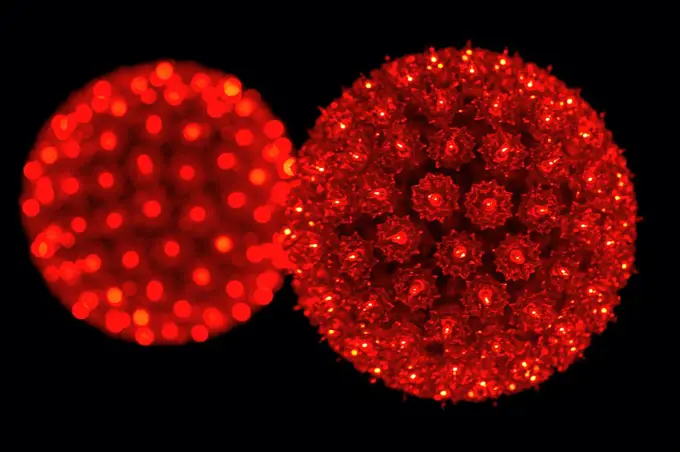
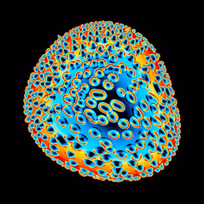
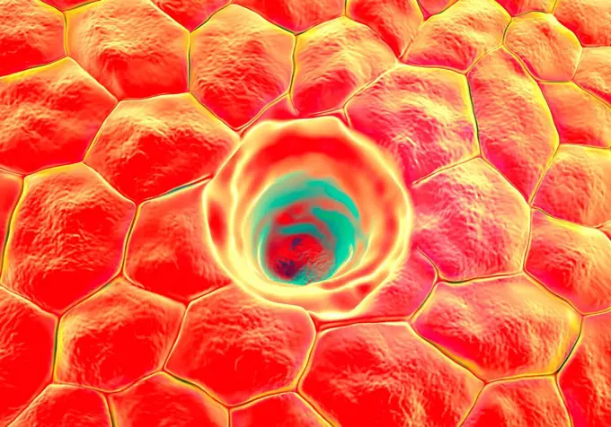
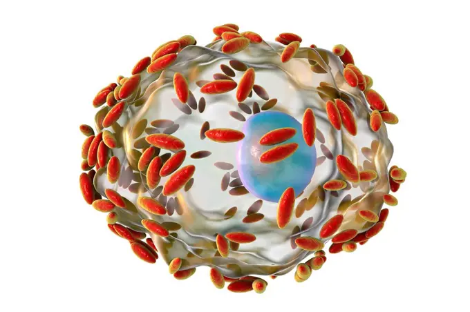
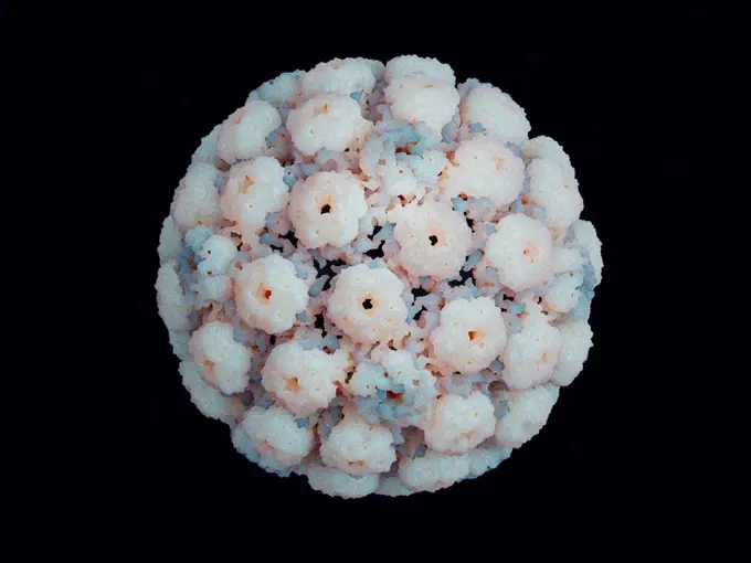
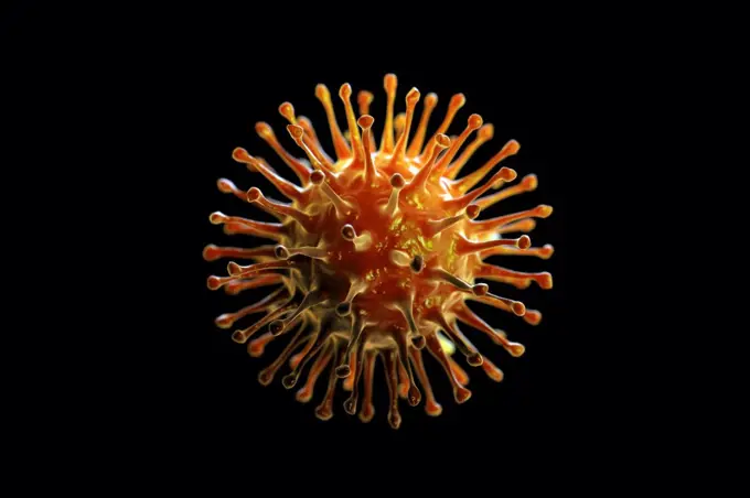
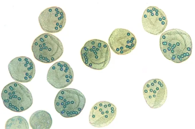
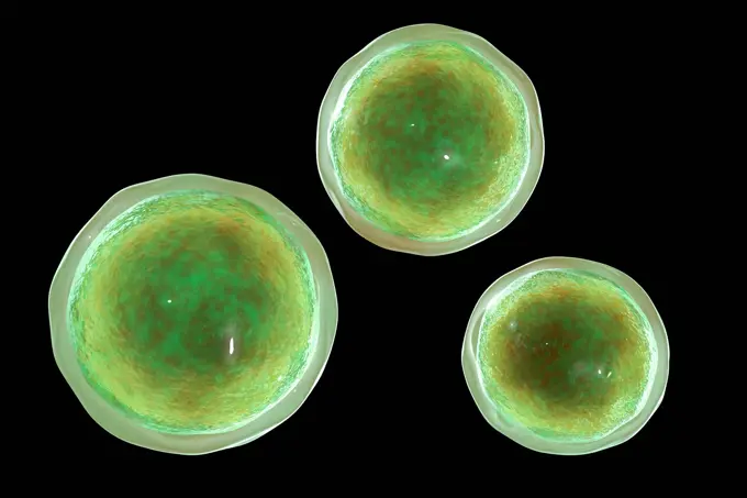
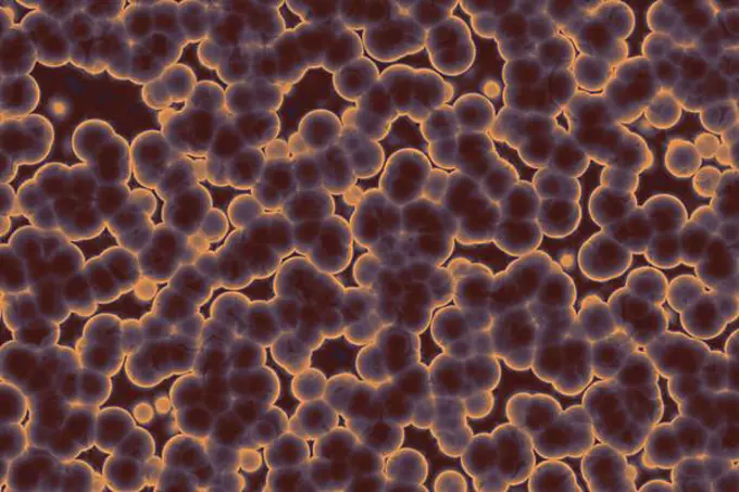
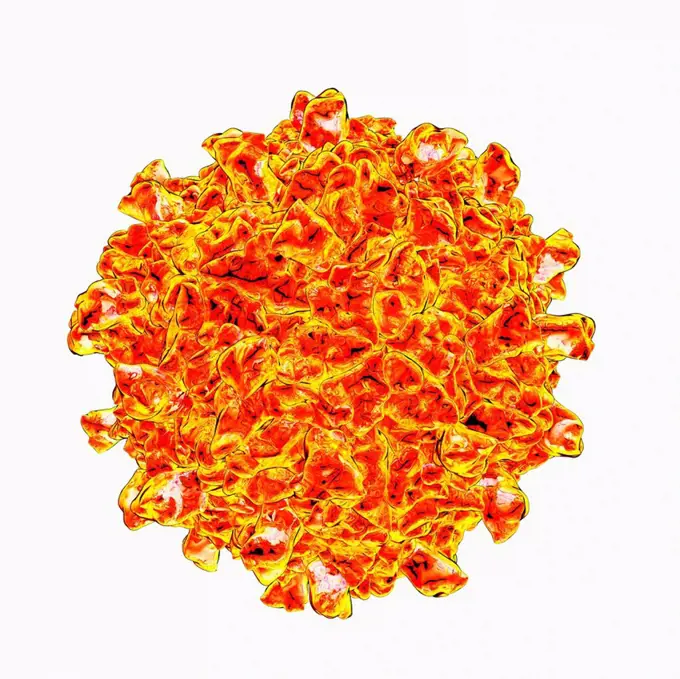
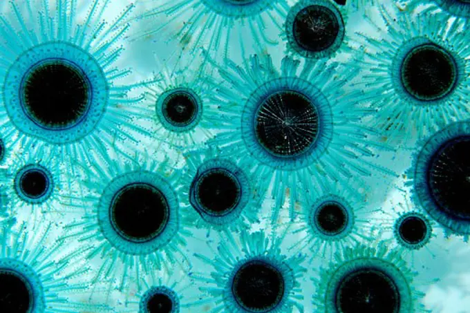
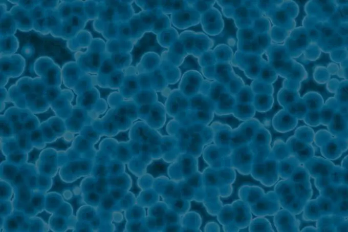
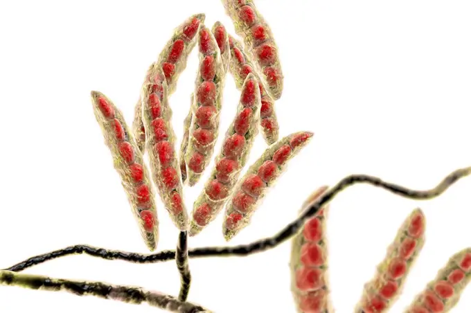
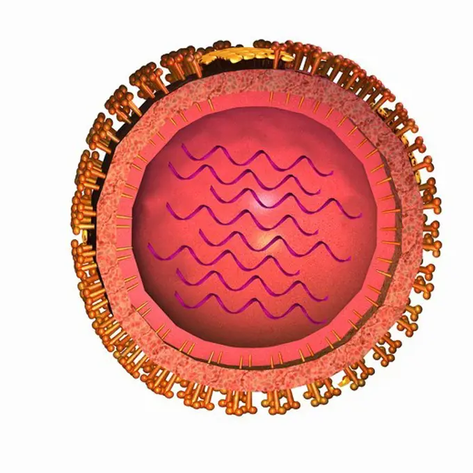
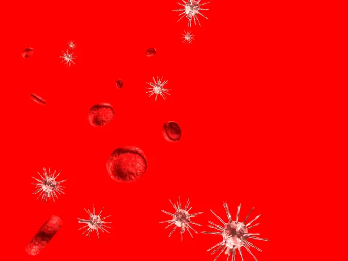
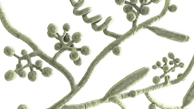
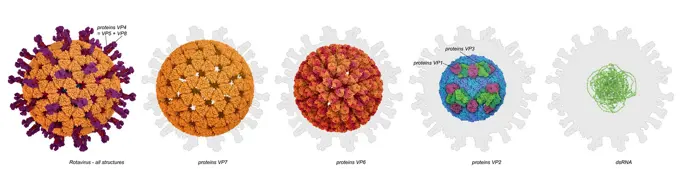
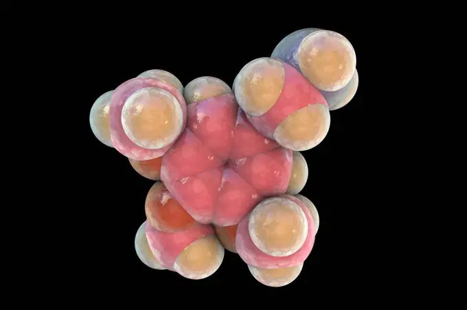
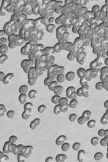
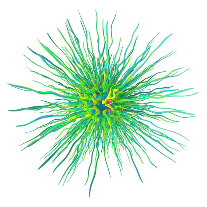
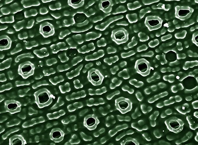
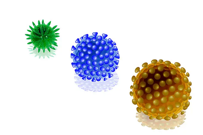
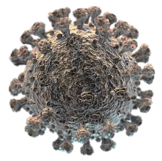
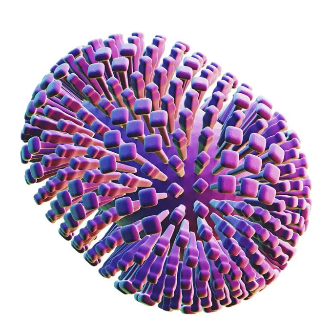
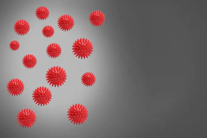
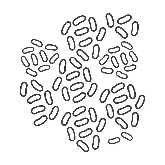
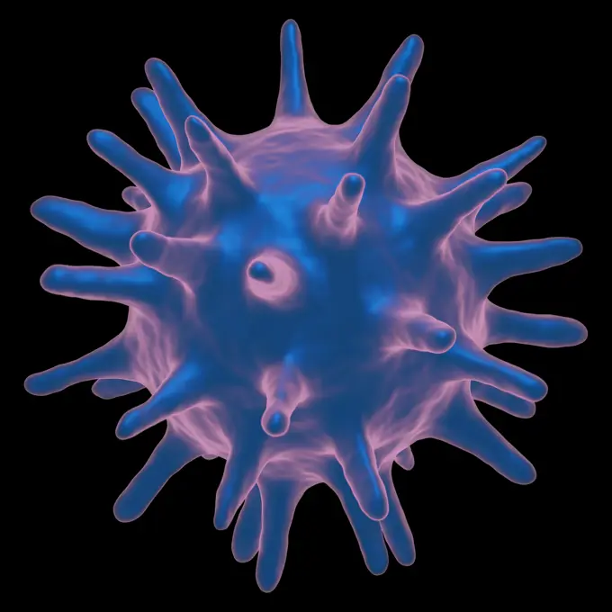
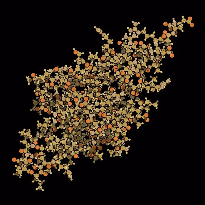
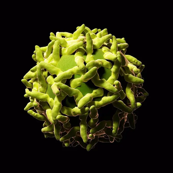
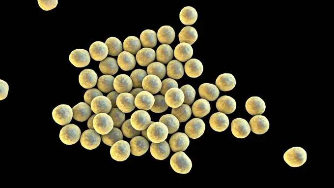
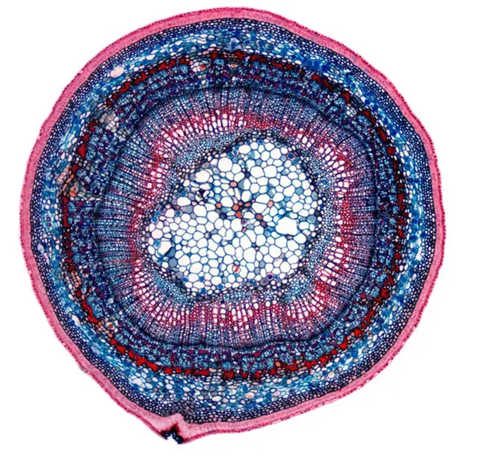
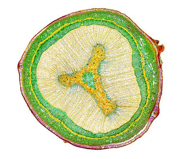
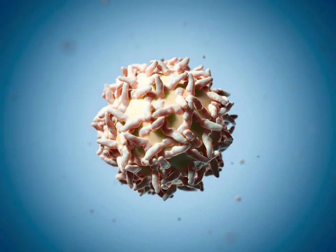
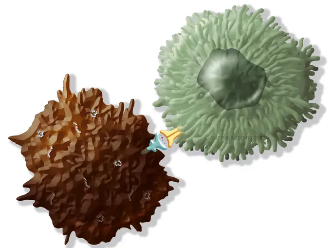
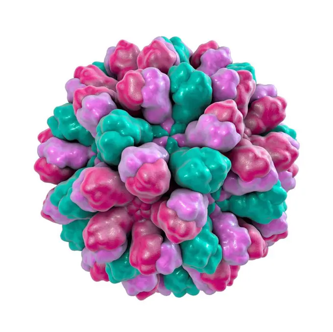
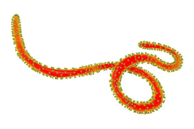
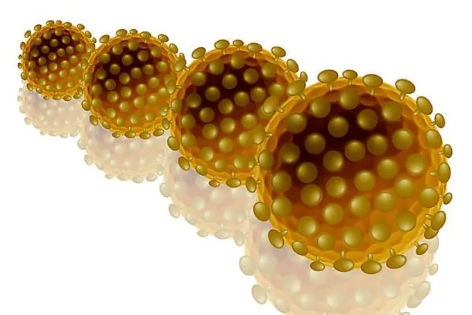
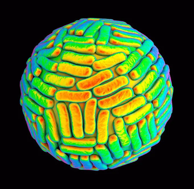
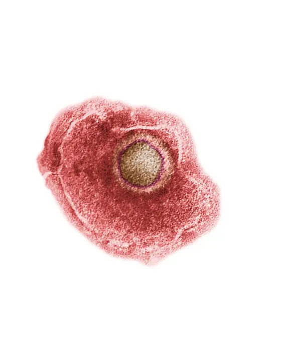
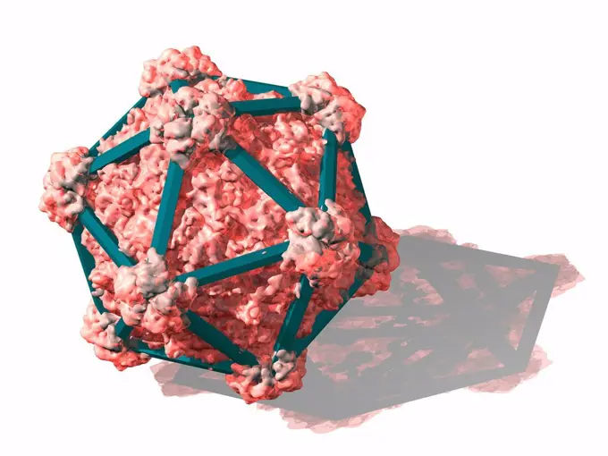
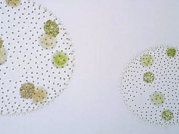
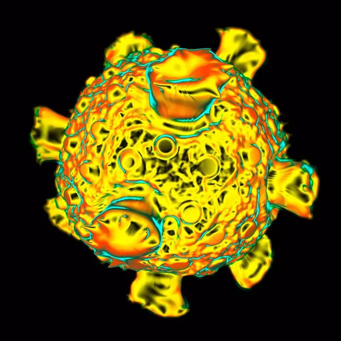
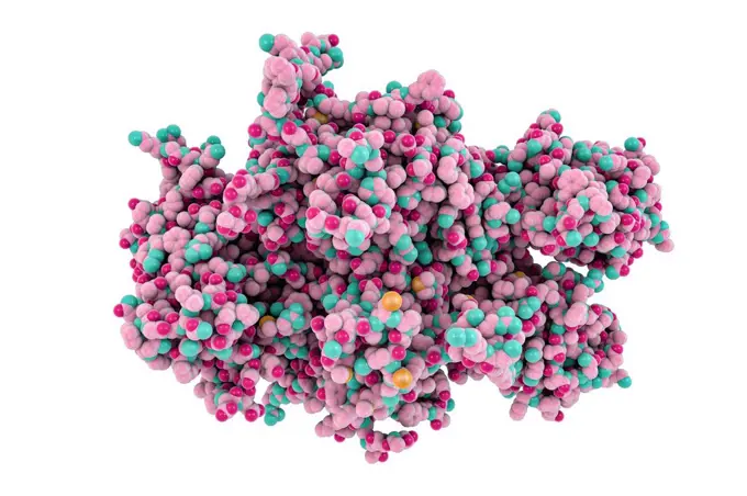
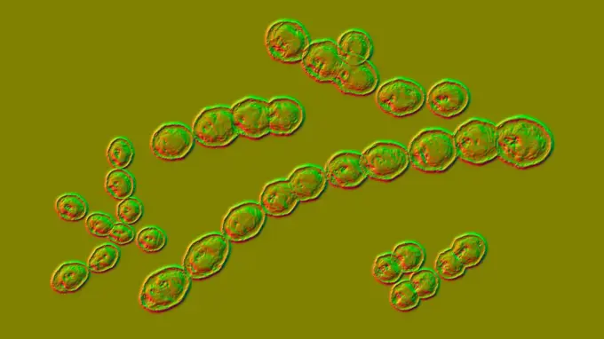
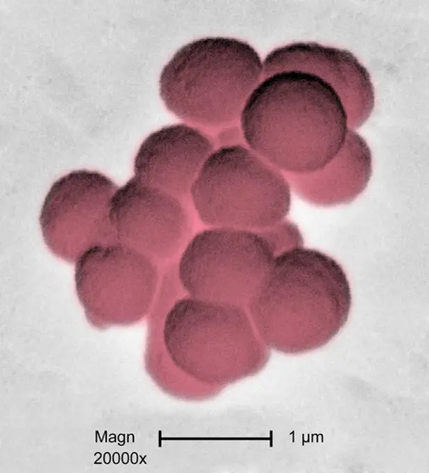
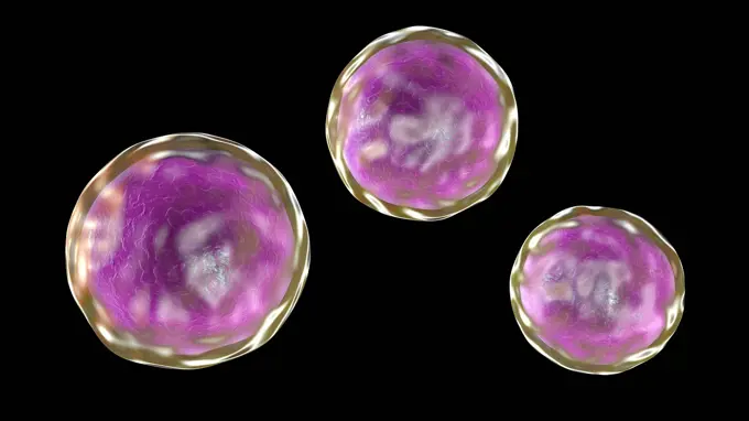
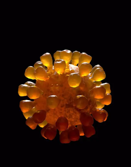
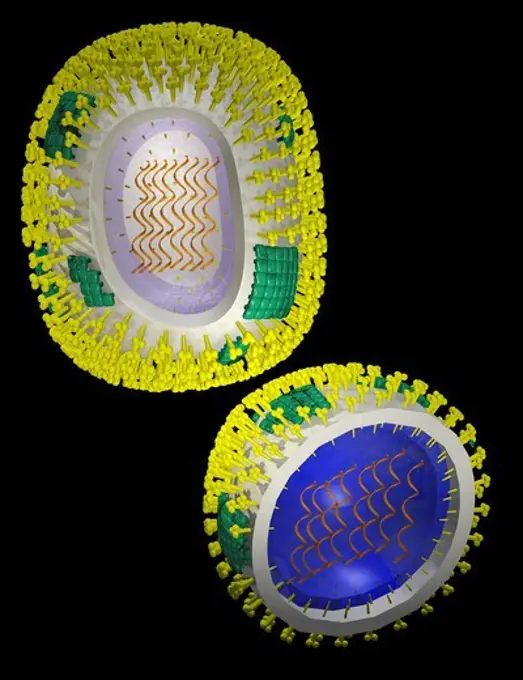
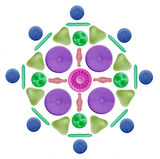
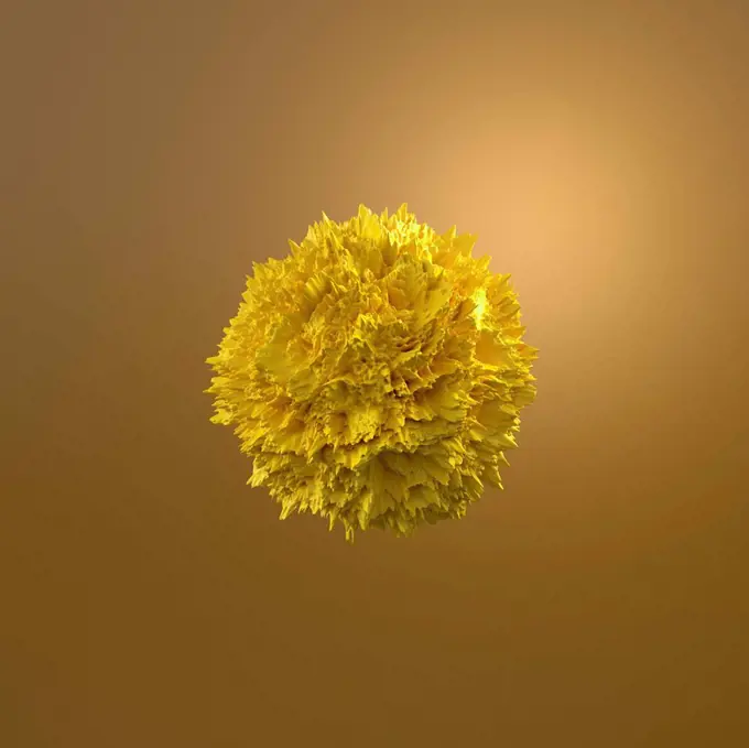
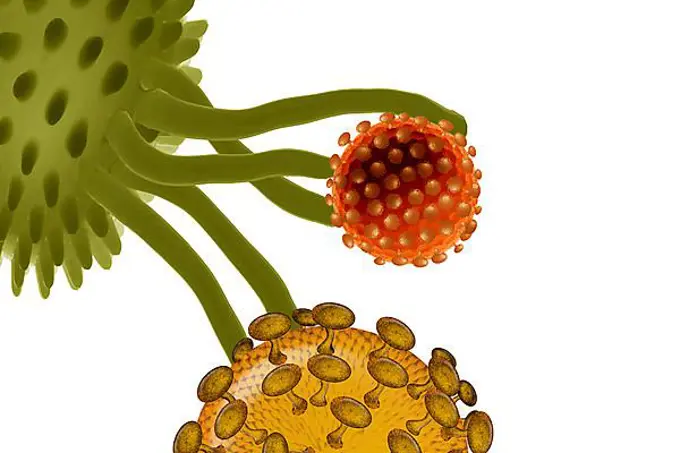
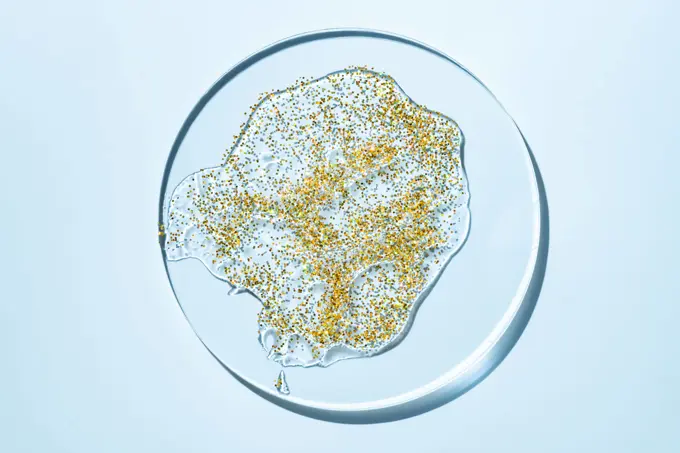
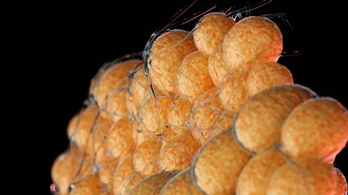
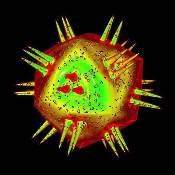
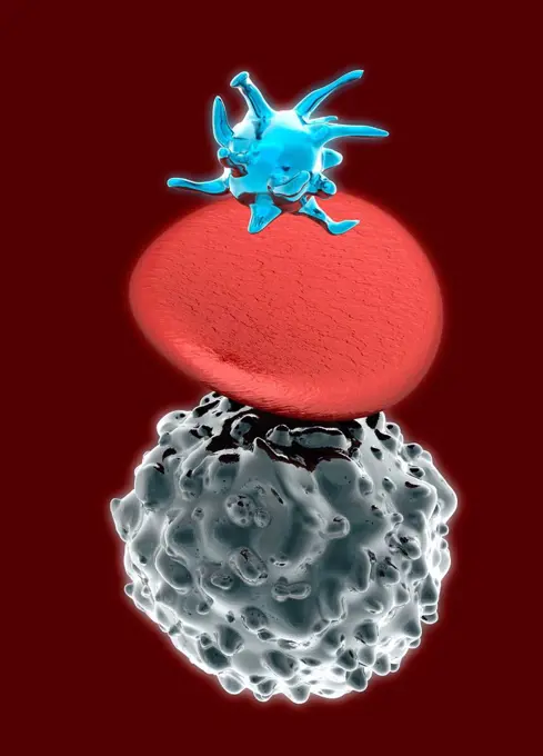
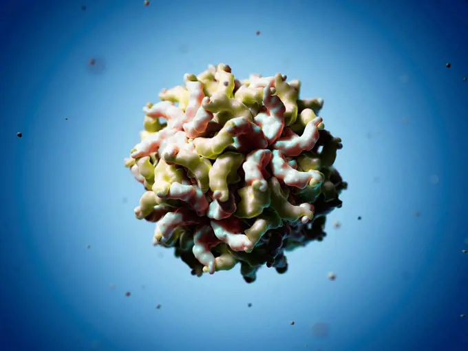
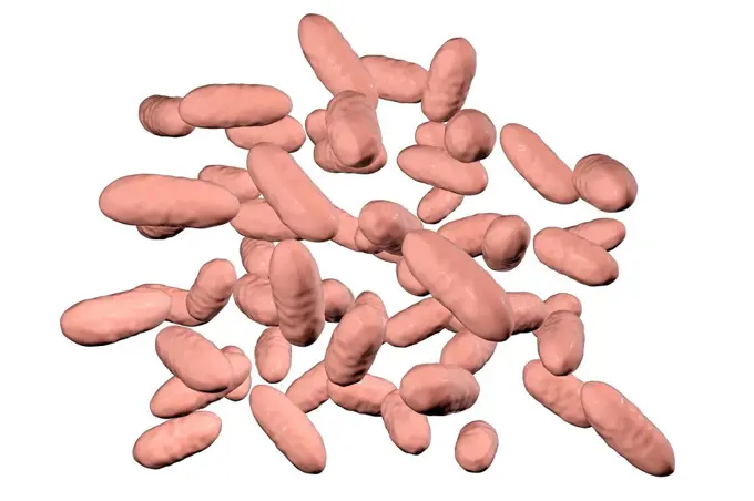
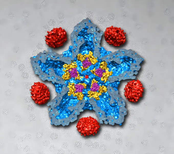
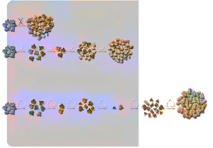
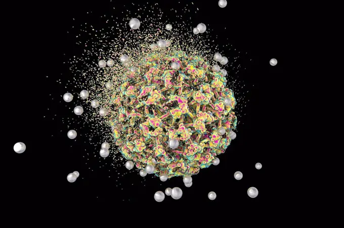
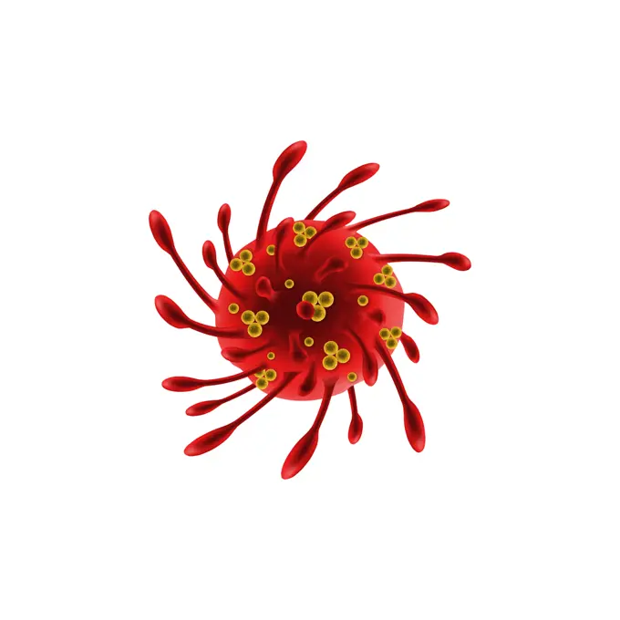
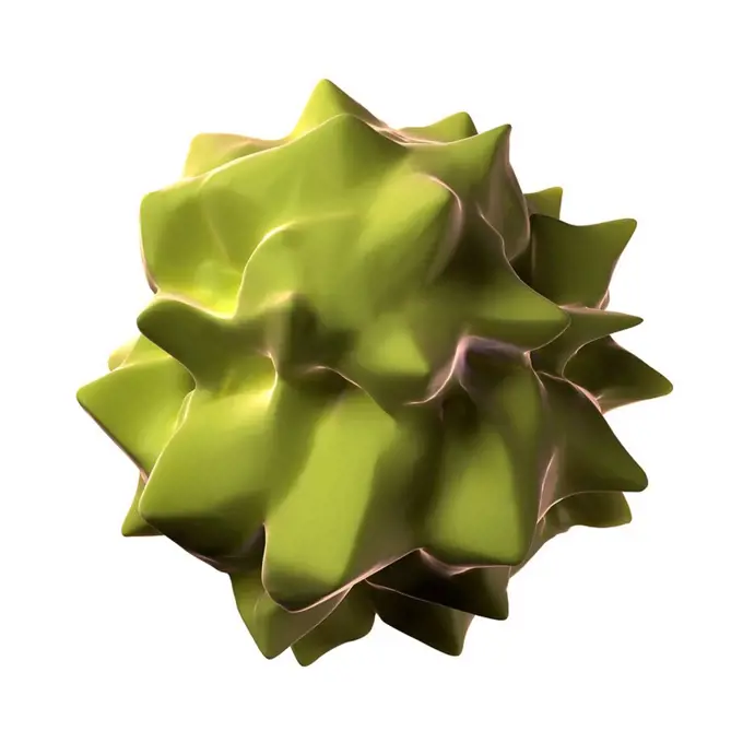
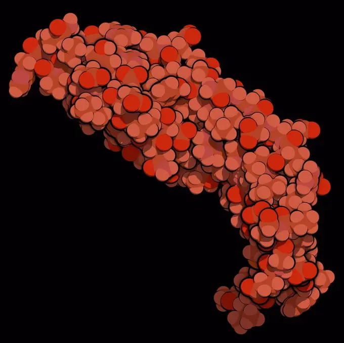
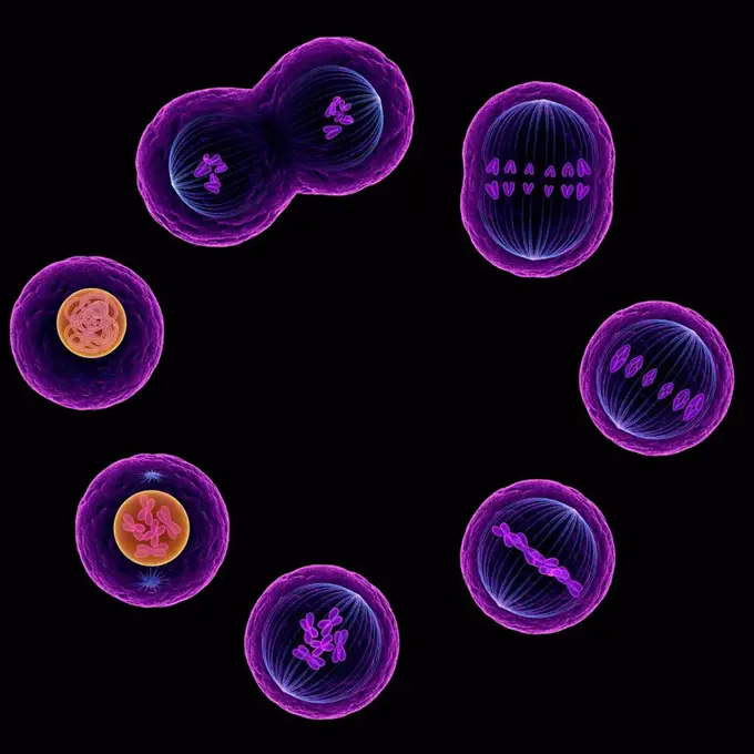
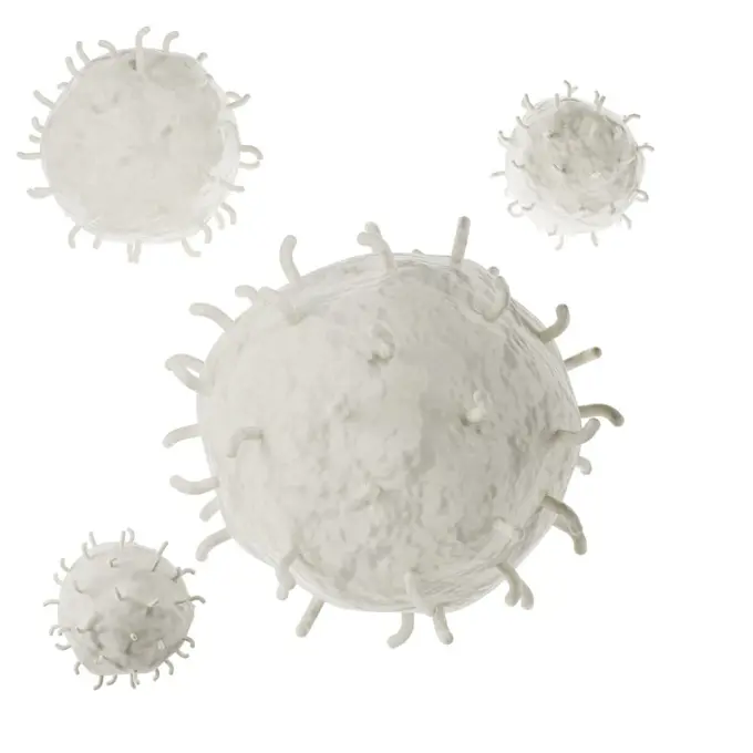
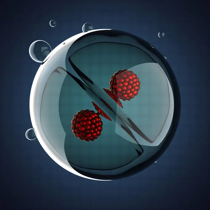
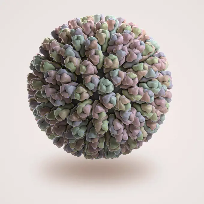
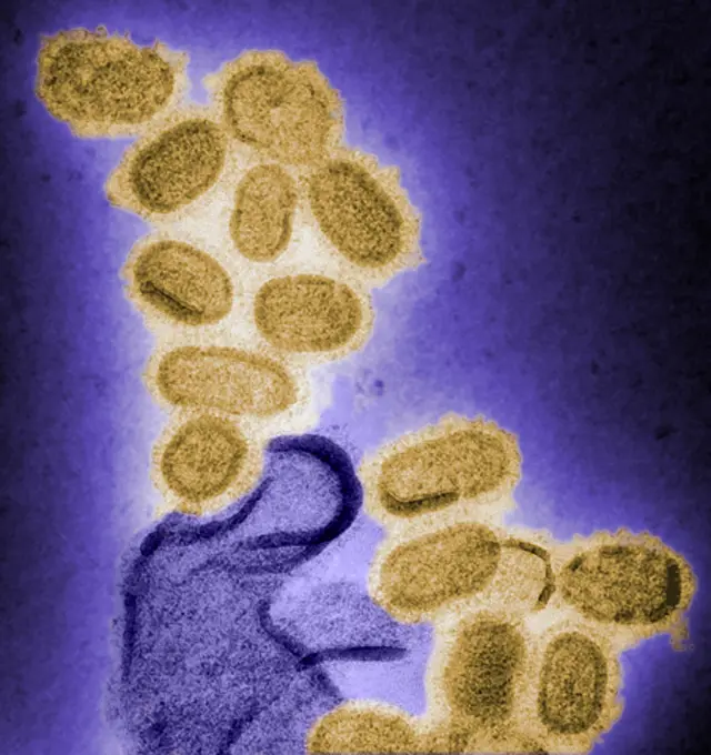
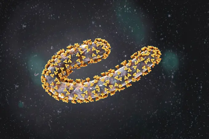
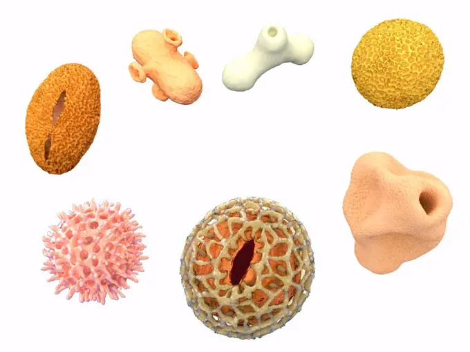
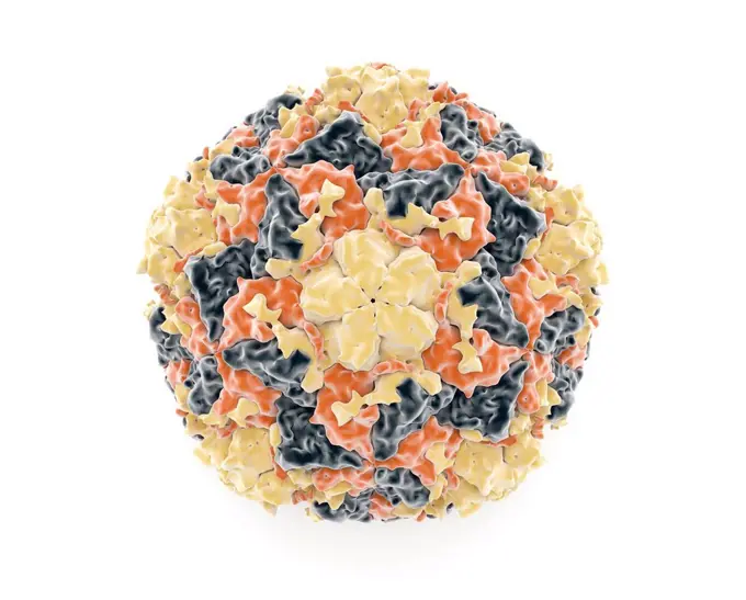
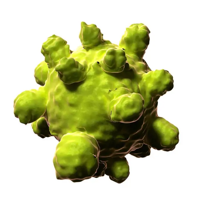
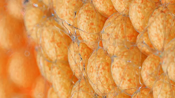
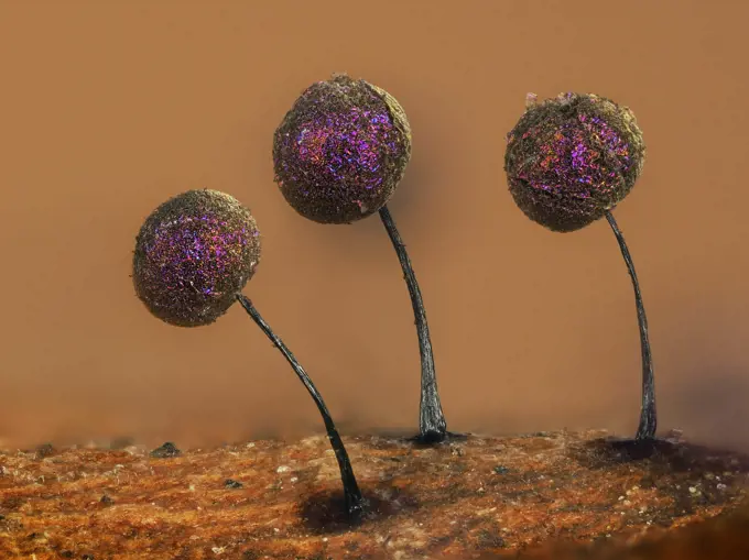
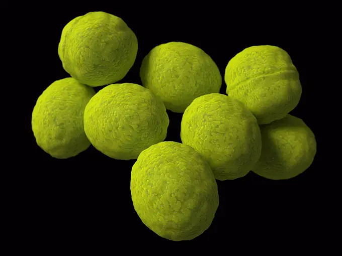
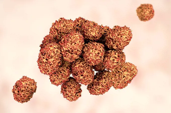
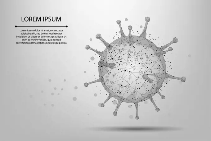
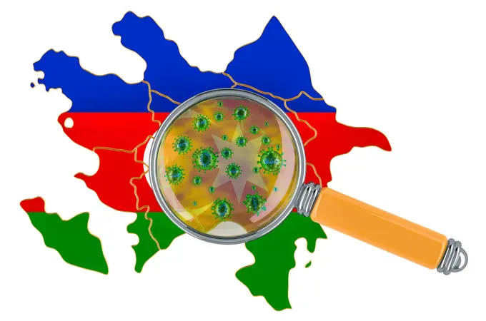
Virus Particles

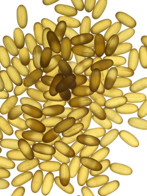
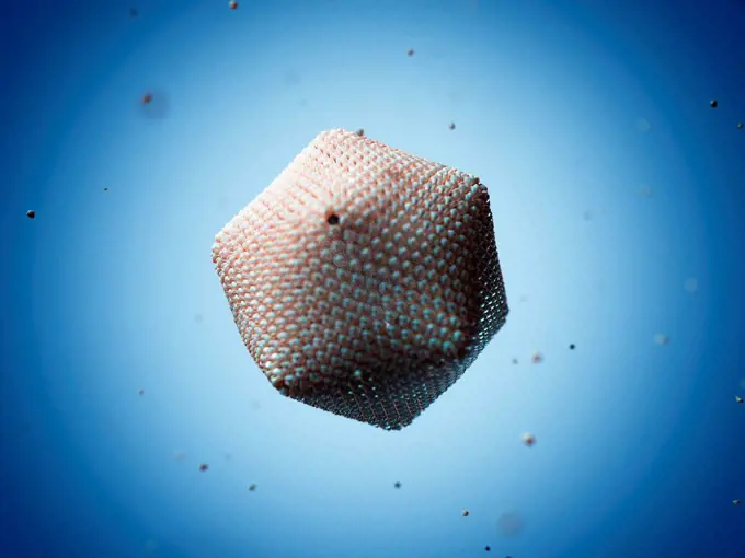
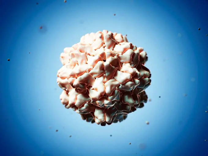
Computer-generated images of various virus particles, including HIV and rotavirus, depicted in bright colors against a contrasting background to highlight their structures.

Rotavirus particle, illustration

Assets in this Story

4128-19358330
Molecule of patulin toxin, computer illustration. Patulin is a mycotoxin produced by mould fungi Aspergillus, Penicillium and Byssochlamys, found in r...
4128R-14057569
Rotavirus particle, illustration
4128R-14060591
Ross River viruses, illustration
4128-20239435
Reovirus, illustration
4128R-13619857
Destruction of Human Immunodeficiency Virus (HIV), computer illustration. Conceptual image for HIV-infection and AIDS treatment and prevention showing...
1525-27165110
Concept image with many viruses around the earth for coronavirus disease COVID-19 pandemic
4128-15816279
Virus, fractal illustration.
1899-61461209
Meningococcus or meningococcal disease or Neisseria meningitidis, is a bacteria involved in meningitis. These bacteria are the cause of mild pharyngitis and only one person in approximately 400 becomes the victim of a serious meningococcal infection, most often in the form of acute purulent meningitis. Image produced from optical microscopy
1525-27149790
3d rendering. Dangerous Red Covid-19 corona virus sign symbol with flowing many cells as background.
4128-19052628
Coronavirus, illustration
4128R-11474201
Rice yellow mottle virus, computer artwork.
4128R-13023071
Zika virus. Computer illustration showing the protein spikes in the outer viral envelope. This is an RNA (ribonucleic acid) virus from the Flavivirida...
4128R-13817891
3D fractal. Computer-generated image of a three-dimensional fractal.
1848-18023481
Raspberry (Rubus idaeus), shoot t.s., coloration Wacker 3A
4128-19249568
Mescaline, molecular model. This is a naturally occurring psychedelic alkaloid of the phenethylamine class and a hallucinogenic substance present in t...
6188-55644951
Margarit blossom cross section of a margarit blossom in the dark field under the microscope 100x copyright: xzoonar.com/dr.xnorbertxlangex 13775996
824-63222937
Coronavirus (CoV). viruses of the Coronaviridae family and the Orthocoronavirinae subfamily. It is a pathogen of respiratory syndromes. Viewed from a transmission electron microscopy (TEM) image. Viral diameter 80nm to 100nm.
4128-111493564
Biofilm, conceptual illustration.
4128-28575376
MRSA bacteria, illustration
824-63224460
Coronavirus (CoV). viruses of the Coronaviridae family and the Orthocoronavirinae subfamily. It is a pathogen of respiratory syndromes. Viewed from a transmission electron microscopy (TEM) image. Viral diameter 80nm to 100nm.
5507-46322914
Virus - Coronavirus 2019-ncov, Disease germ, pathogen organism, infectious micro virology
4128R-13619793
Cryptococcus neoformans fungus, computer illustration. C. neoformans is a yeast-like fungus that reproduces by budding. An acidic mucopolysaccharide c...
6188-55546087
Wassertropfen Wassertropfen in Öl Copyright: xZoonar.com/AndreasxNaumannx 10404460
4128R-14057380
Trypsin enzyme surface, molecule, illustration
1848-54925416
Viruses, scienceart, illustration
5507-40134728
DNA molecule picture
4128R-11543355
Human papilloma virus (HPV) particle, computer artwork. HPV particles (virions) consist of a protein capsid enclosing DNA (deoxyribonucleic acid), the...
6188-55582995
Heliansthus stem micrograph Light photomicrograph of Helianthus stem cross section seen through microscope Copyright: xZoonar.com/ClaudioxDiviziax 6580634
4128R-13620525
Ragweed (Ambrosia psilostachya) pollen, coloured scanning electron micrograph (SEM). This pollen is an allergen. Ragweed is a main cause of weed aller...
4201-66305
Foraminiferan (Quinqueloculina sp) SEM close-up view at 42x magnification, that came from a beach in Formentera, Balearic Islands, Spain
1815-16017582
Three dimensional render of red virus cell
4128-19489930
Blastomyces fungus, illustration
1525-23997339
vector abstract health or chemistry blue background with molecules or microbes. epidemic spreading of microorganism microscopic infection of virus, germs or bacteria. eps10 illustration
4128R-15466766
Set of human chromosomes, illustration
1848-50836644
Raspberry (Rubus idaeus), shoot t.s., coloration Wacker 3A
6188-55584698
Black and white Lily anther micrograph Light photomicrograph of Lily anther cross section seen through microscope in black and white Copyright: xZoonar.com/ClaudioxDiviziax 7783588
4128-111493555
Cell, conceptual illustration.
4128R-14057284
Human growth hormone molecule, illustration
1746-21118103
Chrome yellow - crocoite. When the red mineral was crushed it would produce a yellow pigment.
4128-30418002
Stachybotrys mould, illustration
4128R-13940903
Human papilloma viruses. Seamless computer artwork of human papilloma virus (HPV) virions (particles). This virus consists of a protein capsid enclosi...
4128R-13586617
Destruction of a cancer cell, computer illustration. Conceptual image which can illustrate the treatment of cancer by drugs, nanoparticles, antibodies...
824-63191267
Artistic rendering of the surface of a human dendritic cell illustrating the unexpected discovery of sheet-like processes that fold back onto the membrane surface. When exposed to HIV, these sheets entrap viruses in the vicinity and focus them to contact zones with T-cells targeted for infection. These studies were carried out using ion abrasion scanning electron microscopy, a new technology we have been developing and applying for 3D cellular imaging.
6188-55644440
Dangerous pathogens in the body Dangerous pathogens in the body
4128R-14587961
Acanthamoeba amoeba, illustration
4355-522
Two Glowing Christmas Globes
4128R-11473916
Flavivirus, computer artwork.
4128R-12923041
Hair follicle
4128R-14638066
Bacterial vaginosis, illustration
4128-20042812
Polyomavirus, illustration
4378-1193
Single Influenza A virus.
4413-174713
Colonies of Volvox algaes in light microscopy
4128-24796401
Emmonsia fungi, illustration
1815-17635986
Three dimensional render of single blue stem cell
1848-49387928
Virus Viruses can make people sick like hepatitis or aids, illustration, natural science, biology, illustration, natural science, biology, abstract art
1848-61038463
Fake microorganisms. microbiology. Bacteria, cells, viruses, germs, microorganisms
4128R-11474522
Coxsackievirus b3, computer artwork.
1935-1233
Blue button Porpita porpita is a floating hydroid that lives on the water s surface and feeds on small planktonic animals Mexico Pacific Ocean
1848-61038256
Fake microorganisms. microbiology. Bacteria, cells, viruses, germs, microorganisms
4128R-13587453
Computer illustration of conidia (asexual spores) from a Fusarium sp. fungus. Some Fusarium fungi are pathogens of plants and humans.
4297R-1997
Illustration of the structure of a typical human virus
1525-25093391
virus in human blood. 3d
4128-20041290
Athlete's foot fungus, illustration
4128-28767018
Rotavirus structure, illustration
1525-26470263
Illustration of a coral vector
1525-60163793
Variegated String of Hearts. Watercolor potted plant. Houseplant illustration isolated on white. Variegated String of Hearts. Watercolor potted plant. Houseplant illustration isolated on white.
7203-70647335
Night dive at Barrier Reef near Saint Georges Caye, Fluorescence emitted at night, exposed with special UV blocking filters, Belize, Central America
4128-19249570
Mescaline, molecular model. This is a naturally occurring psychedelic alkaloid of the phenethylamine class and a hallucinogenic substance present in t...
6145-44613340
Summer South Polar Cap
4128R-13574976
Dendrimer, computer artwork. Dendrimers are artificially created branched polymers. Due to the high degree of molecular customisation during synthesis...
4220-21334735
Scanning Electron Micrograph (SEM): Stomata of Yew Leaf.     Date:
1848-54933780
Herpes viruses, Science Art
4128-20042850
Marburg virus, illustration
4417-16039631
Illustration of the Corona Virus Muted Color onWhite
4128R-14756
Flu virus, computer artwork.
4128-15666511
Cells, illustration
1525-27196993
Covid19 pattern. Background coronavirus
1525-27183846
Pro biotic bacteria, Good microorganisms concept isolated on white background
6188-56055944
3d render of flu or virus for epidemic disease 3d render of flu or virus for epidemic disease Copyright: xZoonar.com/mtxKANGx 21159930
4128R-14057345
Cell death 1 protein molecule, illustration
4128R-11313338
Yellow fever virus, computer artwork.
4128R-13941197
Nematode egg, illustration.
4128-28575383
MRSA bacteria, illustration
1848-18022821
Coastal she-oak (Casuarina equisetifolia), leaf t.s., staining Wacker 3A, darkfield illumination
4128-18631758
Pollen grains, illustration
4128R-22539
Roundworm ovaries. Light micrograph of a section through ovaries from an Ascaris sp. roundworm. Magnification: x100 when printed at 10 centimetres wid...
4128R-10357
Lime tree stem, light micrograph
4128-15981271
Zika virus, computer illustration.
1525-57135600
White blood cell 3d realistic icon analysis. Leukocytes medical illustration on white background with clipping path.. White blood cell 3d realistic icon analysis. Leukocytes medical illustration on white background with clipping path
1848-50837191
Black alder (Alnus glutinosa), shoot t.s., colouring Wacker 3A
4128R-11313387
Yellow fever virus, computer artwork.
824-63224352
All uses except for pharmaceutical laboratories in Belgium - Cancer, tumor cell identification by a T lymphocyte via surface antigens. T lymphocytes are immune cells designed to defend the body against pathogens. They are also used to defend the body against cancer cells. The cancer cell has tumor antigens (pearl necklace) on its surface that have mutated. We notice that they have a red pearl among the other blue pearls. See illustration - Restriction : All uses except for pharmaceutical laboratories in Belgium from 08/05/2023
4128R-14846650
Norovirus, illustration
4128R-15290701
Ebola virus, illustration
1848-54925415
Viruses, scienceart, illustration
4128R-13048290
Zika virus, computer illustration. This is an RNA (ribonucleic acid) virus from the Flaviviridae family. It is transmitted to humans via the bite of a...
4269-27746
Varicella-zoster virus. Color enhanced electron micrograph of Varicella (Chickenpox) virus. The infectious disease known as Chickenpox is caused by the varicella-zoster virus, a DNA virus of the Herpesviridae.
4128R-13741
Virus structure, conceptual computer artwork.
4128-V58615278
4128-17810024
Volvox green algae, light micrograph
4128R-11474170
Reovirus, computer artwork.
4128R-14939796
Coagulation factor VIII molecule, illustration
4128-19056167
Leuconostoc bacteria, illustration
4297-1429
Scanning electron micrograph revealed a small clustered group of Gram-positive beta-hemolytic Group C Streptococcus spp. bacteria
4128-24796406
Emmonsia fungi, illustration
5514-63777434
Several translucid yellow pills on white
4128R-11313256
Paramecium bursaria Chlorella virus 1 (PBCV-1) virus, computer artwork.
1525-27985882
Vector design of astronomic objects.
4128R-11313404
Turnip yellow mosaic virus, computer artwork.
4128-16102423
Coronavirus artwork
4128R-8948
Paramecium protozoa, light micrograph
4297R-1993
Illustration of the structure of a typical human virus
1525-19822192
Chlorophyll. Plant Cells under the Microscope. Decorative vector illustration.
4128R-13192935
Diatoms and radiolaria. Coloured scanning electron micrograph (SEM) of a circular arrangement of various diatoms and radiolaria. Diatoms are planktoni...
1815-111457367
3D Rendering, Yellow molecule against brown background
1848-54792638
Illustration, macrophage reaching for a virus
1788-20761663
Pollen grains of lily (Lilium sp) stained with methyl green and observed on a dark background.
6188-66540784
Golden sparkling glittering gel in a petri dish made of glass with shadow on a blue background for skin care and beauty concepts, laboratory sample golden sparkling glittering gel in a petri dish made of glass with shadow on a blue background for skin care and beauty concepts, laboratory sample
4128-19053702
Fat cells, illustration
1525-20392584
Science Atom and Chemical Formula as Concept. Project Management
4128R-11543539
Virus, conceptual illustration.
4128R-13844290
Computer artwork of an icosahedral virus capsid, derived from protein data of a minivirus, a huge icosahedral virus with over 1000 protein chains. The...
4128R-14637611
Blood cells, illustration
4128R-11313553
Partitivirus particle, computer artwork.
4128R-14587964
Aggregatibacter aphrophilus bacteria, illustration
1838-4439
Fingerprints
824-63227109
Bacteriophage Phi-6, which serves as a model system for rotavirus, is the most common cause of diarrheal disease in infants and young children. An understanding of how bacteriophage assemble can help scientists develop new ways to treat rotavirus infections. Shown here is a cryo-electron microscopy image of the protein shell of Phi-6, or procapsid, opened to show the different protein types: P1 (blue), P4 (red), P7, yellow, and P2 (purple).
4128-20042993
Different bacterial shapes, illustration
4128-19052522
Blood clot, illustration
4128R-13844631
Lymphocyte white blood cell, illustration.
824-63164505
Why use triple therapy to try to stop the development of the disease of AIDS.
4141-111428901
Light micrograph smear of a Common frog (Rana) red blood cells X900, UK. Non-mammalian red blood cells are oval in shape and contain a black nucleus.
1525-56509877
Mold samples on white background. A petri dish with colonies of microorganisms for bacteriological analysis in a microbiological laboratory. Close up view of mould. Mold samples on white background. A petri dish with colonies of microorganisms for bacteriological analysis in a microbiological laboratory. Close up view of mould.
4128R-11284867
Restriction enzyme. Compute artwork of a restriction enzyme (orange) complexed with DNA (deoxyribonucleic acid, blue). Restriction enzymes, also known...
1815R-11525
Viola cornuta, elevated view
824-63224354
All uses except for pharmaceutical laboratories in Belgium - PD-L1 protein, the expression of the cancer cell to escape the T lymphocyte. T lymphocytes (in green) are immune cells designed to defend the body against pathogens. They are also used to defend the body against cancer cells (in brown). Apart from this threat, cancer cells escape this attack by expressing proteins on their surface, the PD-L1 proteins. PD-L1 works like a stop sign and inactivates T cells before they attack. PD-L1 (here in red) works by binding to a receptor called PD-1 (here in turquoise blue) located on T lymphocytes. Binding of PD-L1 with the PD-1 receptor leads to inactivation of the T lymphocyte. So the latter cannot destroy the cancer cell. - Restriction : All uses except for pharmaceutical laboratories in Belgium from 08/05/2023
4128R-13619895
Destruction of human papilloma virus (HPV), computer illustration. Conceptual image for papilloma virus treatment and prevention. HPV causes warts, wh...
1525-27218992
Bacteria virus cell isolated red gem causing infections. Vector bacterium with tentacles. Red bacterium with tentacles isolated virus cell
4128R-20555
Ilavirus particle
4128R-14057300
Endothelial growth factor A molecule, illustration
6123-28555889
Many 3D rendered red and blue gel pill tablets contained in frame over white background.
4128R-16121
Mitosis nuclear division, computer artwork.
1525-57135601
White blood cell 3d realistic icon analysis. Leukocytes medical illustration on white background with clipping path.. White blood cell 3d realistic icon analysis. Leukocytes medical illustration on white background with clipping path
4128R-13162697
IgG2a monoclonal antibody (immunoglobulin). Many biotech drugs are antibodies.
1848-56111440
Skeleton of a hard coral
4128-28575337
Staphylococcus aureus bacteria, illustration
1525-24940854
Water
6188-58133305
A micro cell division process A micro cell division process illustration Copyright: xZoonar.com/OxanaxRishnyakx 6225903
4128-30421537
RAGE receptors in membrane, illustration
1848-49315558
Melethallia, gregarious algettes, green algae, art forms of nature, Historic, c. 1840, digitally restored reproduction of a 19th century original
4128R-13818521
Petri dish with bacteria.
4378-3038
Structure of the Rotavirus (3GZU). Rotavirus is the most common cause of severe diarrhoea among infants and young children and is one of several viruses that cause infections often called stomach flu.
824-63194494
This colorized negative stained transmission electron micrograph (TEM) shows recreated 1918 influenza virions that were collected from supernatants of 1918-infected Madin-Darby Canine Kidney (MDCK) cells cultures 18 hours after infection. To separate these virions, the MDCK cells are spun down (centrifugation), and the 1918 virus in the fluid is immediately fixed for negative staining. The solid mass in lower center contains MDCK cell debris that did not spin down during the procedure.
1746-21118101
Chrome yellow - crocoite. When the red mineral was crushed it would produce a yellow pigment.
4128-30420565
Marburg virus, illustration
824-63223174
Colorized scanning electron micrograph of Marburg virus particles (yellow) both budding and attached to the surface of infected VERO E6 cells (blue). Image captured and color-enhanced at the NIAID Integrated Research Facility in Fort Detrick, Maryland. Credit: NIAID.
1525-28196448
Bacteria cells banner with empty space for text. Microorganism collection. Vector doodle style composition.
4128R-11543204
Illustration of pollen grains from seven different species of plants
1525-28044447
Microscopic analysis of chalk, according to Ehrenberg, vintage engraved illustration. From Natural Creation and Living Beings.
4128R-11284877
Rhinovirus particle, computer artwork. The rhinovirus infects the upper respiratory tract and is the cause of the common cold.
4128R-11313758
Measles virus, computer artwork.
4128-19053708
Fat cells, illustration
1773R-22460
A shirt made out of flowers on a line
4070-28602142
Slime mould (Lamproderma scintillans) super close up of 1mm tall sporangia on a beech leaf, Buckinghamshire, England, UK, February Focus Stacked.
4128R-14181675
Aspergillus, illustration
6188-55583254
Light micrograph Micrograph of from top left onion, cotton, vicia, bamboo, corn tilia, cucurbita, helianthus, lily, lily pine, mulberry, pine leaf, silver berry, girospyra stoma, moss, corn root, onion root, leaf Copyright: xZoonar.com/ClaudioxDiviziax 6653491
824-63225837
CCL-81 cells heavily infected with SARS-CoV-2 virus particles. The small spherical structures in the center of the image are SARS-CoV-2 virus particles. The string-like protrusions extending from the cells are cell projections or pseudopodium. Image captured at the NIAID Integrated Research Facility (IRF) in Fort Detrick, Maryland.
1525-25313186
Aquarium
6188-67687615
Schutz von Luxemburg vor dem Coronavirus. 3D-Rendering Protection of Luxembourg from coronavirus. 3D rendering isolated on white background Copyright: xZoonar.com/natatravelx 23146008
4128R-11286178
Streptococcus bacteria, computer artwork.
4128-111581084
Toxic mould spores (Stachybotrys chartarum), computer illustration. Several strains of this fungus (S. atra, S. chartarum and S. alternans) are synony...
1525-27042237
Abstract line and point Virus cell. Low poly Immunology, new strain epidemic, infection pathogen concept vector illustration. Abstract polygonal image.
6188-68094124
Aserbaidschanische Karte mit Coronavirus unter der Lupe, 3D-Rendering Azerbaijani map with coronavirus under magnifier, 3D rendering isolated on white background LicenseRF Copyright: xZoonar.com/alexlmxx 23163721
6188-67684888
Georgische Karte mit Coronavirus unter der Lupe, 3D-Rendering Georgian map with coronavirus under magnifier, 3D rendering isolated on white background Copyright: xZoonar.com/alexlmxx 23149717
6188-67666852
Schutz Ägyptens vor dem Coronavirus. 3D-Rendering Protection of Egypt from coronavirus. 3D rendering isolated on white background Copyright: xZoonar.com/natatravelx 23091678