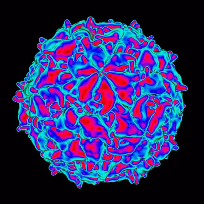
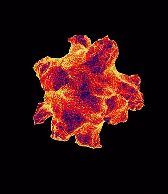
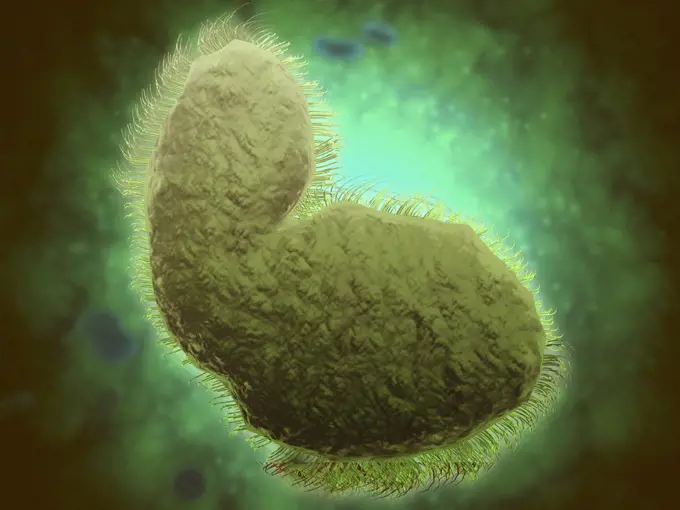
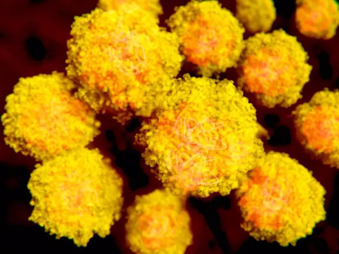
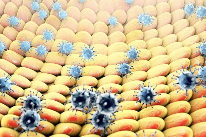
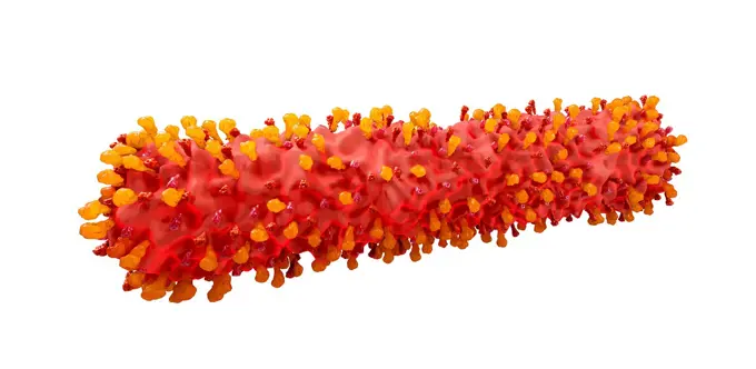
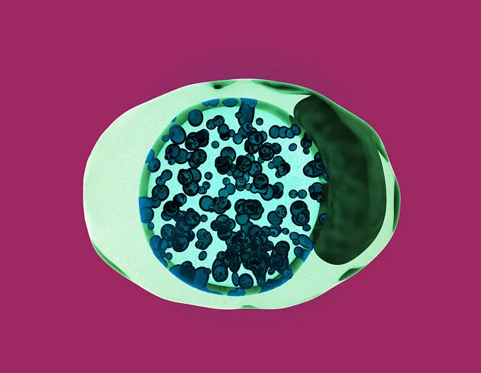
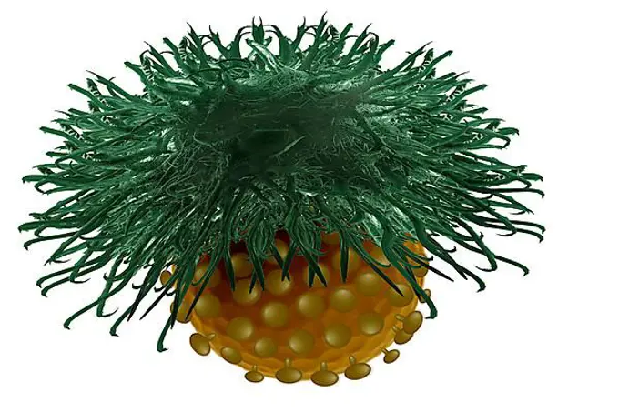
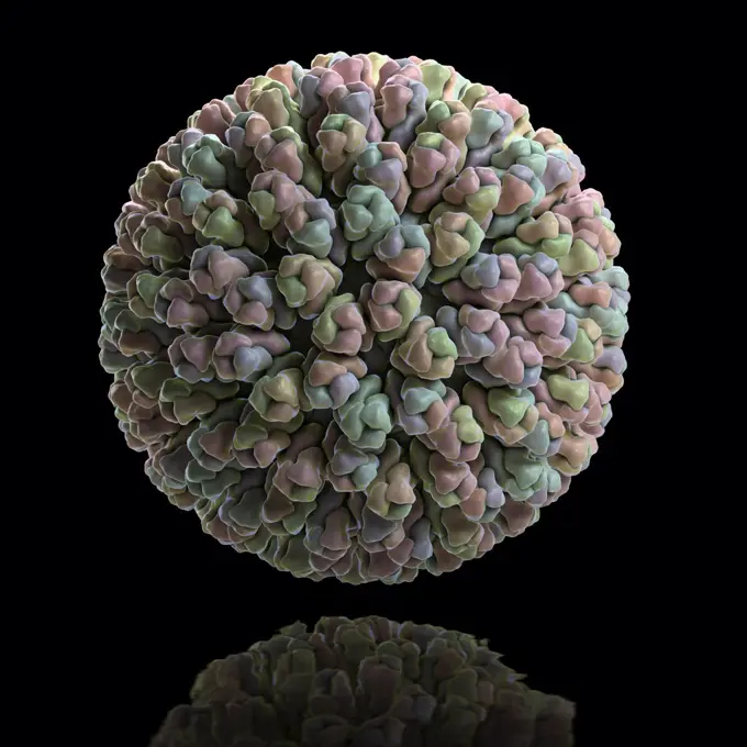
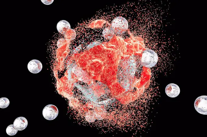
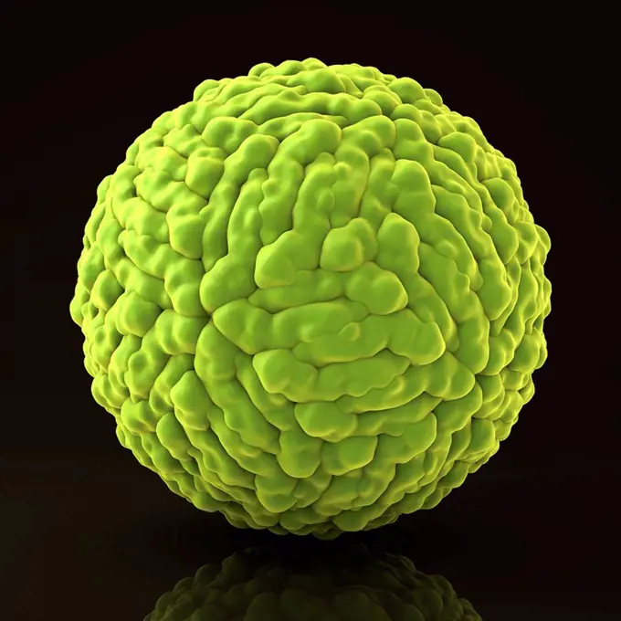
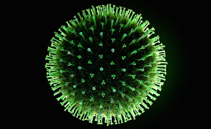
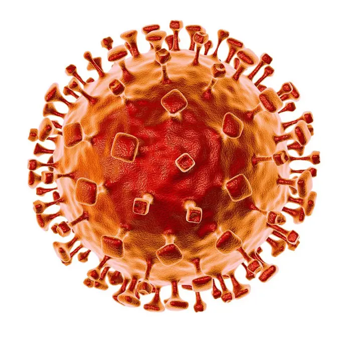
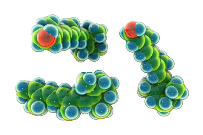
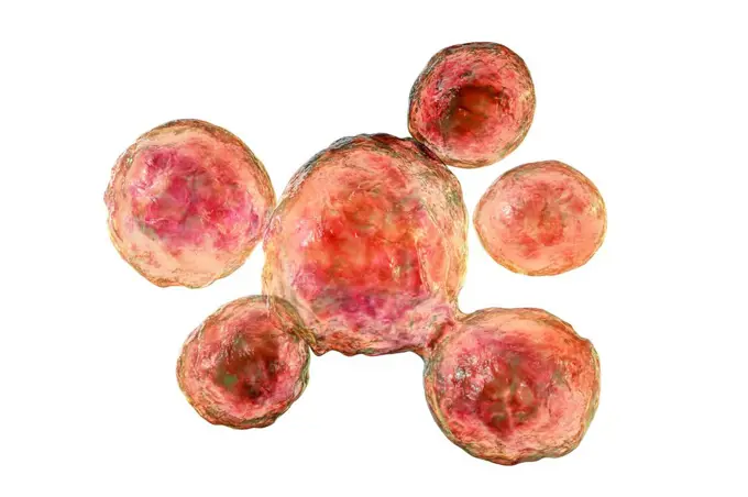
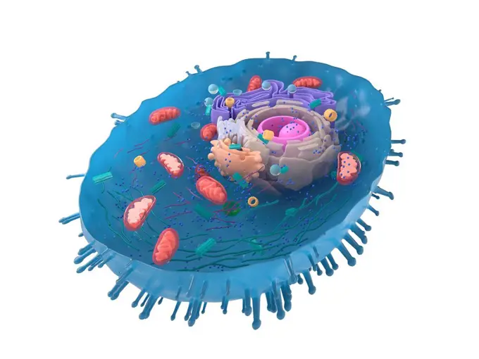
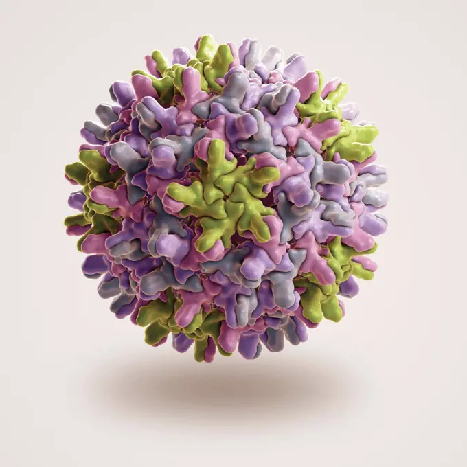
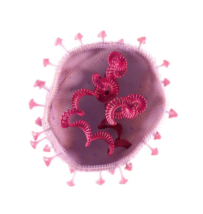
Fine ArtVintageFootageSubscriptionBoardsFree ResearchPricing
  • Fine Art
  • Vintage
  • Footage
  • Subscription
  • Boards
  • Free Research
  • Pricing

Virus and Molecular Structures Artwork

Computer-generated artworks of various viruses including the herpes virus, showcasing vibrant colors and detailed textures on a dark background.

Mengo virus, computer artwork.
269 assets in this story
4128R-10075
Influenza virus, artwork
4128R-14846755
Egg fertilisation, illustration
4128R-14057277
Epidermal growth factor molecule, illustration
4269-24598
Pollen. Pollen of wattle SEM (Scanning Electron Microscope)
4239-18641403
Microscopic view of respiratory syncytial virus (RSV). RSV is a common virus that leads to mild, cold-like symptoms in adults and children.
4128R-13019077
Zika virus particles, computer illustration. This is an RNA (ribonucleic acid) virus from the Flaviviridae family. It is transmitted to humans via the...
4128R-13376628
Viruses infecting cells, illustration
4378-978
Scanning electron microscope styled structure of a typical virus.
4128R-13575618
Meningitis bacteria (Neisseria meningitidis), computer illustration. N. meningitidis are gram-negative diplococci (spherical bacteria arranged in pair...
6188-60959050
Variegated String of Hearts. Watercolor potted plant. Houseplant illustration isolated on white.brVariegated String of Hearts. Watercolor potted plant. Houseplant illustration isolated on white. Variegated String of Hearts. Watercolor potted plant. Houseplant illustration isolated on white Variegated String of Hearts. Watercolor potted plant. Houseplant illustration isolated on white Copyright: xZoonar.com/LiubovxPonomarchukx 21833870
6188-55651374
Desire, infection, immune system, immune system, defense, defensive, defense ** defense, infection, immune system, boxing defending, defenses, infections, immune systems 2OH-GQB
6145-46756480
Skiopticon image from the Department of Photography at the Royal Institute of Technology. Use by Professor Helmer Bäckström as teaching material. Bäckström was Sweden's first professor in photography at the Royal Institute of Technology 1948-1958. Optische Anstal C. P. Goerz, Berlin.
4128R-11312869
Herpes virus, computer artwork.
1525-27143781
Coronavirus Logic. Viral Epidemic series. 3D Illustration Coronavirus particles and micro space elements  for subject of virus, epidemic, infection, disease and health
6188-60022972
3D rendering of a Coronavirus Covid-19 cell a health risk outbreak influenza or flu dangerous pandemic medical health risk concept disease 3D rendering of a Coronavirus Covid-19 cell a health risk outbreak influenza or flu dangerous pandemic medical health risk concept disease on empty background isolated Copyright: xZoonar.com/HugoxKurkx 15106081
4128-111801842
Herpes virus, illustration. Herpes simplex virus.
4128-V58566938
1848-49174300
Spices, saffron crocus (Crocus sativus), dried saffron threads in bundles
6145-44075882
Plant study with inflorescences
4128-30419949
Filamentous respiratory syncytial virus, illustration
4141-9877
sunstar crossaster papposus (formerly solaster papposus)
4128-19357232
Kidneys, conceptual image.
4128R-14181676
Autoimmune thyroiditis, conceptual illustration
824-57660048
Staphylococci (Staphylococcus pyogenes aureus), bacteria responsible for food poisoning, localized suppurative infections, nosocomial infections and in certain extreme cases, septicemia. Image produced from optical microscopy.
4297-1652
Illustration of swine flu virus showing the structure of the influenza virion including the hemagglutinin and neuraminidase proteins on the surface of the particle
4128-20045725
Lipoproteins and red blood cell, illustration
824-65830691
Colorized electron micrograph of the herpes simplex virus (HSV).
1899-65662418
Illustration depicting a chlamydia-infected cell. Chlamydia trachomatis bacteria (represented by small dark spherical shapes colorized blue) undergo development within a membrane-enclosed &quotinclusion body.&quot The dark green shape on the right of the cell denotes the nucleus.
1848-54746144
Dendritic cell of the immune system detecting a bacterium, illustration
4378-3076
Structure of the Rotavirus (3GZU). Rotavirus is the most common cause of severe diarrhoea among infants and young children and is one of several viruses that cause infections often called stomach flu.
4128R-13586608
Destruction of a cancer cell by nanoparticles, computer illustration. Conceptual image which illustrates the potential of nanoparticles in the treatme...
4128R-13022941
Zika virus, computer illustration. This is an RNA (ribonucleic acid) virus from the Flaviviridae family. It is transmitted to humans via the bite of a...
1525-23805075
Golden paint cloud with glitter in water isolated on black background
1849-66221384
Close up of flu virus
4128R-13372982
Lymphocytes, computer artwork. T- and B-lymphocytes are part of the immune system. B cells mature in bone marrow and are responsible for humoral immun...
4128R-15302854
Nipah virus particle, illustration
1525-27040239
Bright sun shining in the universe plenty of stars
4408-11992
Jellyfish: Pectyllis Arctica, Jelly Fish
4128-20044493
Vitamin A molecule, illustration
4128-19056208
Fungi Paracoccidioides lutzii, illustration
4128R-15516382
Illustration of a human cell cross-section
6145-54410374
Project 26 - Operation Dominic (Johnston Island/Christmas Island/Maui, Hawaii) Detonation. Checkmate cloud, aerial view. Photographs of Atmospheric Nuclear Testing at Pacific Island and Nevada Test Sites.
4378-3520
Structure of the Hepatitis B virus (HBV) (PDB 1QGT), a species of the genus Orthohepadnavirus, which is part of the Hepadnaviridae family of viruses.
4128-16008600
Coronavirus particle, illustration
4128R-13575617
Meningitis bacteria (Neisseria meningitidis), computer illustration. N. meningitidis are gram-negative diplococci (spherical bacteria arranged in pair...
824-63223110
3D print of a spike protein on the surface of SARS-CoV-2 also known as 2019-nCoV, the virus that causes COVID-19. Spike proteins cover the surface of SARS-CoV-2 and enable the virus to enter and infect human cells. For more information, visit the NIH 3D Print Exchange at 3dprint.nih.gov. Credit: NIH
4378-3642
Atomic structure of Adeno-Associated Virus (PDB 1LP3), a vector for human gene therapy.
1848-54509521
Horned melon (Cucumis metuliferus)
6123-28556560
A 3D rendering of a realistic microscopic bacteria cell with spores over a black background and clipping path included.
4128-20041467
Retina damage from diabetes, illustration
4128R-13928447
Dust mites (Dermatophagoides pteronyssinus) on human skin, computer illustration. This mite is eight legged. Its rounded, unsegmented body has a few t...
4128-19249585
Psilocybine, molecular model. This is a naturally occurring psychedelic compound found in psilocybin mushrooms. The effects include euphoria and hallu...
4128R-12699226
Cancer cell, illustration.
824-63224353
All uses except for pharmaceutical laboratories in Belgium - Destruction of the tumor cell by the T lymphocyte, apoptosis. A T lymphocyte is activated when it recognizes a tumor cell by the tumor antigen. At this point, granules filled with toxic substances are transported from the T lymphocyte to the tumor cell. These cytotoxic granules release their lethal contents (red beads) into the target cell. The tumor or cancer cell dies by apoptosis. - Restriction : All uses except for pharmaceutical laboratories in Belgium from 08/05/2023
1746-19663788
Keplers Supernova Remnant: A View from Chandra X-Ray Observatory. Each top panel in the composite shows the entire remnant. Each colour in the composite represents a different region of the electromagnetic spectrum, from X-rays to infrared light. The X-ray and infrared data cannot be seen with the human eye. Astronomers have color-coded those data so they can be seen in these images. The bottom panels are close-up views of the remnant. In the bottom, centre image, Hubble sees fine details in the brightest, densest areas of gas. The region seen in these images is outlined in the top, centre panel.
6139-30021888
Adipose cell 3d model. 3D rendered illustration of the white fat cell on a light background with free space for text.
4128-111493749
Computer generated illustration of the moment a bacteriophage lands onto the surface of a bacterium.
4128-19056130
Cysts of Entamoeba coli protozoan, illustration
4269-25462
Pollen of wattle. Scanning Electron Microscope
1525-27050598
Coronavirus outbreak and coronaviruses influenza background as dangerous flu strain cases
5507-31849272
Human hands with monkeypox virus
824-63205878
Illustration of the seasonal flu virus. There are 3 types of virus that all have a nearly identical morphology : Type A and B viruses have 2 types of envelope glycoproteins or viral surface proteins (Neuraminidase et hemagglutinin). Type C virus doesnt have surface Neuraminidase. The lipid bilayer is visible, then the internal part of the envelope made up of M proteins. In the centre, in green, is the ribonucleoprotein (RNP).
4128-18929658
Coronavirus particles behind a fence, illustration
4128R-13818098
MRSA bacteria. Computer artwork of methicillin-resistant Staphylococcus aureus (MRSA) bacteria.
4298-20535933
Bean root with nitrogen nodule  Magnification 40x
1525-27168733
Pet virus as Coronavirus infection in a cat with 3D illustration elements.
1525-19789682
Heliansthus stem micrograph. Light photomicrograph of Helianthus stem cross section seen through microscope
4128R-27213
Tablet computer showing conceptual artwork of an avian influenza (flu) virus. A virus is a tiny pathogenic particle comprising genetic material enclos...
4128-19490005
Pullulan molecule, illustration
PREVIOUS
of 3
NEXT
2401 S. Ervay, Suite 206
Dallas, Texas 75215
United States
Browse
CategoriesGalleriesFree Research
For Creators
License Your ContentSuperStock For StudentsFrame of Mind BlogContributor PortalNew Image SpotlightLicense AI Imagery
Resources
About UsLicensingTerms & ConditionsAPI AccessPrivacyPricing
Contact
+1 866 236 0087help@superstock.com Contact form
©2026 SuperStock. All Rights Reserved. -A
This site is protected by reCAPTCHA and Google Privacy Policy and Terms of Service apply.