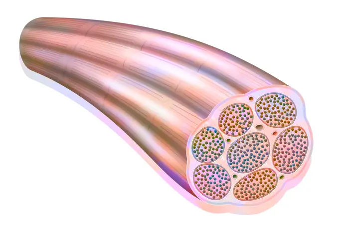
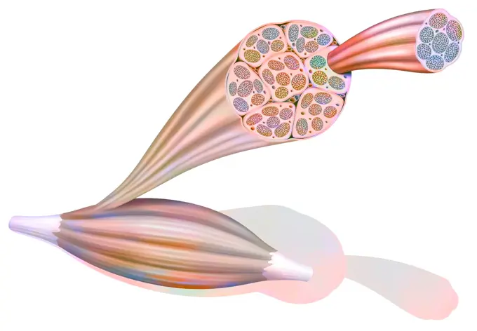
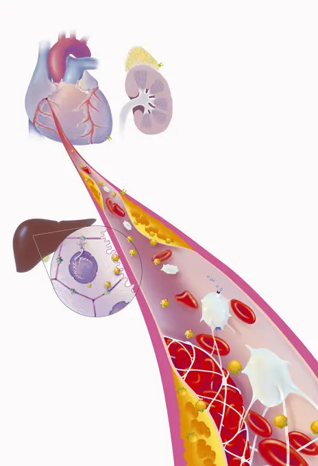
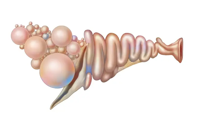
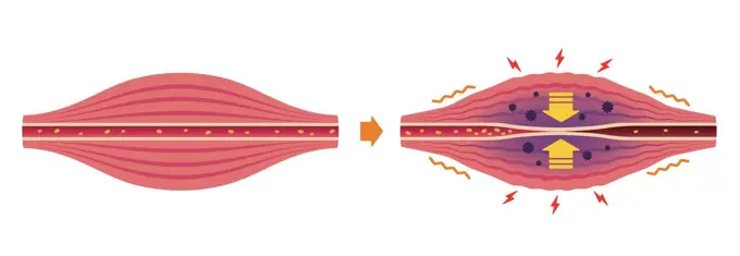
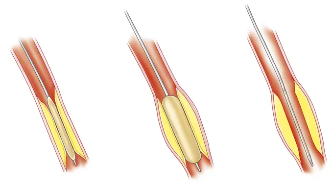
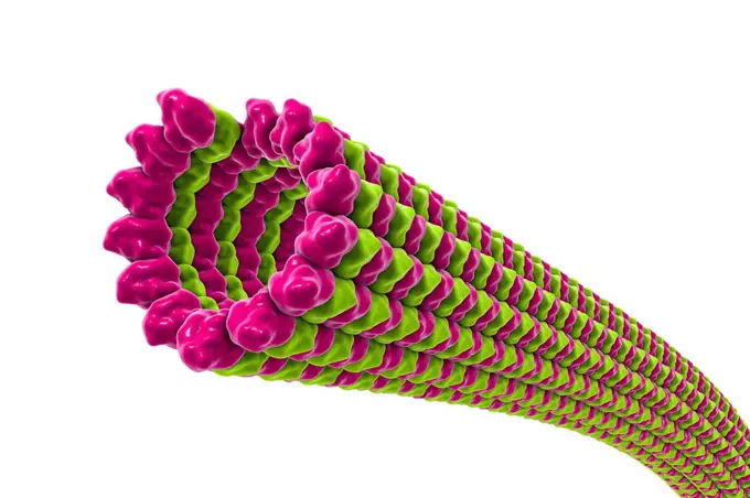
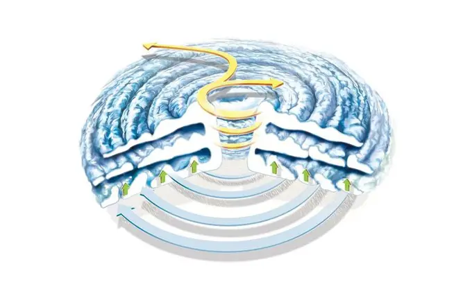
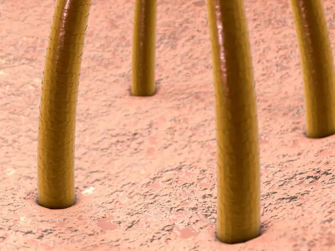
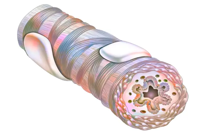
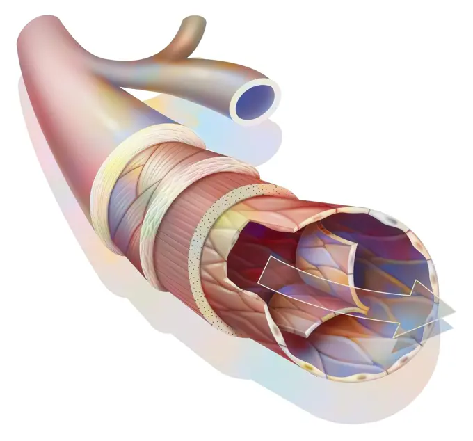
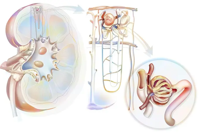
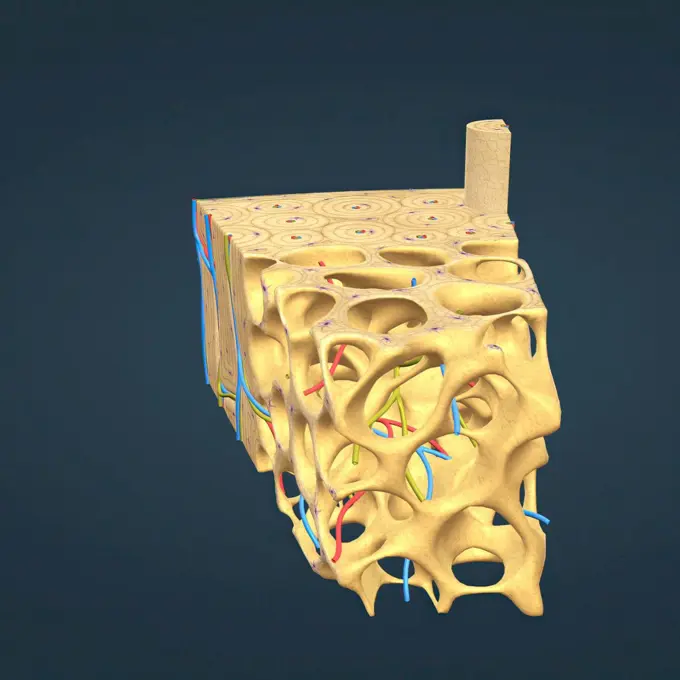
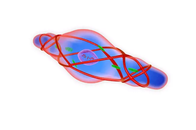
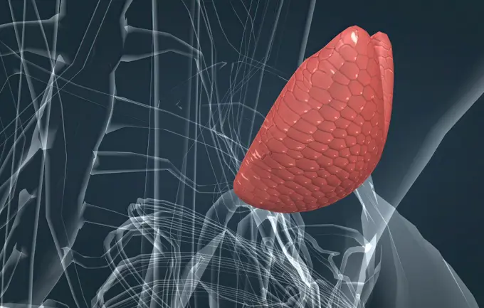
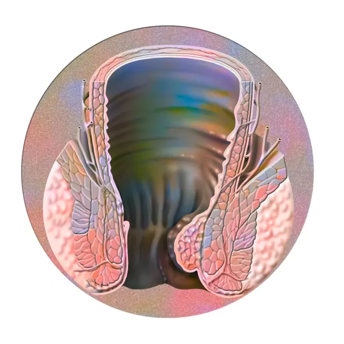
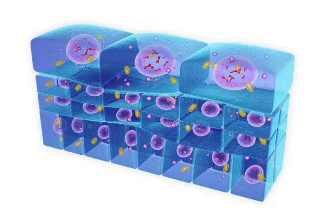
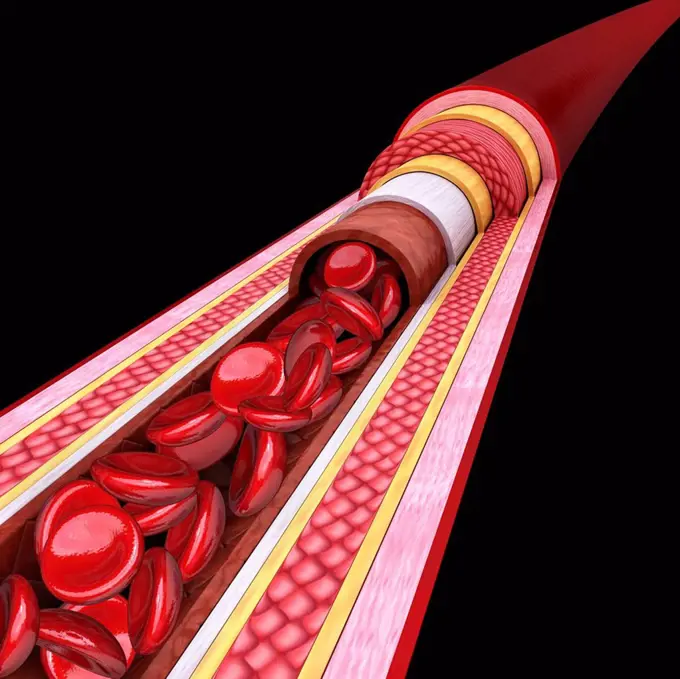
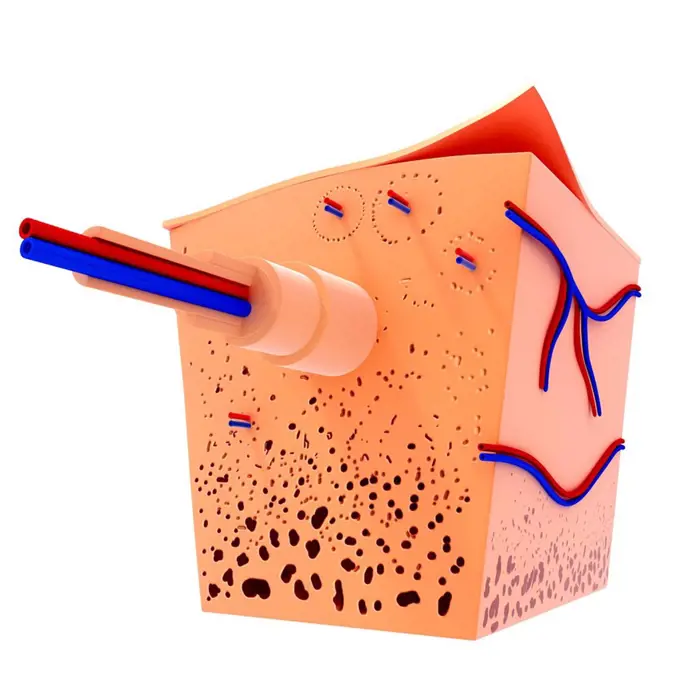
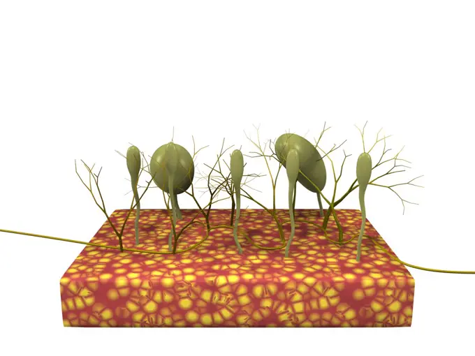
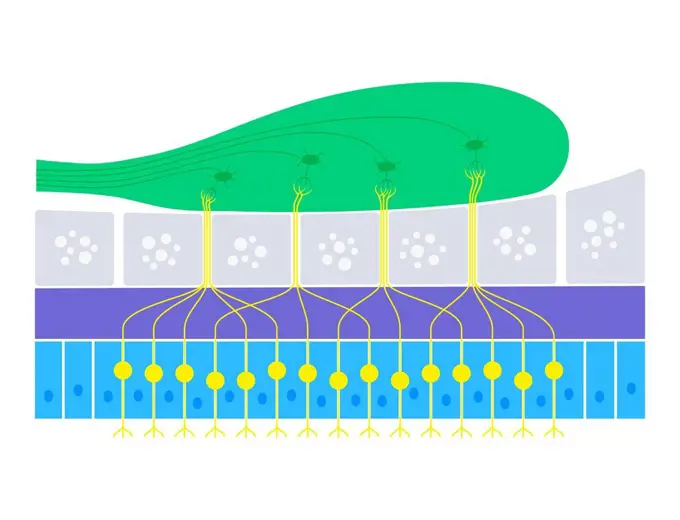
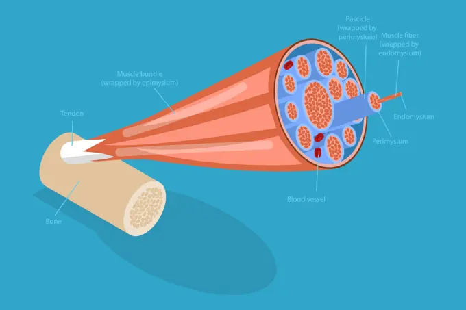
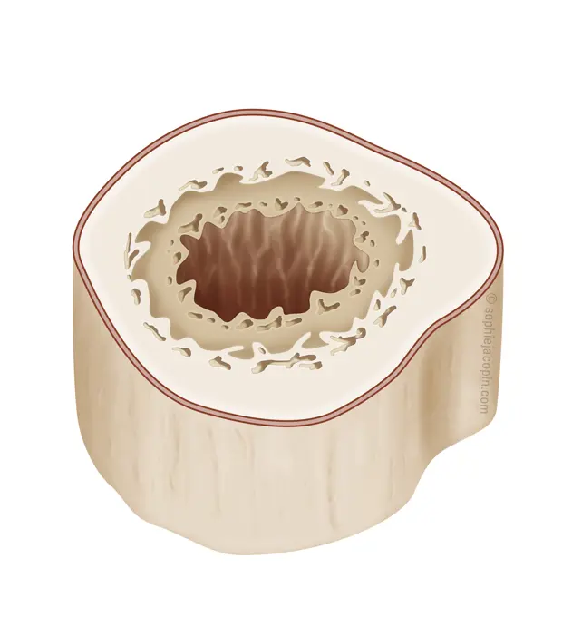
Muscle Fiber and Structure

Illustrations of muscle fibers, myofibrils, and muscle components including membranes and blood vessels, showcasing a scientific style with vibrant colors.

Blood vessels: suture between two arteries during a transplant.
Muscle fiber showing the myofibrils made up of myofilaments (actin and myosin).Muscle fiber showing the myofibrils made up of myofilaments (actin and myosin).Depolarization: phospholipid membrane with NA + and K + ion channels.From muscle to muscle fiber: tendon, muscle, muscle fiber.Blood vessels: suture between two arteries during a transplant.Myocardial infarction, coronary artery with thrombus, atheromatous plaques, treatments. The atheromatous plaques (yellow) in the wall of the coronary artery cause coronary disease. in the zoom of the coronary vessel, a thrombus forms at the bottom right, in contact with the blood by platelet aggregation. Treatments act on platelets to prevent platelet aggregation, on the secretion of aldosterone by the kidneys and adrenals, but also upstream, on the liver by limiting the synthesis of cholesterol at the origin of atheromatous plaques.3D Isometrische flache konzeptionelle Illustration von Nasenschleimhautzellen, medizinisches Lehrdiagrammbr3D-Isometrische flache konzeptionelle Illustration von Nasenschleimhautzellen, medizinisches Lehrdiagramm 3D Isometric Flat Conceptual Illustration of Nasal Mucosa Cells, Medical Educational Diagram 3D Isometric Flat Conceptual Illustration of Nasal Mucosa Cells, Medical Educational Diagram Copyright: xZoonar.com/AnastasiiaxTorianykx 22973347Cut of skin with its different components such as follicles, hairs.Crohn's disease: small intestine with inflammation of the lining.Cross-section of the epidermis of a dark phototype skin with many grains of melanin.Anatomy of the hen's reproductive system with ovary, follicles, infundibulum and cloaca.Illustration of skin tanning under the effect of the suns ultraviolet rays. Melanocytes (green cells in the epidermis) produce melanin, a pigment that absorbs ultraviolet rays. Melanin travels in melanosomes that migrate towards keratinocytes, placing themselves on their nucleus to protect their DNA and prevent genetic mutations due to the sun. Melanin makes the skin browner, thicker and more resistant to ultraviolet rays. (Photo by: BSIP/UIG via Getty Images)On a cholinergic synapse, the nerve impulse is transmitted between the two neurons thanks to acetylcholine.Retina at the level of the bowl drawn by the fovea.Illustration of skin tanning under the effect of the suns ultraviolet rays. Melanocytes (green cells in the epidermis) produce melanin, a pigment that absorbs ultraviolet rays. Melanin travels in melanosomes that migrate towards keratinocytes, placing themselves on their nucleus to protect their DNA and prevent genetic mutations due to the sun. Melanin makes the skin browner, thicker and more resistant to ultraviolet rays.Blood brain barrier.Internal hemorrhoidal plexus and location of internal hemorrhoids.Illustration of the signs of ageing in skin, dulling of the epidermis, brown spots, skin slackening and wrinkles. In the centre of the image: a cross-section illustration of young skin enabling a comparison with different types of ageing: -Top left illustrates the dulling of the epidermis, the skin is dry and cutaneous microrelief is irregular, causing the skin to become permeable to pollutants and dehydration. Superficial microcirculation is less good; skin cells become less oxygenated. -Top right illustrates spots. If they are brown it is due to melanin deposits, when red it is due to superficial microcirculation vessels, which are dilated. -Bottom left illustrates the loss of firmness due to diminished activity of the fibroblasts (blue). Fibroblasts are responsible for skins elasticity, producing elastin and collagen -Bottom right illustrates wrinkles due to the deterioration of collagen (white vertical) and elastin (pink horizontal) fibers.The organ of olfaction with the olfactory bulb, supporting cells.Penetration and duplication of the AIDS virus in the host cell.Eye, cataract, phacoemulsification: consists of breaking the lens with a probe.Spermatogenesis, male genitals and the formation of sperm in the testes.Spermatogenesis: male genitals and the formation of sperm in the testes.Illustration of fertilisation, the journey of the embryo in the fallopian tube and its transformation, until its implantation in the uterine lining.Process of muscle's stiffness and pain / circle type illustrationProstatic adenoma, drawingCut of skin of the palm of the hand, armpit and the rest of the body.Ganglion in section: support for illustration on immunity and immune reactions.A cross-section illustration of bone showing its structure.Structure of a healthy dorsal vertebra with spongy and compact tissue.Illustration comparing healthy skin and skin affected by eczema. In the case of eczema, keratinocytes are destroyed which leads to red patches (inflammation), along with itchiness, peeling and sometimes blisters. The causes of eczema are : -irritants -emotional disturbance -changes in temperature or humidity -skin infectionsAtherosclerosis, illustration.Development of cancerous tumor: The tumor continues to grow.Illustration of lumbar vertebrae.Colorized normal lateral abdomen x-ray of a 9 day old girl. The spine has been highlighted in yellow, the lungs in blue, and the abdominal cavity in pinkA cross-section illustration of an artery showing the introduction of a balloon before placing a stent.Microtubules, illustrationStructure of a hurricane.Human hair, computer illustration.Lymph node anatomy, illustrationStraight hair strand structure, illustrationStratified cuboidal epithelium, illustration.Human vertebrae, illustrationCoxofemoral joint and femoral neck.Bronchus of an asthmatic, wall of the bronchus thicker.Anatomy of a medium caliber vein with the outer tunica.Blood vessels: suture between two arteries during a transplant.3d dentitionHuman stomach. Internal organ of the body. Food and medicine route with red direction arrow. Medical care. X-ray of the belly. Digestive problems. Cartoon flat illustration. Human stomach. Internal organ of bodyIllustration of an IVF (In Vitro Fertilization). On the left, a classic IVF: the spermatozoa and occytes are placed in an embryo culture environment that favors their survival. Spontaneous fertilization takes place at 37° during incubation. On the right: IVF ICSI: having taken and prepared the sperm and ovules in a favorable embryo culture environment, a spermatozoon is injected into the ovules cytoplasm. After a 48-hour incubation period, the fertilized egg becomes an embryo which can be transferred to the uterus. Several embryos are be obtained, two or three of which will be transferred to the uterus. The others are frozen and transferred later if the couple so wishes.Breast ultrasound, conceptual illustration.Urinary system from kidney to glomerulus with structures of kidney and ureter.long bone microscopic view anatomy 3d illustration. long bone microscopic view anatomySpore-forming parasite (Microsporidia), computer illustration.Synapse with neurotransmitters isolated on white background.Functioning of the macule: organ of static balance (position of the head).Regeneration of nerves following a transplant.Bronchial mucosa with chorion, ciliated epithelial cells.Illustration of both types of diabetes. On the left, glucose absorption in a healthy individual : insulin (yellow) latches into an insulin receptor (green), which opens the glucose transporter (pink), and allows the cell to absorb the glucose (purple). In the middle, type 1 diabetes : insulin is not produced so cells cannot open their glucose transporters. They dont absorb the glucose, lack sugar to function and blood sugar levels increase. On the right, type 2 diabetes : cells become resistant to insulins action which is no longer recognised by insulin receptors, limiting glucose absorption which then accumulates in the blood causing hypoglycemia.Illustration of the anatomical forms of congenital esophageal atresia and congenital tracheoesophageal fistula. From left tp right : -total cordonal atresia -segmental atresia -segmental atresia with cranial fistula -segmental atresia with caudal fistula -segmental atresia with cranial and caudal fistulas -esophagotracheal fistula without atresia, known as H-type fistulaIllustration of a bronchial blocker, deflated (left) and inflated (right). It is an endotracheal probe to which a detachable tube is fixed, containing a balloon, which can be pushed into one of the two stem bronchi. The inflated balloon obstructs the chosen bronchus.Contracted smooth muscle cell, illustration. This muscle lines the blood vessels, digestive tract and genitourinary tract.The thyroid gland is an endocrine gland found in the neck in humans and consists of two interconnected lobes 3D illustration. The thyroid gland is an endocrine gland found in the neck in humans and consists of two interconnected lobes.Internal hemorrhoidal plexus and location of internal hemorrhoids.Epithelium transitional, illustration.Human bone, illustrationAngioplasty, illustration.Brain anatomy, computer artwork.Micturition neural control, illustrationA person's breath to his lungs and respiratory systemHuman artery, cut-away computer artwork.Blastocysts, illustration.Illustration of an bronchial blocker, inflated in the left bronchial tube. It is an endotracheal probe to which a detachable tube is fixed, containing a balloon, which can be pushed into one of the two stem bronchi. The inflated balloon obstructs the wrong bronchial tube.Female chest, cross section, illustration. Human body.Cytokeratin Wide Spectrum: Immunoperoxidase staining of formalin-fixed, paraffin-embedded mouse skin showing cytoplasm staining in the epidermis.The Eyeball, vintage engraving. The Eyeball, vintage engraved illustration. Copyright: xZoonar.com/PatrickxGuenettex 10656323Simple squamous epithelium, illustration.Phlyctene (blister, blister), bubble under the skin filled with plasma.Cell types according to their shape and size.Human bone structure, computer artwork.Illustration of spinal cord anatomy.DNA double helix with blue nodes and lines situated above a blue-ribbon gift box. Dashed arc line connecting both elements. Ideal for genetics, biology, research, birthdays, surprises, science. DNA Helix Structure Above a Gift Box with Dashed Arc Line ConnectingTiny air sacs at the end of the bronchioles . The alveoli are where the lungs and the blood exchange oxygen and carbon dioxide during the process of breathing in and breathing out 3D illustration. The alveoli form clusters, called alveolar sacs, that resemble bunches of grapes.The Eyeball, vintage engraved illustration.A graphic laid atop an image of Enceladus jets taken by NASA's Cassini's imaging cameras shows bubbles in seawater traveling through a passage in the ice crust to feed a geyser. Seawater flows back down to the subsurface ocean through cracks in the ice.Actin and myosin filaments structure, illustrationOsteoporosis, comparison between the structure of a young vertebra and that of an old vertebra.Radiolarian tests (salt water), coloured scanning electron micrograph (SEM). The hard skeletons are composed of silica or strontium sulphate. The Radi...Spine, lumbar vertebrae and spinal cord.Anatomy of the colon: the cecum and the appendix.The humerus also known as the upper arm bone is a long bone that runs from the shoulder and scapula to the elbow. 3d illustration. The humerus also known as the upper arm bone is a long bone that runs from the shoulder and scapula to the elbow.Parts of an earthquake.Virus frozen in ice cube, 3D rendering Virus frozen in ice cube, 3D rendering isolated on white background Copyright: xZoonar.com/natatravelx 22971287Phlyctene (blister, blister): bubble under the skin filled with plasma.The Adrenal Cortex Hormone Release 3D rendering. The Adrenal Cortex Hormone ReleaseCross section of dermal layer of skin tissue exposing touch sensory nervesOlfactory system, illustration3D-Isometrische flache konzeptionelle Illustration der Skelettmuskeln, anatomische Strukturbr3D Isometric Flat Conceptual Illustration of Skeletal Muscles, Anatomical Structure 3D Isometric Flat Conceptual Illustration of Skeletal Muscles, Anatomical Structure 3D Isometric Flat Conceptual Illustration of Skeletal Muscles, Anatomical Structure Copyright: xZoonar.com/AnastasiiaxTorianykx 22973431Horizontal section of a child's bone, bone structure, pediatrics. This illustration shows a horizontal section through the shaft of a long bone in children. The medullary cavity in the center is narrow. It is surrounded by resorbed bone tissue (dark beige). Then this tissue is surrounded by new bone tissue (light beige). Bone progression goes from the resorbed tissue to the outside of the bone. The bone reaches its final ossification between the ages of 18 and 25. Finally, the bone is covered on the outside by the periosteum (pinkish brown). (Photo by: JACOPIN/BSIP/UIG)