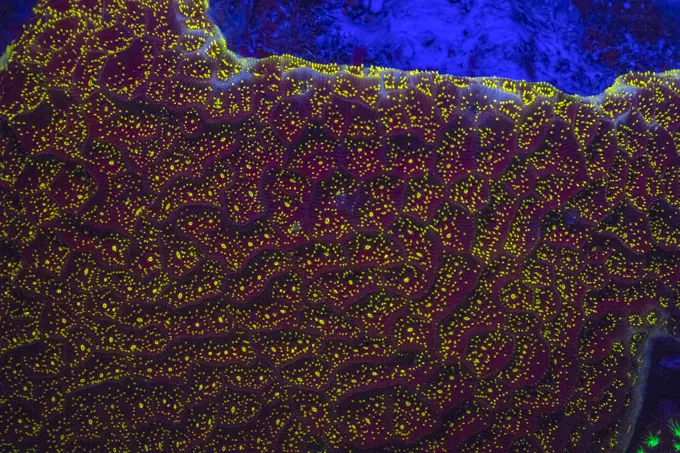
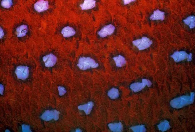
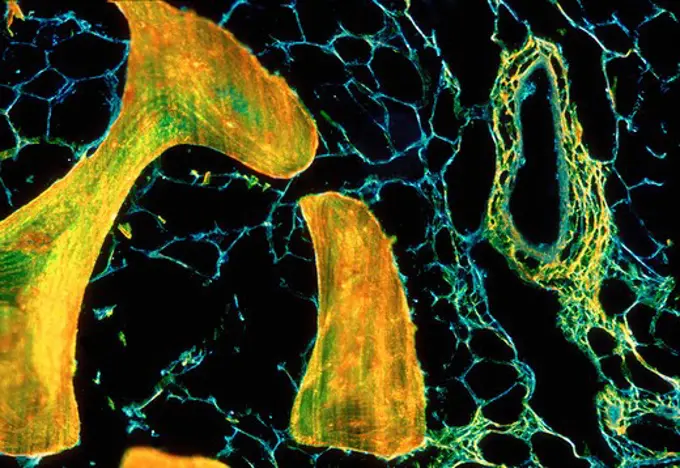
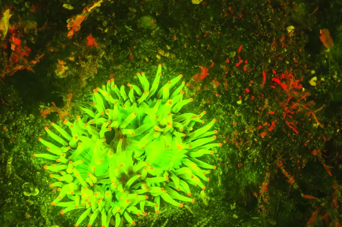
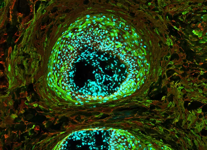
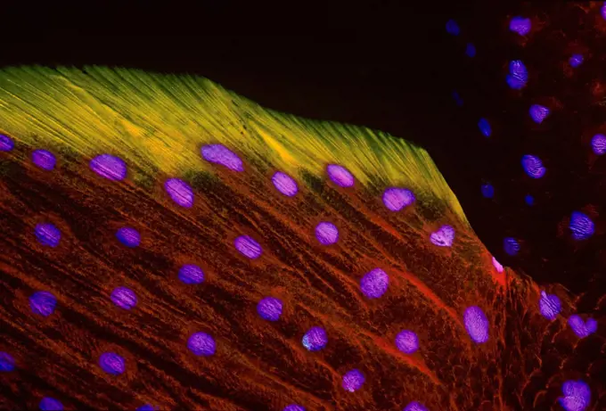
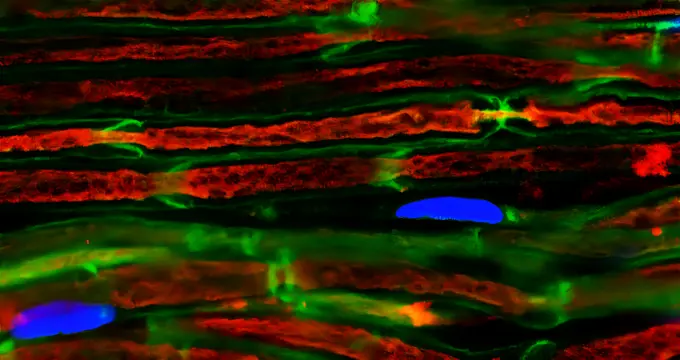
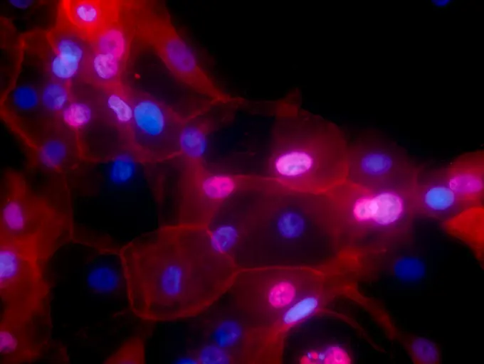
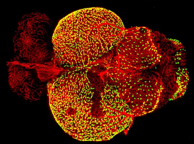
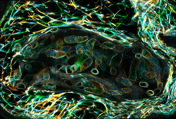
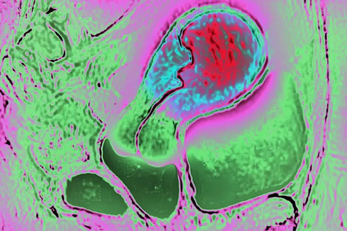
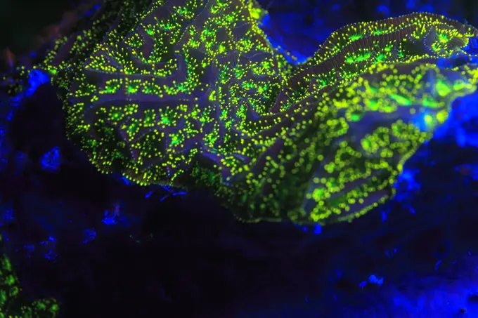
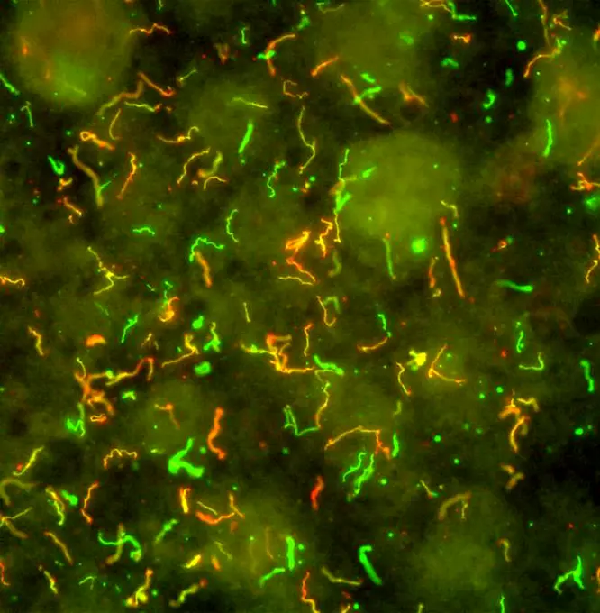
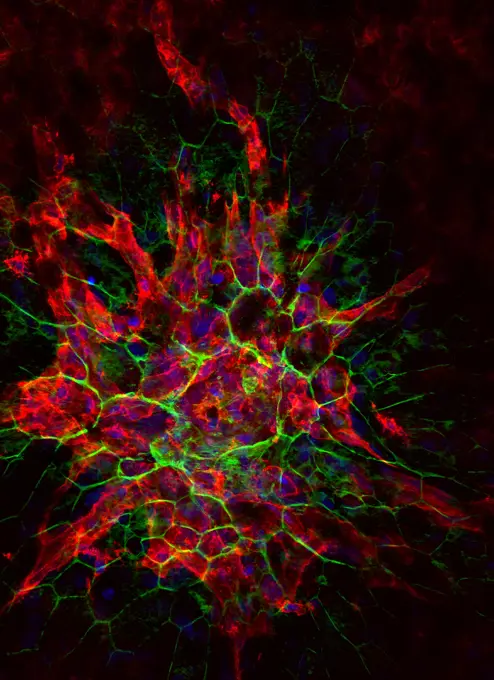
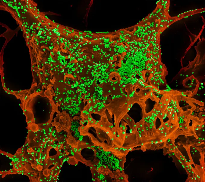
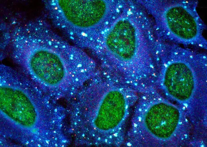
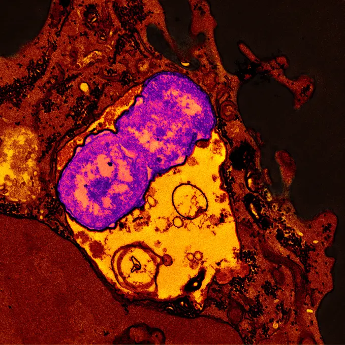
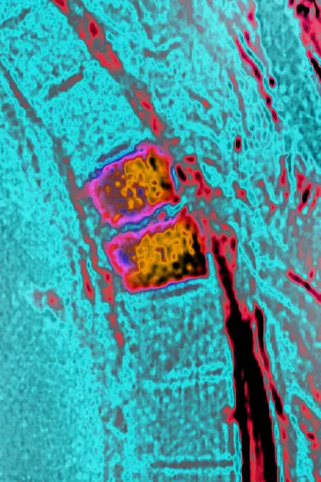
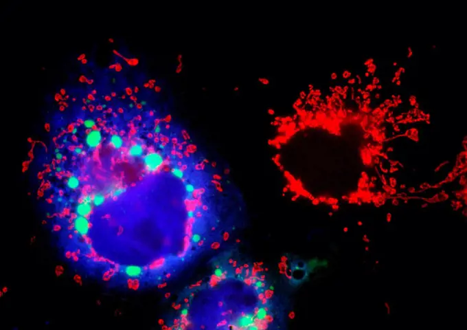
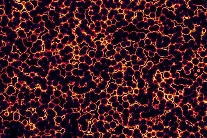
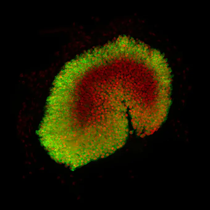
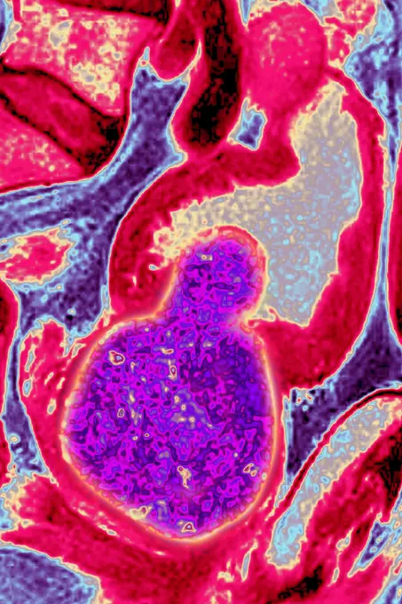
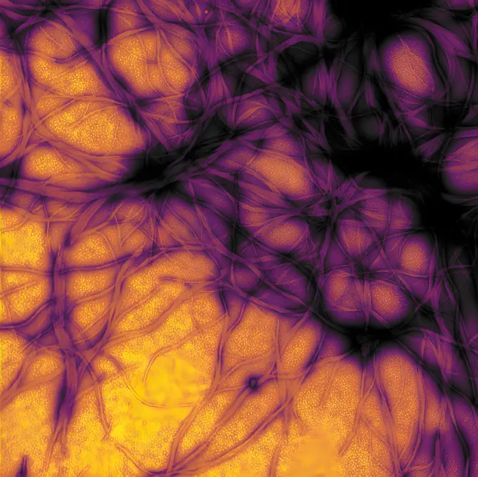
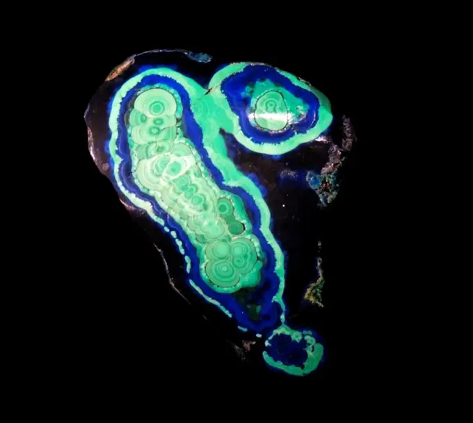
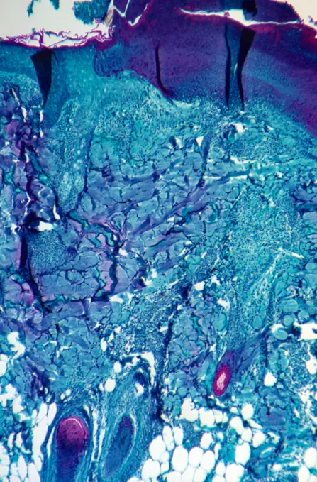
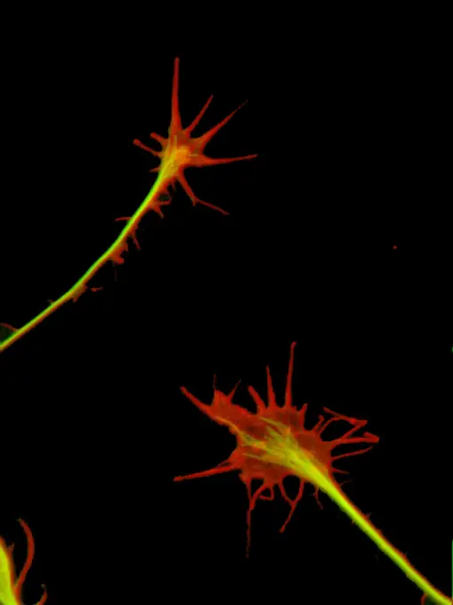
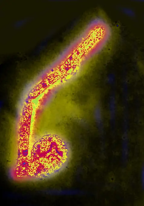
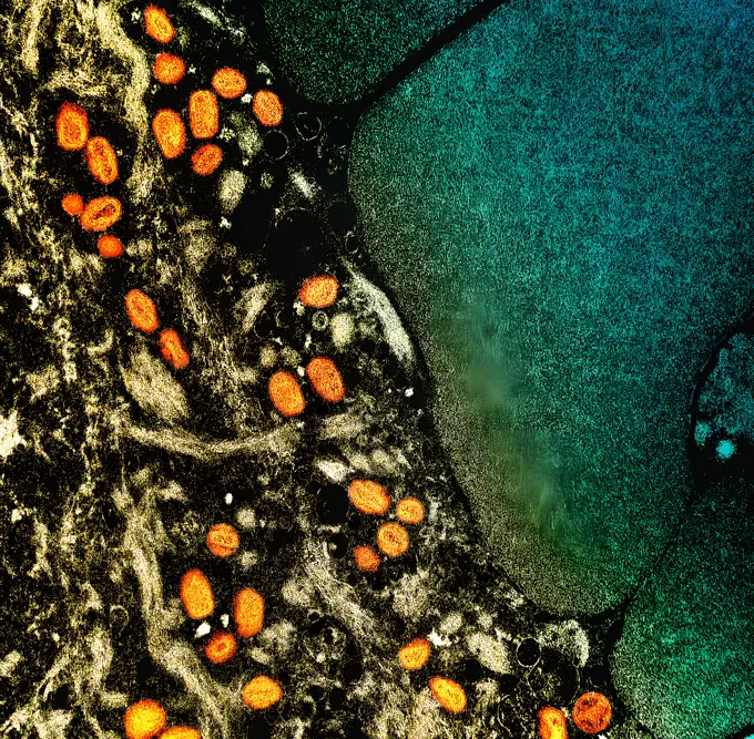
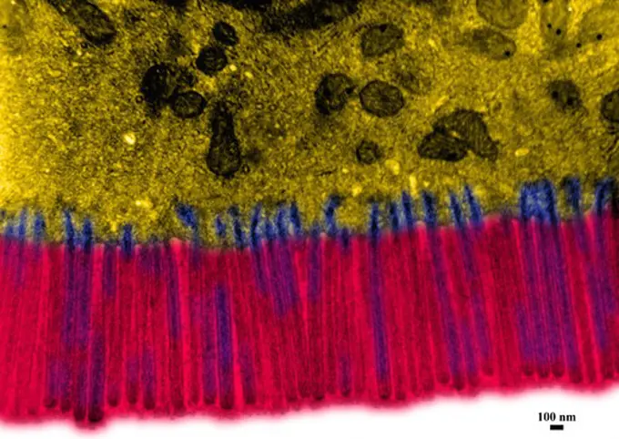
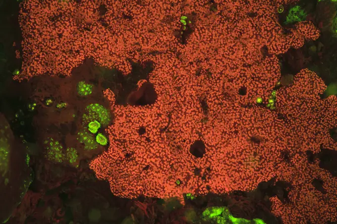
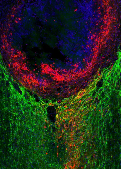
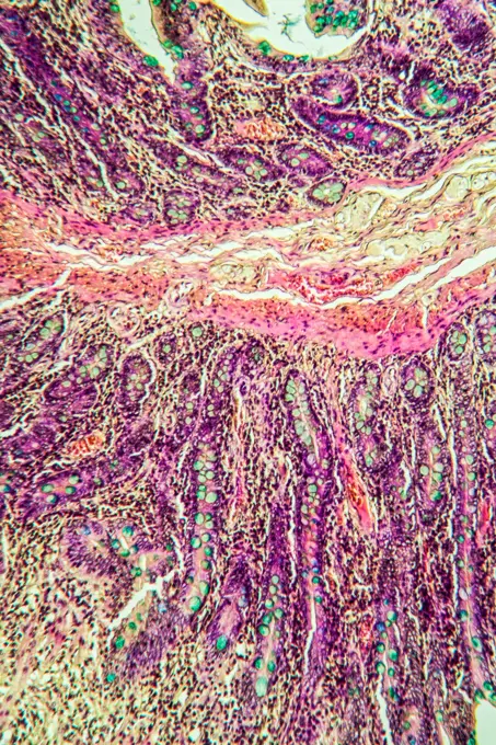
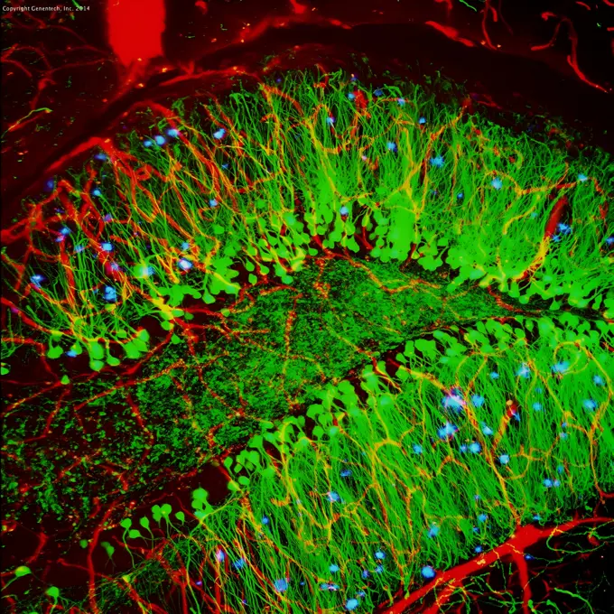
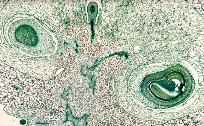
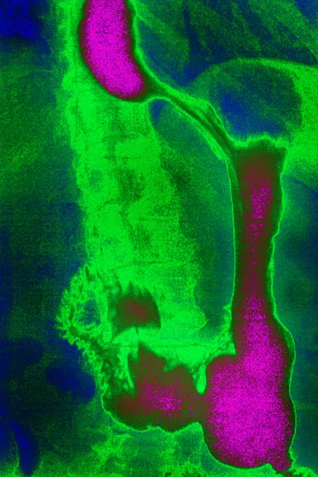
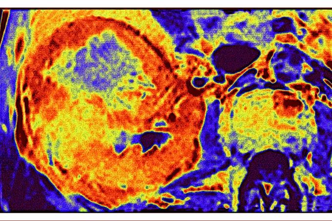
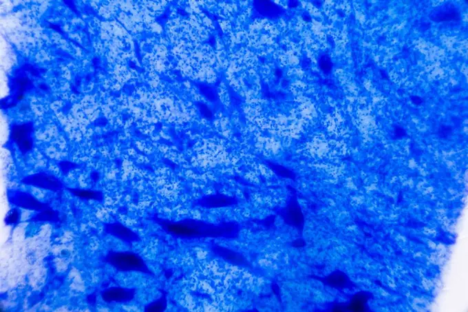
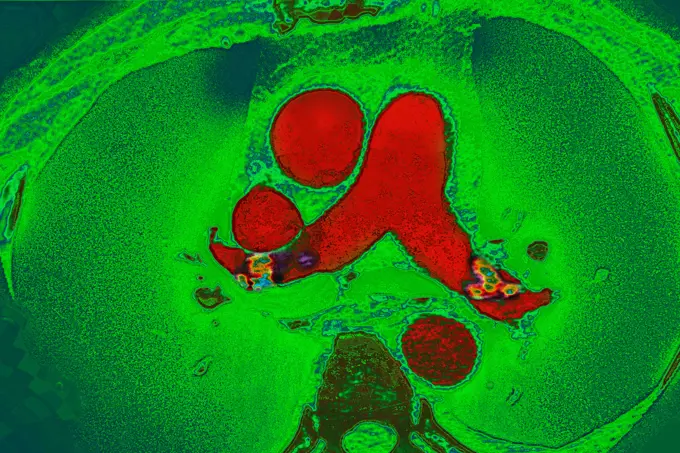
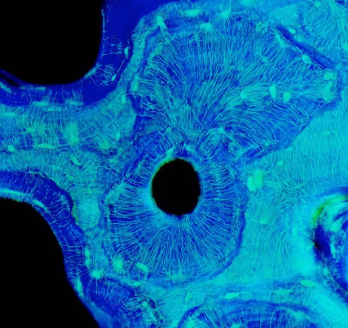
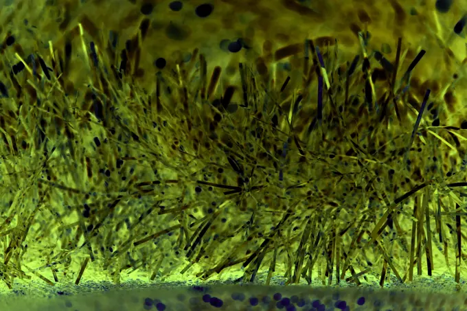
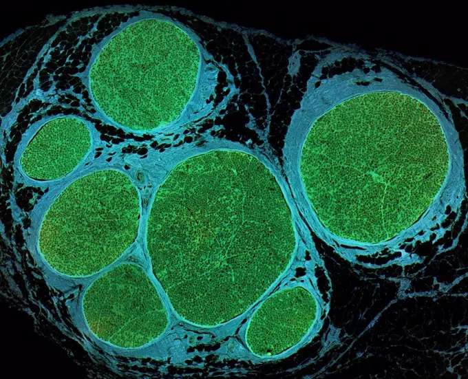
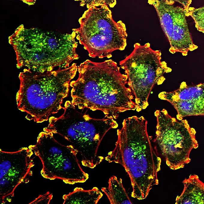
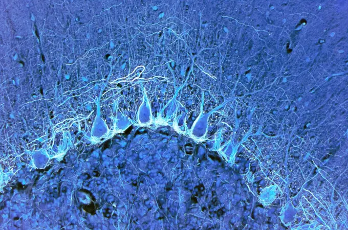
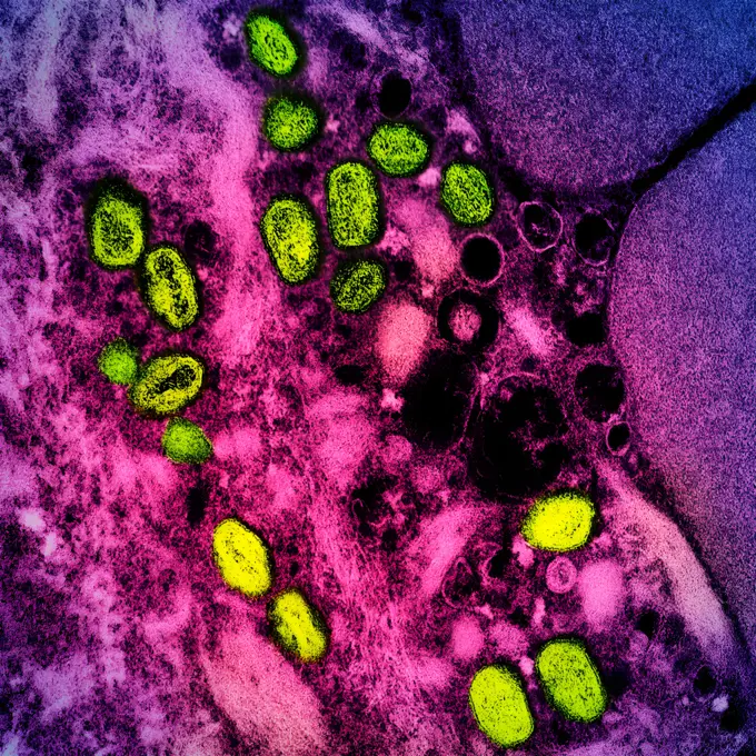
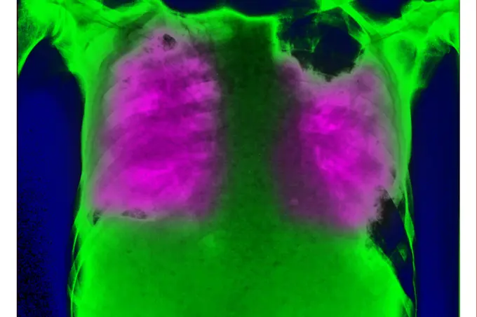
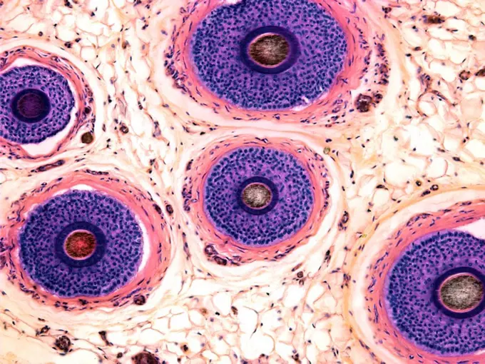
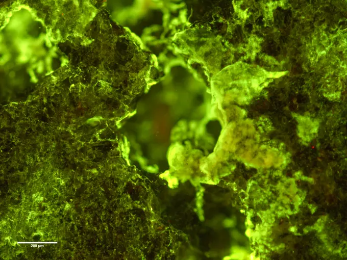
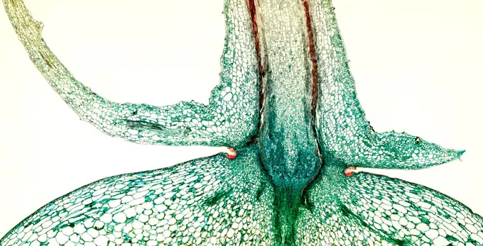
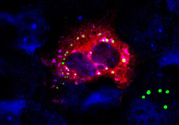
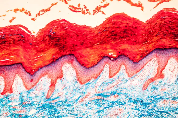
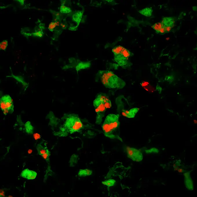
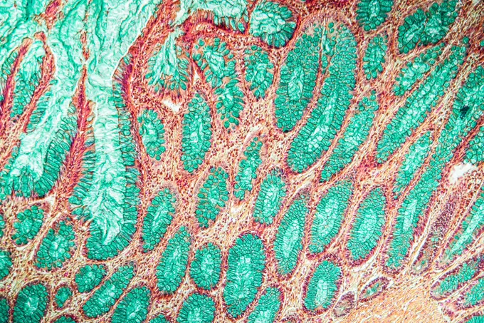
Fine ArtVintageFootageSubscriptionBoardsFree ResearchPricing
  • Fine Art
  • Vintage
  • Footage
  • Subscription
  • Boards
  • Free Research
  • Pricing

Microscopic Tissue Analysis

Close-up microscopic images of various tissues, including skin and cancerous cells, revealing intricate textures and colors.

Scroll Coral, Night Fluorescing. Bonaire, Caribbean
210 assets in this story
4128R-13192203
Microscopic image of skin tissue.
824-63227351
As shown here, pancreatic cancer is associated with an extensive desmoplastic reaction in which the connective tissue around the tumor thickens and scars. Tests similar to the one described here can distinguish between and ampquotgood and ampquot and and ampquotbad and ampquot stromal reactions. The researchers hope this could enable the design of therapies that will reprogram the tumour-stroma interaction. Cancer cells are in red nuclei are in blue stroma / desmoplasia is in cyan and stroma-specific markers are yellow and green while marker overlays appear white.
4413-75202
Close up of Jewel Grouper skin Red Sea in Egypt
1773R-87681
Bone spicules, marrow and adipose tissue
7203-70646342
Natural occurring fluorescence in underwater red fluorescing sponge (Leucetta) and sea anemone (Macrodactyla), captured by using special UV blocking barrier filters, Night dive at Kalabahi Bay, Alor Island, Indonesia
824-63227248
This image shows the expression of keratin 5 (red) and keratin 1 (green) in skin tumors. Keratins are fibrous structural proteins found in epithelial structures, such as skin, hair, and nails. They form filaments that give strength and tenacity to structures. Keratins are often used to aid in the diagnosis of squamous cell carcinoma, a form of skin cancer.
7203-70646500
Scroll Coral, Night Fluorescing. Bonaire, Caribbean
4413-121090
Pectoral fin of a Lunartail Grouper Red sea Egypt
824-63227388
Microscopy image of mouse sciatic nerves showing axons (red) enveloped by Schwann cells (green) with their nuclei shown in blue.
824-63227127
The image shows a cell culture of conditionally reprogrammed human breast cancer cells. The fluorescent red color represents MHC-I and the nuclei are shown in blue.
824-63227183
A new study in rats shows that stem cell secretions, called exosomes, appear to protect cells in the retina, the light-sensitive tissue at the back of the eye. The study was conducted by researchers from the National Eye Institute (NEI), which is part of the National Institutes of Health.
824-63227099
This image, winner of the Federation of American Societies for Experimental Biology's 2017 BioArt competition, shows the brain of an adult zebrafish, a popular organism for studying brain function. It captures dense networks of blood vessels (red) lining the outer surface of the brain. Next to many of these vessels are previously little-studied cells called fluorescent (yellowish-green) granular perithelial cells. Researchers now believe that these cells, often abbreviated as FGP, carry a lot like garbage cans that constantly absorb and store waste to keep the brain clean and functioning properly.
824-63227256
The parasitic worm that causes schistosomiasis hatches in water and grows into a freshwater snail, as shown here. Once mature, the worm returns to water, where it can infect people through skin contact. Initially, an infected person may have a rash, itchy skin, or flu-like symptoms, but the real damage is done over time to internal organs.
824-63224686
Angiography of myocardial infarction caused by atheromatous plaque in the right coronary artery. Angiographic visualization is produced by injection of contrast product.
824-63191265
The cerebellum from a GAD65-eGFP (green) transgenic mouse stained for calbindin to show Purkinje cells and DAPI (blue) to show nuclei.
6214-V62574655
6188-55645074
Zellkern vergrößert Zellkern unter dem Elektronenmikroskop 10.000x Copyright: xZoonar.com/Dr.xNorbertxLangex 13785168
824-63179021
Cervical cancer. Squamous cell metaplasia seen by the microscopy of a histological section of the endocervical epithelium.
1899-53511518
Uterine cancer visualized by sagittal MRI.
7203-70647315
Night dive at Barrier Reef near Saint Georges Caye, Fluorescence emitted at night, exposed with special UV blocking filters, Belize, Central America
824-63227422
The image above shows a small section of the trachea, or windpipe, of a developing mouse. Although only the diameter of a pinhead at this stage of development, the mouse trachea has much in common structurally with the much wider and longer human trachea.
7203-70646321
Natural occurring fluorescence in underwater red and green fluorescing hard coral (Oxpora lacera) and orange fluorescing Ostracods, captured by using special UV blocking barrier filters, Night dive at Kalabahi Bay, Alor Island, Indonesia
1899-53511906
Pulmonary metastases (secondary to kidney cancer), visualized by chest CT scan in axial section. (Photo by: CAVALLINI JAMES/BSIP/UIG)
7203-70646174
Natural occurring red fluorescing sponges (Leucetta sp.) and green hard coral (Acropora sp.). Night dive at Wetar Island, Banda Sea, Indonesia
6214-V61192524
4298-1025
Lily ovary, megaspores, magnification 400x, image enhanced
1525R-78401
Close-up of a pattern
824-63194428
Lyme Disease Bacteria, Borrelia burgdorferi. The Lyme disease spirochete, Borrelia burgdorferi, is an obligate parasite that cycles between ticks and vertebrate hosts. B. burgdorferi alters the proteins expressed on its outer surface, depending on the state of each host. Here, we used immunofluorescent antibodies to identify spirochetes that express outer surface protein D (yellow and red) and merged the image with an image of all the spirochetes labeled with an anti-B. burgdorferi antibody (green).
824-63227153
Confocal microscope image of laser-induced choroidal neovascular complex (CNV) 14 days after laser injury. RPE-choroid flat mount (brown brown rat). The IB4 lectin (red) labels endothelial cells in the CNV complex. Phalloidin (green) labels f-actin in RPE junctional complexes and stress fibers. Cell nuclei are labeled with DAPI (blue).
824-63227271
Color-enhanced scanning electron micrograph of Vero E6 cells infected with Middle East respiratory syndrome coronavirus (MERS-CoV).
4341-106
Stressed Over-Oxygenated Cells, Microscopic View
824-65830678
A human neutrophil (red) containing ingested Klebsiella pneumoniae (purple). NIH Investigates Multidrug-Resistant Bacterium Emerging in Community Settings
6214-V60916329
1899-61460148
Vertebral osteitis (inflammation of the bone tissue of two dorsal vertebrae, caused by a bacterial infection). sagittal radiograph of the dorsal vertebrae
7203-70646209
Natural occurring fluorescence in underwater hard coral (Diploastrea heliopora). Night dive near Lewolin Village, Illi Api Island, Selat Boleng Strait, Indonesia
1525-27217140
Far being shone nebula and star field against space. Elements of this image furnished by NASA . 3D rendering.. Far being shone nebula and star field. 3D rendering
824-29057234
Influenza virus of the Orthomyxoviridae family (respiratory viral infection). Transmission electron microscopy, viral diameter 80 to 120 nanometers.
4341-117
Comparing Mutant Cell and Normal Cell, Microscopic View
1848-51348766
Shape of bacterial cell: cocci, bacilli, spirilla bacteria background
824-63227240
Hair cells (red) and associated supporting cells (green) in the sensory patch of a mouse utricle, part of the balancing apparatus of the inner ear.
1848-61033048
Ebru marbling handmade art, traditional Ebru technique. abstract background of marble liquid ink art
824-63227305
This image shows mouse muscle cells seen under a microscope. The cells have fused to form myotubes which have many nuclei (stained blue). The cells produced from mouse skeletal muscle stem cells with a harmless virus that made them glow green. The green color remained when the stem cells fused into myotubes. Some myotubes are stained red for a protein involved in muscle contraction (myosin heavy chain), a characteristic of mature muscle fibers. Researchers should use the same viral delivery system to genetically modify cells and assess how altering cell fusion impairs myotube growth. This image was the 2017 winner of the Federation of American Societies for Experimental Biology (FASEB) BioArt competition.
824-65830978
Colorized transmission electron micrograph of two mature mpox virus particles (yellow) attached to the surface of an infected VERO E6 cell (purple/pink).
824-63225948
Large prostatic adenoma, visualized by frontal x-ray of the pelvis.
1899-54027985
The human coronavirus causes respiratory infections (colds), and gastroenteritis. Image made from a transmission electron microscopy view. Approximate viral diameter: 80 to 160 nm.
824-63227357
Prion protein shown in E. coli, purified and fibrillated at pH 7. Recombinant proteins such as the prion protein shown here are often used to model how proteins respond to each other and sometimes polymerize in neurodegenerative disorders.
1746-19691587
The optical properties (color, intensity) of minerals such as azurite and malachite are explained in the context of conventional electronic spectroscopy of coordination complexes. Relatively detailed descriptions are proided by ligand field theory.
824-63227034
Dendritic cells entering the hair follicle. Credit: NIAID
1525-28464128
Air and oil bubbles inside water base form patterns
824-63227401
Researchers study star-shaped brain cells: NIH-funded researchers have used 3D collections of brain tissue from human cells to study star-shaped astrocytes in the brain.
824-63179017
Cancer of the uterus. Adeno-acanthoma (glandular tumor). Cancer of the endometrium develops from the membrane lining the uterus. Microscopy of a histological section.
824-63227035
Transmission electron micrograph of Ebola virus nucleocapsids (small orange circles) and virus particles (larger orange filamentous shapes) in infected African green monkey kidney cells. Focused image at the NIAID Integrated Research Center in Fort Detrick, Maryland.
4341-101
Prion-like Aggregating Protein in a Cell, Microscopic View
824-57657600
Under 50X magnification, this image shows a section of skin tissue, taken from a lesion on the skin of a monkey infected with monkeypox virus. The specimen was obtained on day 8 of rash development.
6145-29255542
Dorsal Root Ganglion neuron growth cones
7099-70352885
Italy, Sicily, Mt Etna Lava. The dark of night intensifies the dramatic color of flowing lava from Mt. Etna on Sicily in Italy.
824-63178730
Avian influenza H5N1 virus
1899-65662470
Colorized transmission electron micrograph of mpox virus particles (orange) found within an infected cell (brown and green), cultured in the laboratory.
824-57656860
Arteritis of the lower limbs (disease affecting the arteries of the legs, it is almost always due to a deposit of cholesterol in the arteries, which limits blood circulation). microscopy on a histological section of a diseased artery.
6188-55644120
Kiefer Kiefer Stamm quer 100x Copyright: xZoonar.com/Dr.xNorbertxLangex 13693821
6188-55644147
Small intestine small intestine pig crossing 100x copyright: xzoonar.com/dr.xnorbertxlangex 13693893
824-63191243
Cells in the hilus of the dentate gyrus of the hippocampal formation from an Nr4a1-eGFP (green) transgenic mouse stained for calretinin (red) and DAPI (blue) to show nuclei.
824-63225940
Rotavirus-infected cell revealing numerous viral factories in the cytoplasm.
824-63227258
This image captures the many layers of nerve cells in the retina. The top (green) layer is made up of cells called photoreceptors that convert light into electrical signals to send to the brain. The two most well-known types of photoreceptor cells are rod-shaped and cone-shaped. Rods help us see in low light conditions, but can and
6214-V61192451
4297-1252
Transmission electron microscopic image of microvilli from a human epithelial cell in the jejunum of the small intestine
7203-70646254
Natural occurring fluorescence in underwater orange fluorescing tunicates (Botryllus) and green hard coral (Acropora), captured by using special UV blocking barrier filters, Night dive at Wetar Island, Banda Sea, Indonesia
824-63227325
An NIH-funded mouse study suggests scar formation may help, not hinder, nerve regrowth.
6145-29295570
Cell death in limb bud of a webbed foot
6188-55644113
Dünndarm Dünndarm Schwein quer 100x Copyright: xZoonar.com/Dr.xNorbertxLangex 13693679
824-63227122
In addition to blood vessels (red) and nerve cells (green), this mouse brain has abnormal clumps of proteins called plaques (blue). These plaques multiply in the brains of people with Alzheimer's disease and
4141-111428926
Light micrograph cross section through the young fruit of a Tomato plant (Lycopersicum esculentum) CS, UK
824-63204728
Frontal upper gastrointestinal (GI) series.
824-63207421
Cancer of the liver, seen in a radial cross-section abdominal scan
824-63227297
Microscopy of a spinal cord sample from a mouse model of neurodegeneration. The following fluorescent stains are used: DAPI is blue, TDP-43 is green, and GFAP is red. Researchers from the Developmental Neurodegeneration Unit at the Eunice Kennedy Shriver National Institute of Child Health and Human Development (NICHD) are using these models to study the early events that underlie the onset and progression of neurodegenerative diseases, such as amyotrophic lateral sclerosis and Parkinson's disease.
1525-25708012
Nerve cell under the microscope - Abstract blue dots on white background.
824-63215964
Pulmonary embolism in the left and right pulmonary arteries, seen on a radial section chest CT scan.
4128R-22540
Compact bone, light micrograph. Compact bone consists of concentric layers of collagen_containing matrix lamellae, dark blue, around Haversian canals ...
6188-64760471
tinsel for decorating a Christmas festive tree. computer processing, inversion. tinsel for decorating a Christmas festive tree. computer processing, inversion Copyright: xZoonar.com/OvchinnikovaxOlgax 11565235
4128R-13573034
Nerve fibres. Light micrograph (LM) of a transverse section through a bundle (fascicle) of nerve fibres. Within each fascicle are many myelinated nerv...
6162-76258107
Aerial view of molten lava flowing like a fiery river through the dark, rugged volcanic landscape, Reykjavík, Reykjavíkurborg, Iceland.
824-63227276
The ability of cancer cells to move and spread depends on central actin-rich structures such as podosomes (yellow) shown here in melanoma cells. Cell nuclei (blue), actin (red) and an actin regulator (green) are also shown.
1899-53513323
Ebola virus isolated in November 2014 from patient blood samples obtained in Mali. The virus was isolated on Vero cells.
1848-50935785
Ebru marbling handmade art, traditional Ebru technique. abstract background of marble liquid ink art
4128-18929670
Purkinje nerve cells, LM
824-63170802
This sagittal MRI image of the thoracic spine reveals a spondylodiscitis.
824-57657570
Colorized transmission electron micrograph of monkeypox particles (green) found within an infected cell (pink and purple), cultured in the laboratory.
6188-55645201
Mushrooms and bacteria destroy emulsion films under the microscope 100x mushrooms and bacteria destroy emulsion film under the microscope 100x copyright: xzoonar.com/dr.xnorbertxlangex 13803108
824-63215948
Allergic bronchopulmonary aspergillosis (ABPA), seen on a frontal chest x-ray.
4128R-10574
Hair follicles, light micrograph
824-57657692
Colorized transmission electron micrograph of monkeypox virus particles (green) found within an infected cell (orange), cultured in the laboratory.
6145-45209974
jsc2019e039825 (7/16/2019) --- Preflight Ffuorescence microscopy image of biofilm of Spingomonas desiccabilis growing over and into the surface of a basalt slide as part of BioRock experiment. Organisms are stained with DNA binding sye Sybr Gold. Growth can be seen into the rock cavities. The purpose of the Biorock investigation is to examine the effects of altered gravity on the rock/microbe/liquid system as a whole. (Image Courtesy of: ESA)
4141-111428928
Light micrograph longitudinal section through the fruit pedicel of a Tomato plant (Lycopersicum esculentum) LS, UK.
4341-135
Genetically Engineered Cell, Microscopic View
6188-55643792
unbehaarte Haut unbehaarte Haut Gewebe unter dem Mikroskop 100x Copyright: xZoonar.com/Dr.xNorbertxLangex 13650702
824-63227283
Microscopic image of mitochondria in a single heart cell. Mitochondria highlighted in red were exposed to ultraviolet light.
1899-53511887
Cancer of the right kidney visualized by an abdominal scanner in radial section. (Photo by: CAVALLINI JAMES/BSIP/UIG)
824-63226731
Cancer of the right kidney visualized by an abdominal scanner in radial section.
824-63227281
Microglia, a type of nervous system cell previously suspected of causing retinal damage, has been shown to be beneficial in slowing the progression of prion disease in the brain, and now in the retina. In this image, microglial cells (green) engulf and remove prion-damaged photoreceptors (red), which appears to slow retinal degeneration.
6188-55644909
Colon cancer colon cancer tissue cut under the microscope 100x copyright: xzoonar.com/DR.XNORBERBERTXLANGEX 13772981
PREVIOUS
of 3
NEXT
2401 S. Ervay, Suite 206
Dallas, Texas 75215
United States
Browse
CategoriesGalleriesFree Research
For Creators
License Your ContentSuperStock For StudentsFrame of Mind BlogContributor PortalNew Image SpotlightLicense AI Imagery
Resources
About UsLicensingTerms & ConditionsAPI AccessPrivacyPricing
Contact
+1 866 236 0087help@superstock.com Contact form
©2026 SuperStock. All Rights Reserved. -B
This site is protected by reCAPTCHA and Google Privacy Policy and Terms of Service apply.