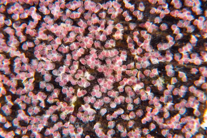
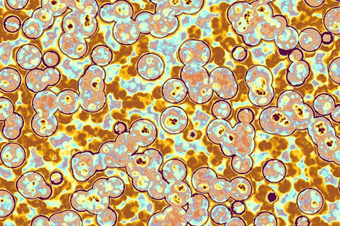
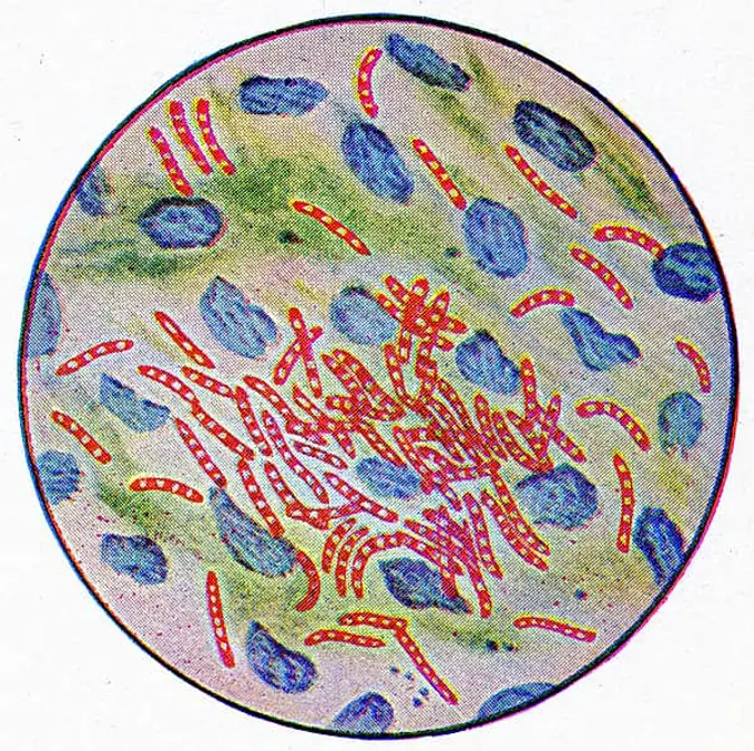
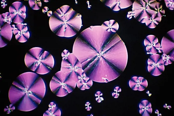
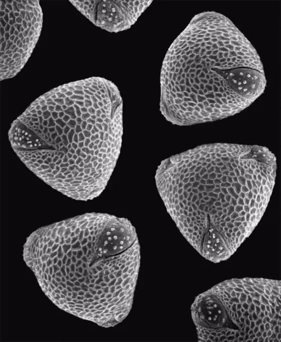
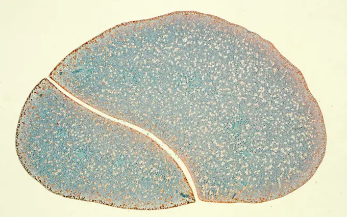
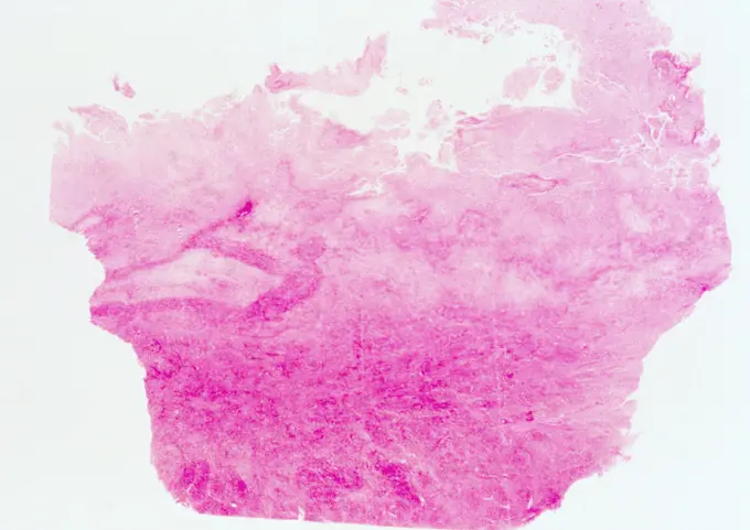
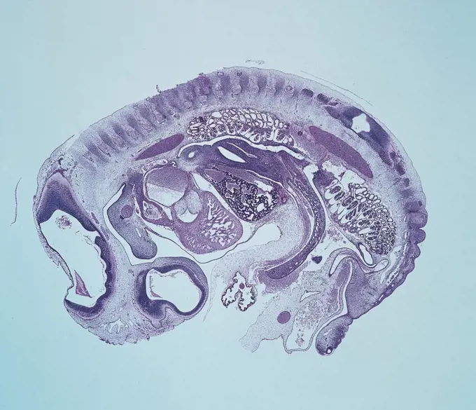
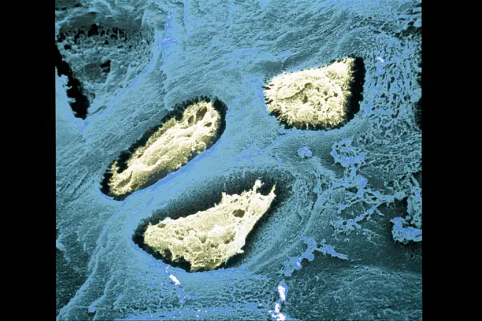
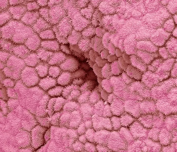
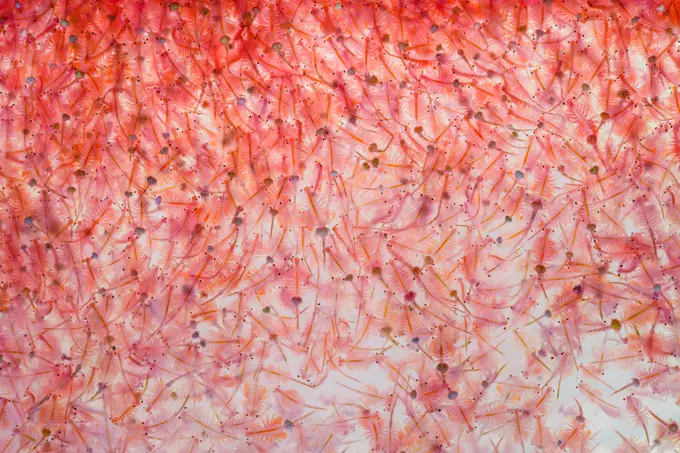
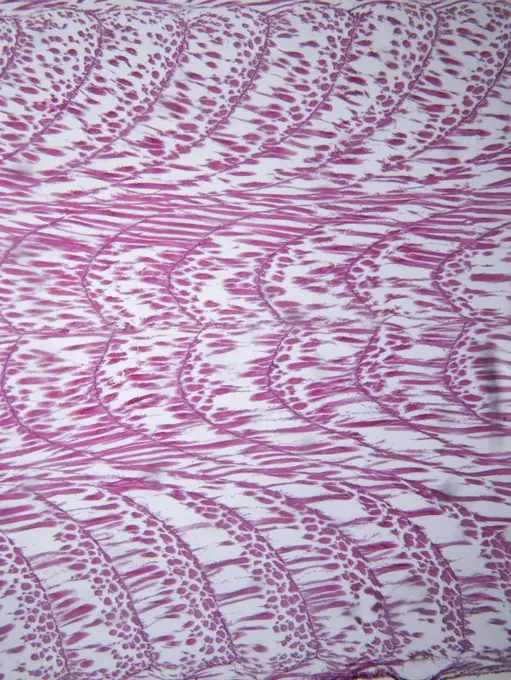
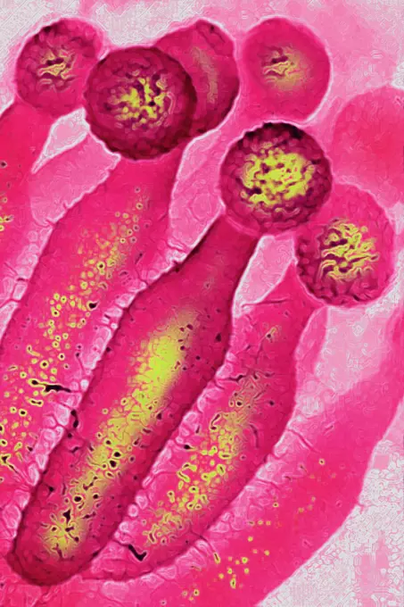
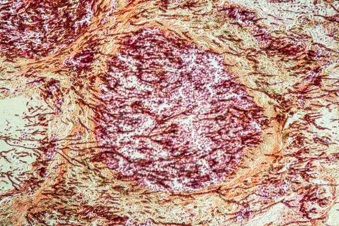
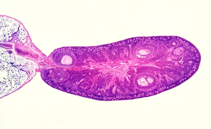
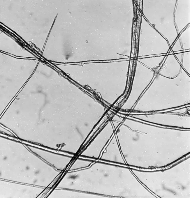
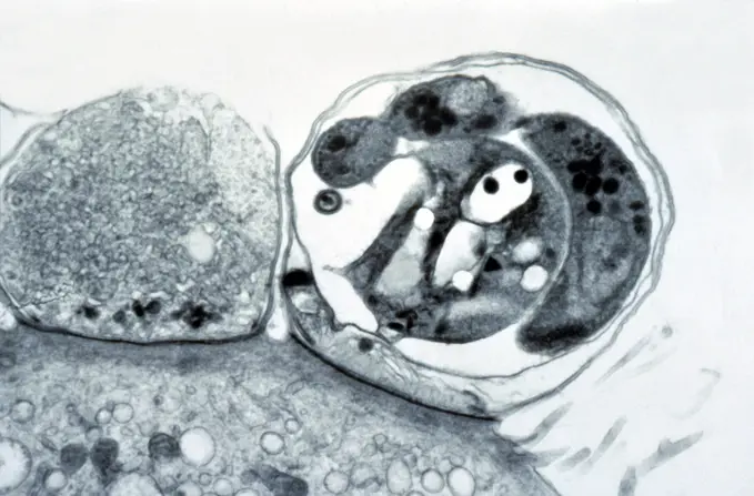
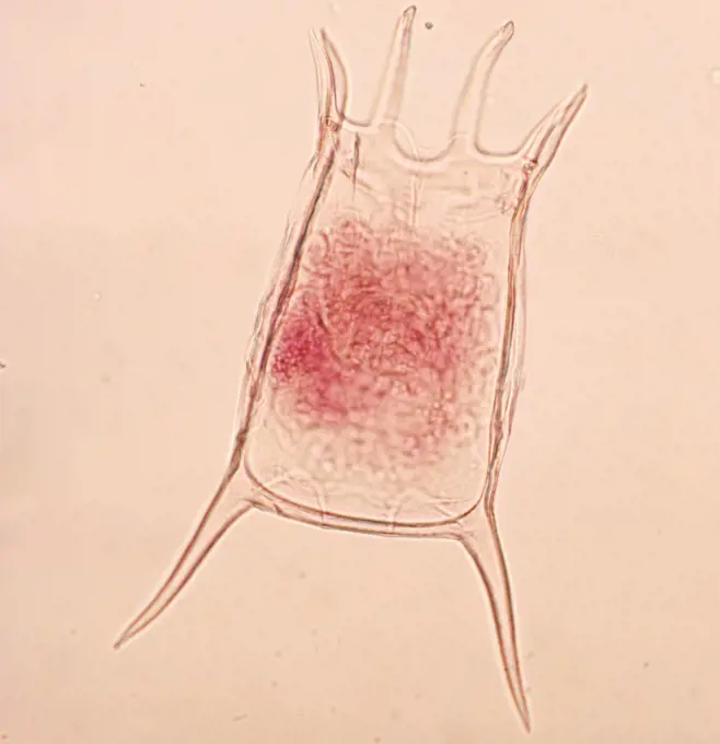
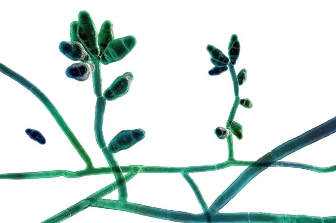
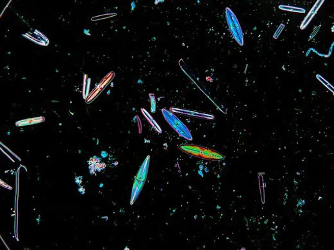
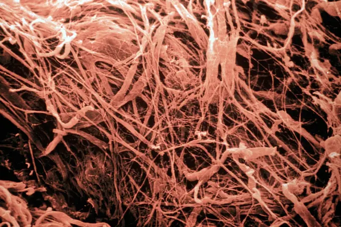
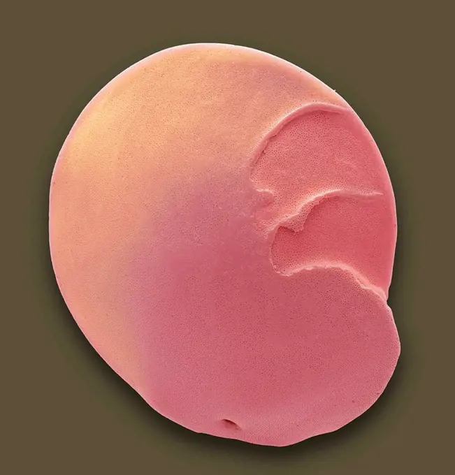
Microscopic Plant Structures

Detailed images of plant tissues viewed under a microscope, showcasing cell structures and colors, offering insights into botanical anatomy and morphology.

Swords swords cross 100x copyright: xzoonar.com/DR.XNORBERBERTXLANGEX 13688506
Computer Generated ImageAcrosymphyton purpuriferum, red algae, Florideophyceae.Staphylococcus. Color enhanced scanning electron micrograph of a Staphylococcus biofilm on the inner surface of a needleless connector.Bubbles of dung in a creekIndo-Pacific Sergeant, Abudefduf vaigiensis, egg brood, Mabul, Sabah, Malaysia, Borneo.Shape of bacterial cell: cocci, bacilli, spirilla bacteriaGerms of tuberculosis, vintage engraved illustrationColoured scanning electron micrograph (SEM) of a freeze fracture of the vagina showing the stratified squamous epithelium and the underlying lamina pr...S-benzyl iso-thiouronium chloride, Crossed Polars, x60flat lay hydroalcoholic gel close upCommon Nasturtium (Tropaeolum majus) SEM close-up view of pollen grains at 1050x magnificationRed blood cells, SEMKosmetische schimmernde Peeling oder Maske smudge gel peeling swatch isoliert auf weiß Cosmetic shimmering scrub or mask smudge gel peeling swatch isolated on white Copyright: xZoonar.com/IrinaxKvyatkovskayax 22736403Light micrograph cross section through the fruit of a Loquat tree (Eriobotrya japonica), UK. This evergreen tree of shrub is also known as Japanese medlar or Japanese plum.Stechpalme Stechpalme Blatt Querschnitt 100x Copyright: xZoonar.com/Dr.xNorbertxLangex 13762695Amoebiasis: amoebic abscess in liverGerms of tuberculosis, vintage engraved illustration.Microphotograph of a small jellyfish, genus Obelia, Coelenterata, at x38 magnification.Rhodochrosite, ArgentinaMicrophotograph of a longitudinal section of a pig embryo, at x6.3 magnification.Winter Jasmine leaf under a microscope (Leaf of Winter Jasmine C.S.) (Jasminum nudiflorum), 100xLiver fluke, light micrographCartilage. Cartilage with 3 holes containing 3 cells ESM (Electron Scanning Microscope)Uterus lining, SEMArtemia plankton Brine shrimpFish muscle tissue, light micrographPigeon blood cells, vintage engraved illustration. From the Universe and Humanity, 1910.Penicillium chrysogenum, a penicillin-producing mold, visualized by scanning electron microscopy.Lungs of an aid patient lung of an aid patient lung tissue 200x copyright: xzoonar.com/dr.xnorberbertxlangex 13735558Light micrograph transverse section through the ovary of a rat showing a range of developing follicles. TS, UKPower cabinet image of flax. Uppmed Plant Fibergiberg enlarged 200 times.Gastrointestinal tract: cryptosporidiosis and HIVThe retina, inside the eye, contains a light-sensitive membranous layer of cells. These are specialized nerve cells: elongated rods (flower like shape...Transmission electron micrograph of SARS-CoV-2 virus particles, isolated from a patient. Image captured and color-enhanced at the NIAID Integrated Research Facility (IRF) in Fort Detrick, Maryland. Credit: NIAID.Vaccinia virus particles, TEMLiver cancer, CT scanSix patches of skin showing types of skin disease. Coloured stipple etching by J. Pass, c. 1822.Liver: bacterial cholangitis and HIVKeratella square, Rotifers, seen under a microscope.Curvularia mould fungus, computer illustration. Curvularia is a group of about 35 species, most of which only infect plants. However, C. geniculata ca...The eye, as seen through a microscope: two figures. Colour lithograph, ca. 1862.Diatoms found in pond water near Grant, NY. USA. Image enhanced to show structure.  Magnification 100xSkin. Derm after action of collagen disease (connectivity) Elastic fibres after destruction of collagen ESM (Electron Scanning Microscope)Vinestain on a white table.CALCETIN DE LANA.This 1973 photomicrograph depicts three Naegleria gruberi, free-living amoebae. CDC/Dr George Healy 1973Sample ca. 1830 French, Mulhausen. Sample 219981Skin. Derm after action of collagenosis Deterioration of the elastic fibres further to destruction of collagen SEM (Scanning Electron Microscope)Italy, Apulia, Castellana Caves, detailThree-dimensional pattern of a cell endoplasmic reticulum. Drawing.Bacteria cells square banners. Microorganism collection. Vector doodle style composition.Foraminiferan. Coloured scanning electron micrograph (SEM) of a foraminiferan microfossil from maldives beach sand. Microfossils' are roughly 0.05 to ...Collection Dutch Heidematschappij. development. undatedFavus. Dr. Selden I. Rainforth (American, 1879 - 1960)