Fine ArtVintageFootageSubscriptionBoardsFree ResearchPricing
  • Fine Art
  • Vintage
  • Footage
  • Subscription
  • Boards
  • Free Research
  • Pricing

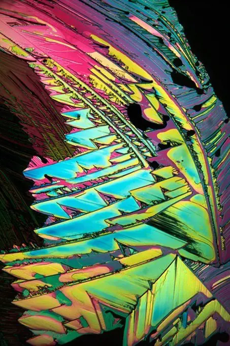
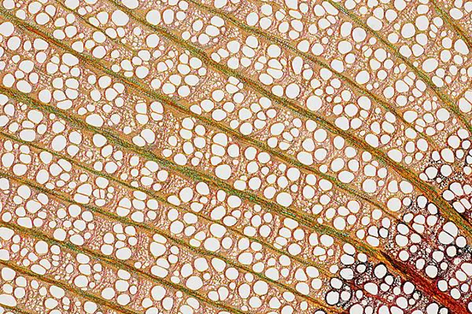
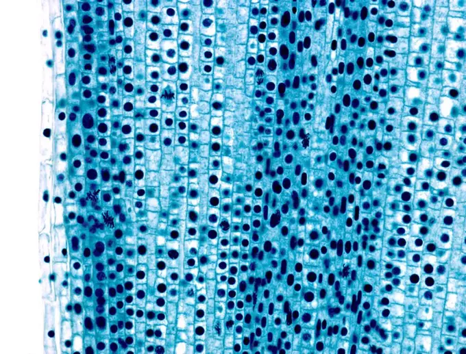
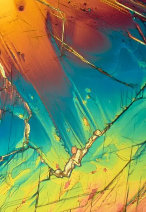
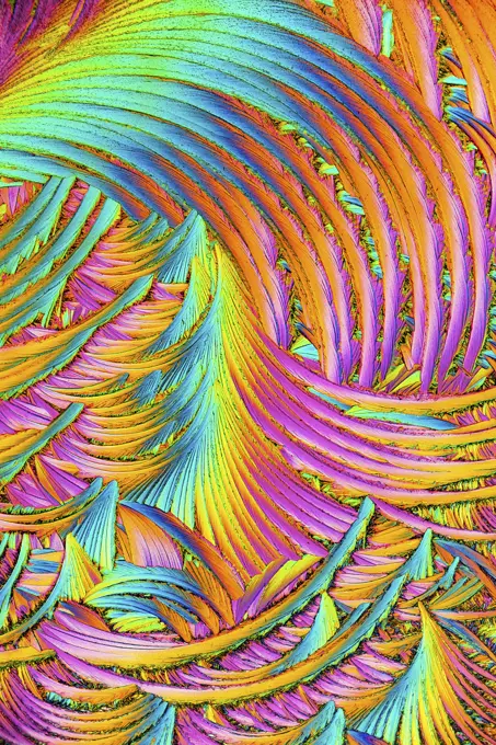
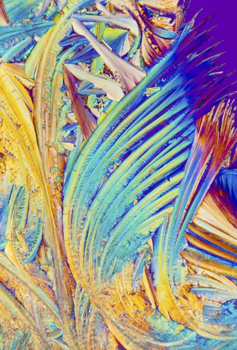
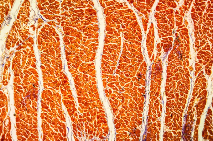
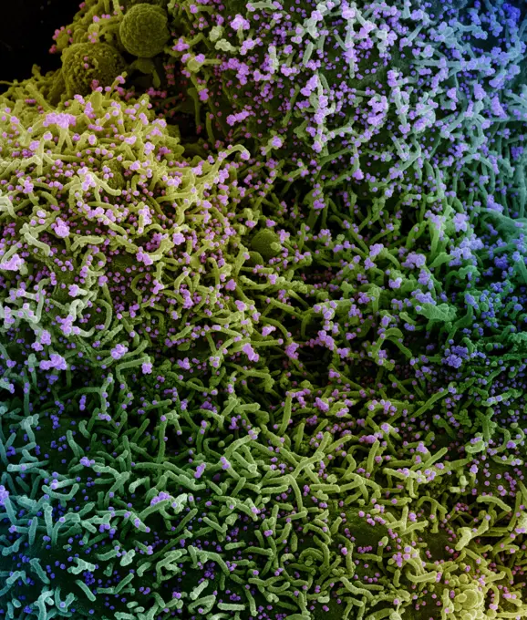
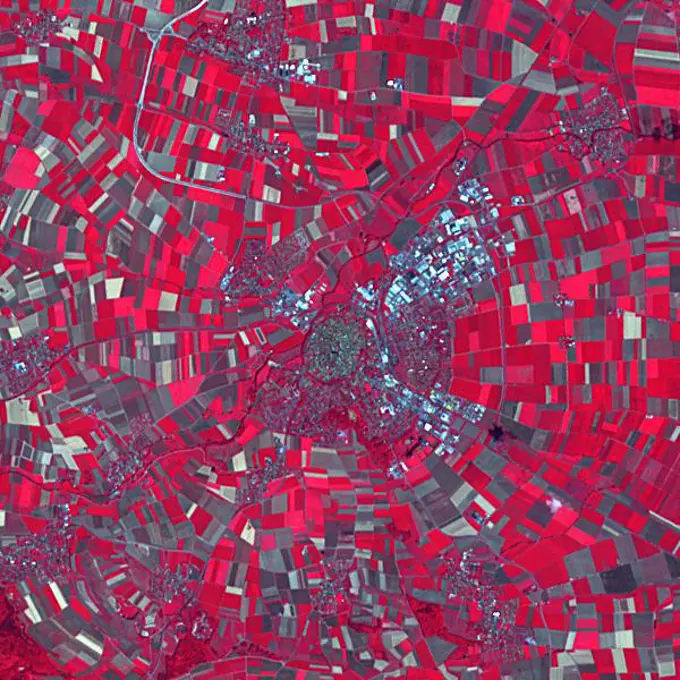
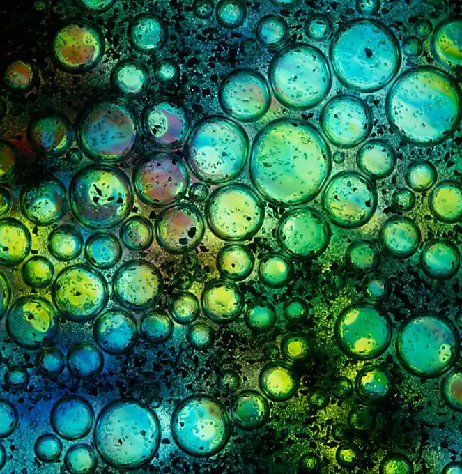
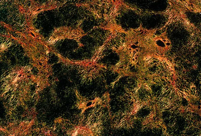
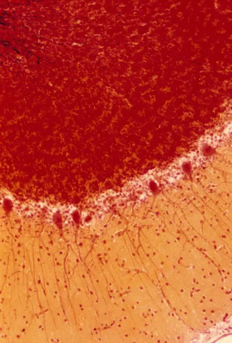
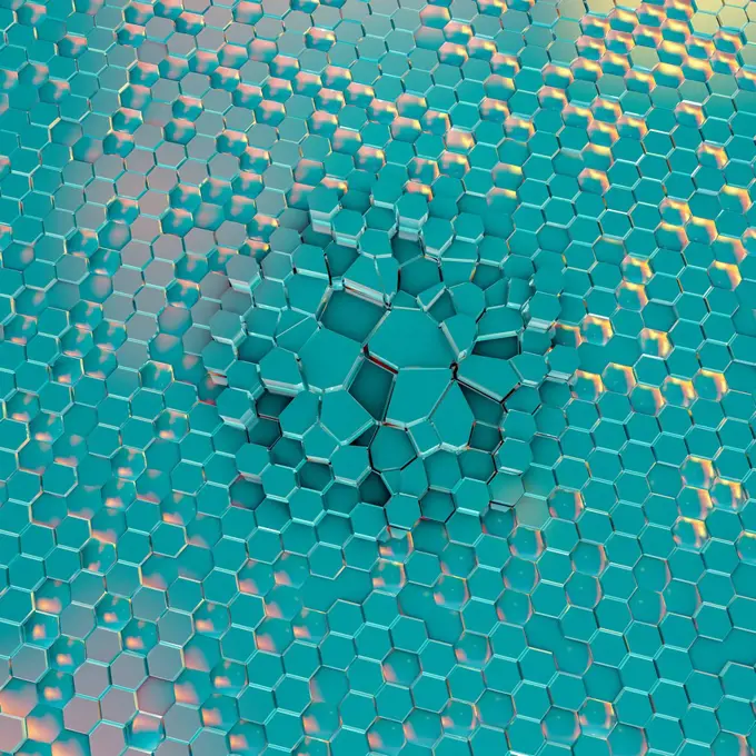
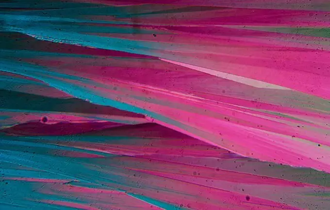
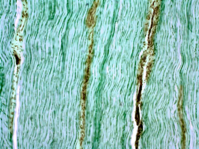
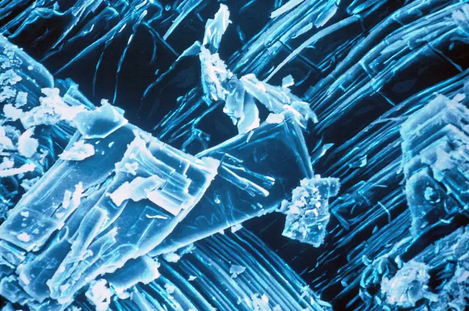
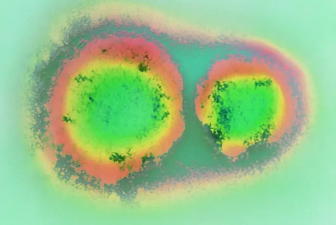
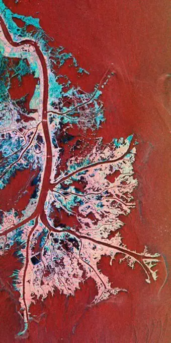
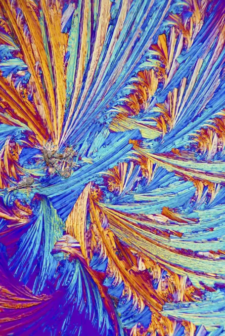
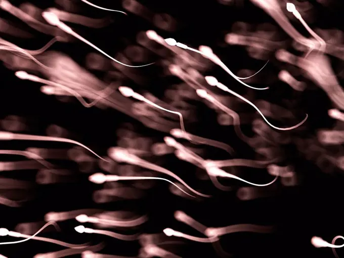
Microscopic Patterns and Textures

Colorful microscopic images showcasing various textures and patterns, including bacteria shapes and vibrant organic materials.

Dinosaur Petrified Bone
267 assets in this story
1525-26835773
Shape of bacterial cell: cocci, bacilli, spirilla bacteria background
7008-69756998
Malachite
4491-V111125320
1848-49830819
Oil bubbles inside water base form patterns
1525-25708033
Yellow pepper under the microscope.
1788-111172696
Microphotograph of agrobacterium tumefaciens (Rhizobium radiobacter), bacteria, responsible for plant tumors.
4128R-12699808
Coloured scanning electron micrograph (SEM) of breast cancer cells growing in culture. These are from the MCF-7 cell line. Magnification: x280 when pr...
1848-49828906
Oil bubbles inside water base form patterns
7008-69756983
Dinosaur Petrified Bone
6145-58971792
A severe drought in the Amazon is disrupting transportation, isolating communities and putting wildlife at risk. The Amazon and its tributaries, including the Rio Negro that joins the Amazon at Manaus (2023 image), fell to their lowest level in over a century. The images were acquired October 9, 2023 and August 15, 2020. They cover an area of 30 by 45 km, and are located at 2.8 degrees south, 60.7 degrees west.
1525-28110021
Shape of bacterial cell: cocci, bacilli, spirilla bacteria background
1525-24681987
Shape of bacterial cell: cocci, bacilli, spirilla bacteria background
4413-150598
Skin of Male panther chameleon
1828R-1508
Asorbic Acid Crystals
7008-69756972
Dinosaur Petrified Bone
824-63227160
This image shows the crimp (ripple) pattern of collagen fibers in a medial collateral ligament, which is the ligament on the inside of the knee. The ligament section was magnified to 200X using polarized light microscopy.
1815-69353039
Colorful microcrystals of urea displaying vibrant patterns
1848-49830310
Oil bubbles inside water base form patterns
824-63170770
This sagital MRI image of the brain shows a hypophyseal atrophy.
1525-24756389
Oil bubbles inside water base form patterns
1815-69353066
Colorful abstract patterns of salicylic acid crystals under a microscope
6188-55643673
Fremdkörper Granulom Fremdkörper Granulom Gewebe unter dem Mikroskop 100x Copyright: xZoonar.com/Dr.xNorbertxLangex 13633559
1525-25701200
Abstract ink spot in many color print on flyer paper under the microscope.
1848-18023449
Ascorbic acid, vitamin C crystallized, polarized light
1848-66232670
Oil bubbles inside water base form patterns
1848-66605182
Shape of bacterial cell: cocci, bacilli, spirilla bacteria background
1848-66573973
Air bubbles inside water base form patterns
6188-55643807
Froschnest frog funny tissue under the microscope 100x copyright: xzoonar.com/DR.XNORBERBERTXLANGEX 13656583
4491R-22225704
From above textured background of majestic ferrofluid illuminated with pink and blue neon light
1525-28410696
Air and oil bubbles inside water base form patterns
4491-V111125324
4128-18800112
Fungal growth on biological material
1848-50836643
Hazel root (Asarum europaeum), petiole t.s., colouring Wacker
7008-69762916
Banded Agate, Quartzsite, AZ.
4070-16279651
Polyps on gorgonian fan coral.  West Papua, Indonesia.
4201-71391
Fire Urchin (Asthenosoma varium) detail showing the colors and pattern of spines on its surface, Indonesia
4201-80003
Sample of sea ice core 1 millimeter thick with crystals frozen under turbulent water conditions, photographed under polarized light, collected in Weddell Sea, Antarctica
1848-50836650
Clematis vitalba, shoot t.s., colouring Wacker W3A
4128R-13622323
A blade of grass with a serrated edge (Paspalum sp), coloured scanning electron micrograph (SEM). Plants evolve specialized physical and chemical char...
1788-111175391
Powdery apple mildew (Oidium farinosum) seen under a microscope, at x75 magnification.
4128R-13620443
Light micrograph (LM) of Acyclovir crystals. Acyclovir (aciclovir), marketed as Zovirax, is one of the main antiviral drugs used in the treatment of h...
4491R-22225709
From above textured background of majestic ferrofluid illuminated with pink and blue neon light
1848-66205944
Oil bubbles inside water base form patterns
6019-V19393495
4128R-10361
Mitosis, light micrograph
4286-54419
Dichlorobenzene
1815-75609528
Vivid display of salicylic acid crystals under a microscope
1848-51021418
Oil bubbles inside water base form patterns
1815-69224910
Vibrant colors of salicylic acid microcrystals in an abstract pattern
1848-49166513
Full frame dark holi color powder mixed with liquid
6188-55643698
chronische Myokarditis chronische Myokarditis Erkranktes Gewebe 100x Copyright: xZoonar.com/Dr.xNorbertxLangex 13644813
824-63225932
Colorized scanning electron micrograph of a cell (green) infected with a variant strain of SARS-CoV-2 virus particles (purple), isolated from a patient sample. Image captured at the NIAID Integrated Research Facility (IRF) in Fort Detrick, Maryland.
1525-25172359
closeup of green shower gel structure
4128R-13620002
Radiolarian test (marine), coloured scanning electron micrograph (SEM). The hard skeleton surface is composed of silica or strontium sulphate. The Rad...
1525-24505171
Texture of brain tissue, Organic surface and Blood vessels, animation 3D rendering
1889-60420327
Oil-covered surface of water after an oil spill in Barataria Bay.; Barataria Bay, Louisiana, United States of America
6145-45298967
The town of Nordlingen in southern Germany preserves its circular 14th century walls. The circular shape is a result of the town having been built in an impact crater, the 10 million year old Ries crater. Stone buildings in the town contain millions of tiny diamonds, created by the impact of the meteorite. The image was acquired July 18, 2014, covers an area of 9 x 9 km, and is located at 48.8 degrees north, 10.5 degrees east.
1525-25708212
Yellow pepper under the microscope.
1848-57217133
Bacterial culture, pure cultures
1848-53779117
Scales of a butterfly wing from a Meander Prepona (Archaeoprepona meander), upper wing surface, Tambopata, Peru, South America
1525-24174535
Electric flash effect. Thunderbolt light and storm sparks, realistic Energy lighting voltage power charge and electricity strike
1525-27983123
Shape of bacterial cell: cocci, bacilli, spirilla bacteria background
4128-15666508
Cells, illustration
4348-113
2,4-Dichloro-1-naphthol, Transmitted Light, Crossed Polars, x80
4491-V111125307
4128R-13447250
Nasal sinuses. Light micrograph (LM) of the nasal sinuses ( lined by cyan epithelium ) and the supporting cartilages (green). Bone tissue is identifie...
1773R-87678
Liver cirrhosis cells
1788-111177716
Microphotograph of white matter (in yellow) and gray (in red) of the human cerebellum with Purkinje cells, at x100 magnification.
1848-66607638
Oil bubbles inside water base form patterns
4128-24795749
Graphene disorders, conceptual illustration
4141-69759
Sulfur cristals photomicrography with polarized light. Portugal
6019-V19393521
824-65830941
Brian W.J. Mahy, BSc, MA, PhD, ScD, DSc This transmission electron micrograph (TEM) revealed the ultrastructural appearance of a virus particle, or virion, of the measles virus.
1848-53779112
Scales of a butterfly wing from a Meander Prepona (Archaeoprepona meander), upper wing surface, Tambopata, Peru, South America
1525-26250778
blue liquid with beautiful pink foam blobs
1788-111172618
Section of the stem of bamboo (Bambusa arundinacea) seen under a microscope.
824-66066011
Researchers at the National Institutes of Health applied artificial intelligence (AI) to a technique that produces high-resolution images of cells in the eye. They report that with AI, imaging is 100 times faster and improves image contrast 3.5-fold. The advance, they say, will provide researchers with a better tool to evaluate age-related macular degeneration (AMD) and other retinal diseases. Read more:.
1848-49586046
Man drawing with stick stained inks on water surface
1848-61036987
Electric flash effect. Thunderbolt light and storm sparks, realistic Energy lighting voltage power charge and electricity strike
4128R-13620107
Light micrograph (LM) of aspartame crystals. Aspartame is the name for the artificial, non-carbohydrate sweetener, Aspartyl-phenylalanine-1-methyl est...
1848-18023001
Nordmann fir (Abies nordmannina), shoot cross section, colouring Wacker 3A
1848-66606439
Air and oil bubbles inside water base form patterns
1788-111174405
Cross section of human skin from the palm of the hand, seen under a microscope.
1795-16462566
Microscopic view of CMYK printer dots
4348-111
Vitamin C, Transmitted Light, Crossed Polars, x50
6188-60025287
Natural Microscopic Organism Material for Research 4k uhd 3d illustration Background Combination of Red-Green Organic Blood Splatter 4k uhd 3d illustration background Copyright: xZoonar.com/MichaelxFesslerx 16030433
6145-44582532
This radar image, taken by NASA's Airborne Synthetic Aperture Radar (AIRSAR) in 2002, shows Nokor Pheas Trapeng, Cambodia.
824-63227445
This image shows hundreds of living cells from a tiny piece (0.0003348 square inches) of skin on the tail fin of a genetically modified adult zebrafish. Zebrafish are normally found in tropical freshwaters and are a preferred research model for studying offspring development and tissue regeneration. Cells were highlighted with a fluorescent imaging tool called Skinbow. It uniquely encodes cells by causing them to express genes coding for red, green and blue fluorescent proteins.
4348-122
2,2-Dipyridyl, Polarized Light, x200
4128R-8808
Tendon, light micrograph
4269-24635
Magnesium acetate. SEM (Scanning Electron Microscope)
4269-7063
Transmission electron micrograph (TEM) of the Sin Nombre virus (SNV), which are members of the genus Hantavirus, within the family Bunyaviridae. Hantavirus members are responsible for causing the deadly Hantavirus pulmonary syndrome, or HPS, transmitted to man by the deer mouse.
4389-1576
Space Radar Image of the Active Mississippi Delta Front From Shuttle Endeavor
6019-V19393510
6188-56043679
magma image of hot cracking lava or magma Copyright: xZoonar.com/PhilxMorleyx 1225177
1815-69224911
Colorful patterns of salicylic acid microcrystals displayed in an abstract design
1525-24488965
multicolor water with bubbles
4128-15980057
Sperm, computer illustration.
1525-26875164
Shape of bacterial cell: cocci, bacilli, spirilla bacteria background
4348-112
Urea Crystal Melt, Crossed Polars, x50
PREVIOUS
of 3
NEXT
2401 S. Ervay, Suite 206
Dallas, Texas 75215
United States
Browse
CategoriesGalleriesFree Research
For Creators
License Your ContentSuperStock For StudentsFrame of Mind BlogContributor PortalNew Image SpotlightLicense AI Imagery
Resources
About UsLicensingTerms & ConditionsAPI AccessPrivacyPricing
Contact
+1 866 236 0087help@superstock.com Contact form
©2026 SuperStock. All Rights Reserved. -A
This site is protected by reCAPTCHA and Google Privacy Policy and Terms of Service apply.