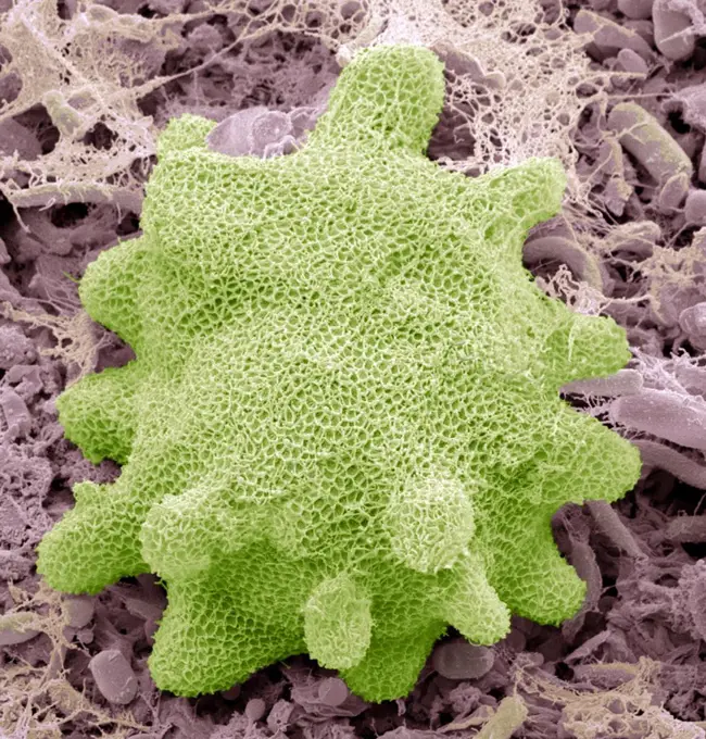
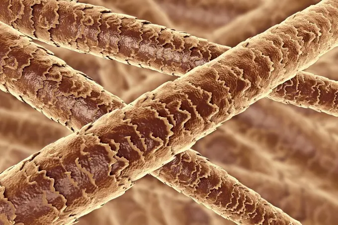
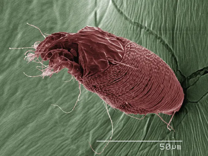
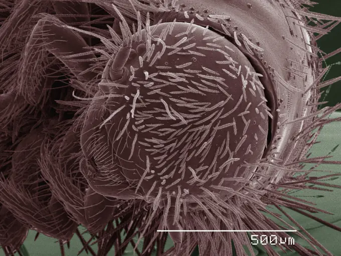
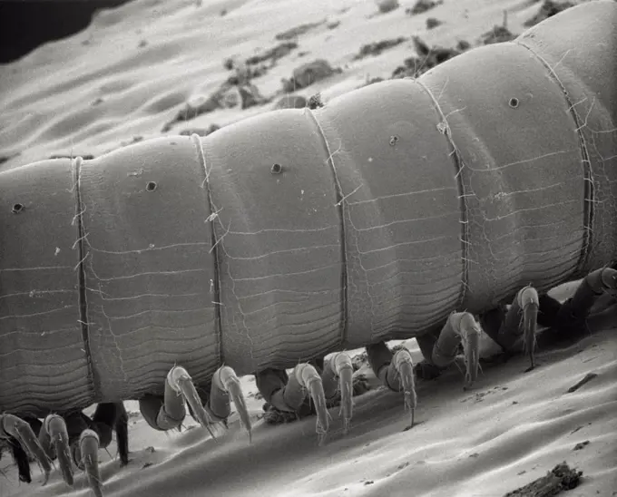
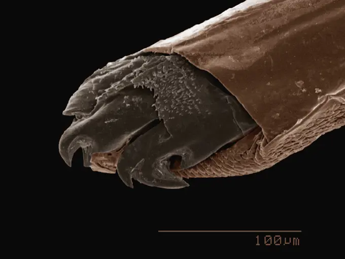
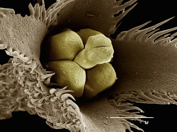
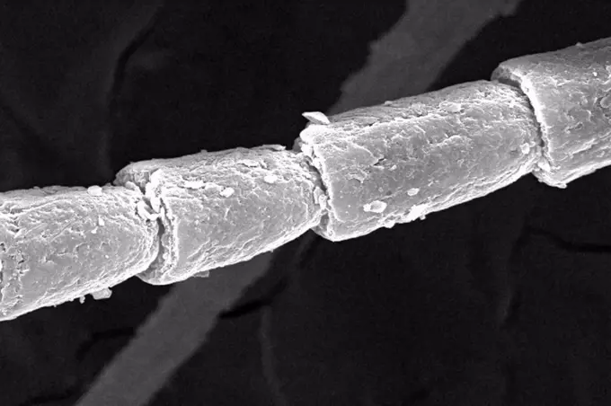
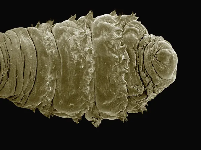
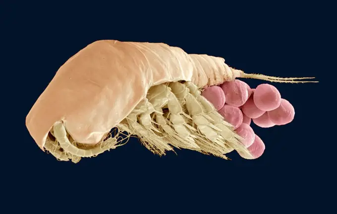
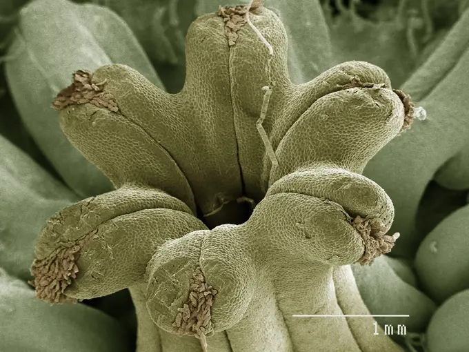
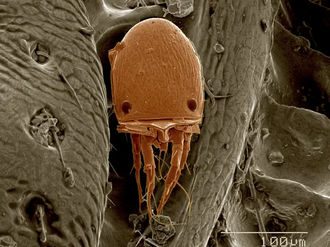
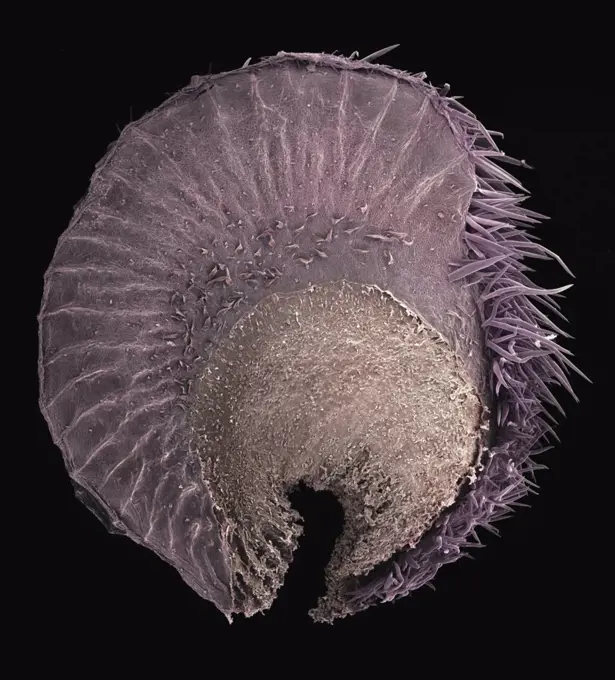
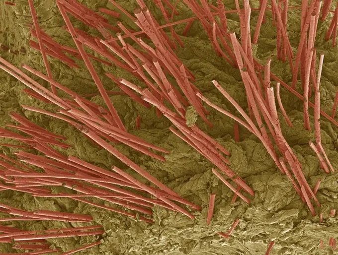
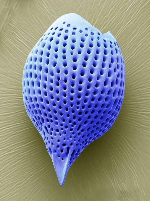
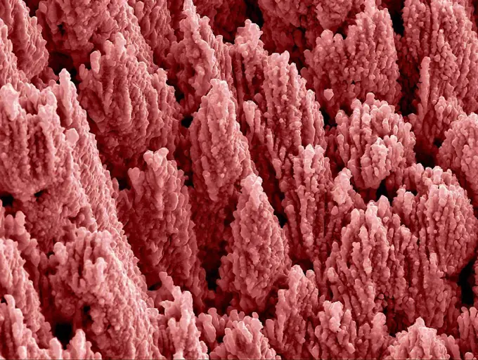
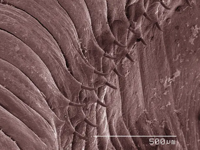
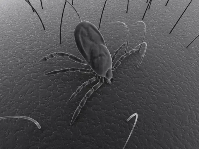
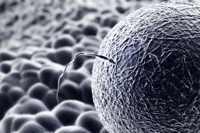
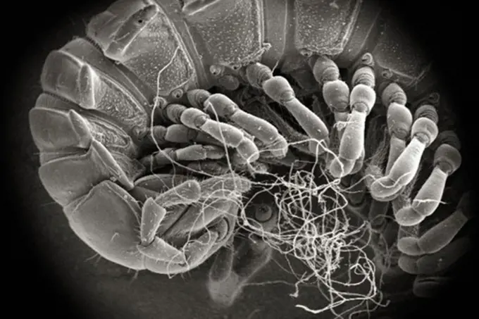
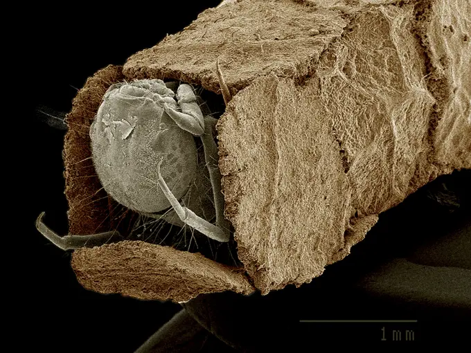
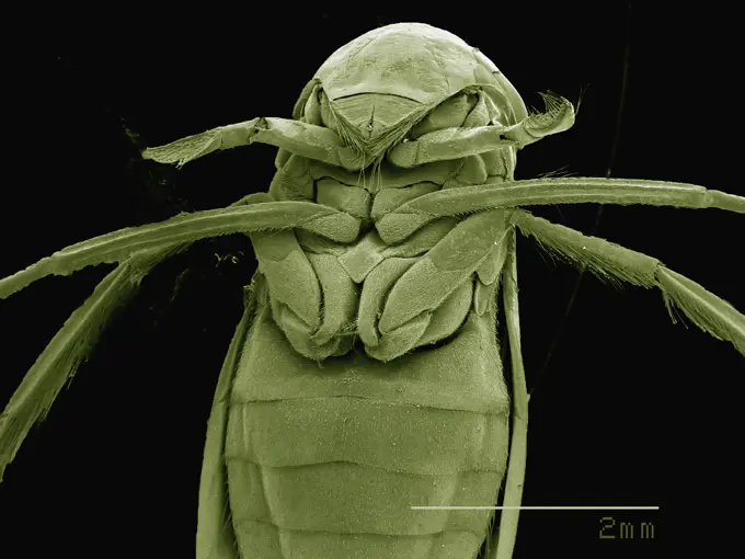
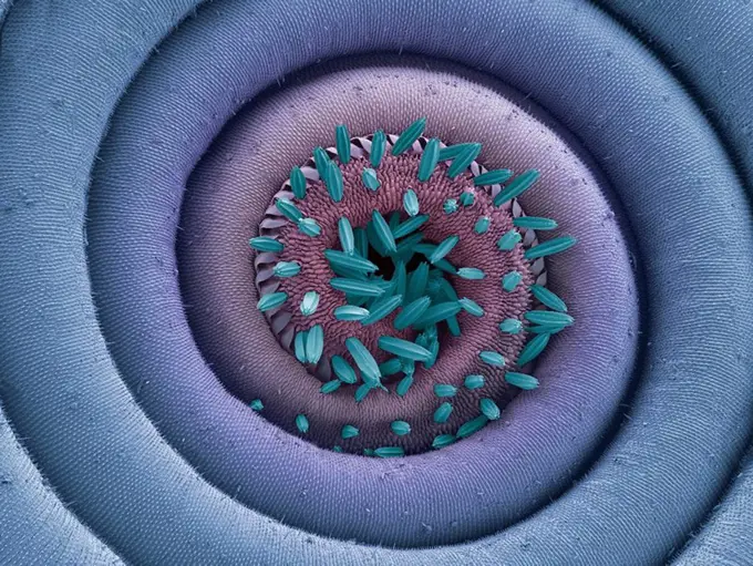
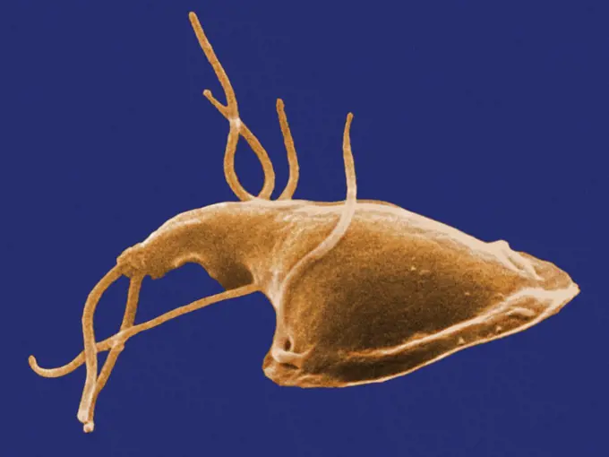
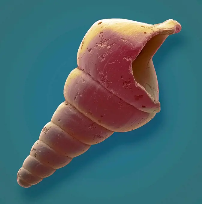
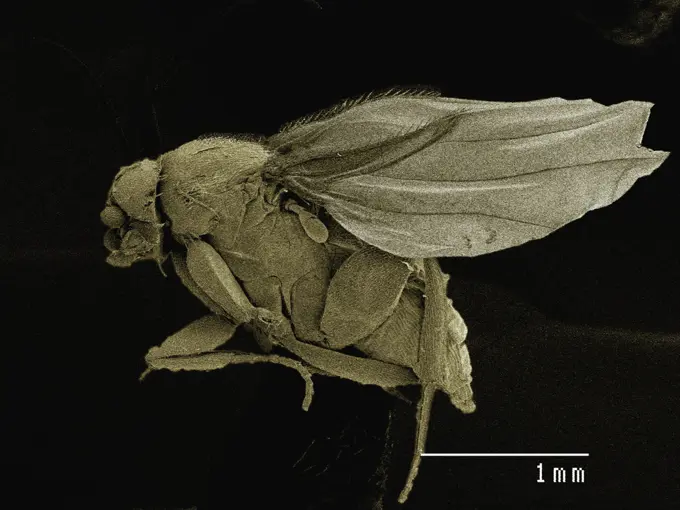
Fine ArtVintageFootageSubscriptionBoardsFree ResearchPricing
  • Fine Art
  • Vintage
  • Footage
  • Subscription
  • Boards
  • Free Research
  • Pricing

Microscopic Organisms

Close-up images capture various microscopic organisms and structures, showcasing intricate details and textures under high magnification.

Extreme closeup of fibrous material texture
175 assets in this story
1439-57940048
Closeup view of a textured surface resembling a serrated edge
4384-311
Chitinous Shell of Ladybug
4384-332
Anal Orifice of Millipede
4128R-8682
Korotnevella. Coloured scanning electron micrograph (SEM) of the scales covering a 'naked' amoeba of the genus Korotnevella. This is an amoeba with fi...
4128R-15366450
Human hair, illustration
1439-57940037
Microscopic mite up close in vivid detail.
1439-57940118
Closeup view of a microscopic organism.
1439-57943292
Closeup of microscopic structures resembling an otherworldly landscape
4201-66241
SEM close-up of a millipede at 70x magnification showing 2 pair of legs per body section, the attribute distinguishing this group from centipedes which have but a single pair of legs per section
1439-57948989
Extreme closeup of an insect leg under a scanning electron microscope
1439-57952393
Closeup of pollen grains captured by an electron microscope
4384-176
Under a moderately-high magnification of 1244x this scanning electron micrograph (SEM) depicted some of the ultrastructural details displayed on the antennal flagellum emanating from the head region of a larval antlion. This larva is sometimes referred to as a doodlebug, because of the trails it leaves in the soft sand as it hunts for prey.  This arthropod undergoes dramatic morphologic changes when it metamorphoses into a beautiful flying antlion lacewing. The antenna is composed of three mai
1439-57952647
Closeup view of a segmented organism on a black background.
4128R-8945
Female copepod, SEM
1439-57952399
Intricate details of a plant under a microscope.
824-63227290
A scanning electron micrograph of mouse tooth enamel after being briefly treated with acid.
1439-57943305
Closeup of a microorganism on a textured surface under a scanning electron microscope
4128-V58558800
4201-66379
Holly Hock (Alcea rosea) SEM close-up view of seed at 14x magnification
4128R-3539
Human hair, SEM
4128R-30468
Fractured tendon. Coloured scanning electron micrograph (SEM) of a fractured flexor tendon of the finger showing the collagen fibres. The parallel arr...
4128-V58560243
4128R-14152
Ruptured venule. Coloured scanning electron micrograph SEM of a ruptured venule running through fatty tissue. Stacked red blood cells rouleaux formati...
4128R-12573035
Radiolarian. Coloured scanning electron micrograph (SEM) of a radiolarian. Radiolarians are amoeboid protozoa that produce intricate mineral skeletons...
4128R-13586653
Nanostructures on titanium. Coloured scanning electron micrograph (SEM) of nanostructures formed on a titanium surface by femtosecond pulses of a lase...
4128R-9306
Water bear, SEM
4128R-8674
Geranium pollen, SEM
4378-1833
Bacteriophage on bacterium.
4128R-6648
Calcareous phytoplankton, SEM
1773-209586
Daisy floret, x900 magnification
4128R-2730
Cat flea head, SEM
4384-175
Under a moderately-high magnification of 633X this scanning electron micrograph (SEM) revealed the ultrastructural features found on the ventral exoskeletal surface of this larval-staged antlions mandibles. This larva is sometimes referred to as a doodlebug, because of the trails it leaves in the soft sand as it hunts for prey.  This arthropod undergoes dramatic morphologic changes when it metamorphoses into a beautiful flying antlion lacewing. Note the presence of numerous hairs covering v
4384-336
Detail of Domesticated Cat's Claw
4128R-8938
Angel´s trumpets pollen, SEM
1439-57949056
Closeup view of a delicate microscopic structure.
4128R-2568
Pubic louse, SEM
1439-57940200
A closeup micrograph showcasing intricate details of a microscopic structure.
4128R-9408
Copepod, SEM
1439-57940050
Closeup view of a microscopic landscape, showcasing the intricate patterns and textures of a biological specimen.
1525-22197338
tick bite
4378-1150
Scanning electromicrograph styled view of the moment of conception.
4384-375
Millipede
4128R-13619955
Human hair with dandruff, coloured scanning electron micrograph (SEM). The outer layer of hair (the cuticle) has overlapping scales of keratin. These ...
1439-57940038
Closeup of a dust mite on a microscopic landscape
1439-57953820
Closeup of a spider under the microscope
4128R-8683
Sutured wound, SEM
1439-57943288
A closeup view of a dust mite on a fibrous surface.
1439-57952635
Scanning electron micrograph of the head of a female backswimmer (hemiptera: corixidae)
4128R-5954
Moth proboscis, SEM
4384-142
This digitally-colorized scanning electron micrograph (SEM) depicted the dorsal (upper) surface of a Giardia protozoan that had been isolated from a rats intestine. Some of the identifying morphologic characteristics include pairs of thread-like flagella that facilitate motility, and a ventolateral flange that appears as a ruffle around the anterior portion of the organism.  Pairs of flagella seen here include an anterior, posterior-lateral, and caudal pairs.  The protozoan Giardia causes the
4384-206
Under a low magnification of 49X, this scanning electron micrograph (SEM) depicted a dorsal view of an unidentified male  tick (Dermacentor  sp.) found upon a cat in the suburbs of Decatur, Georgia, which measured approximately 3.5mm from its mandibulae to the distal abdominal margin. Note that the entire dorsum of this ticks abdomen is covered by its tough scutum, or shield, categorizing it as a male, whereas, in female Ixodid-species ticks, the scutum only partially covers the dorsal abdo
4128R-12571814
Clostridium phytofermentans bacteria. Coloured scanning electron micrograph (SEM) of Clostridium phytofermentans bacteria A gram-positive, anaerobic, ...
4128R-11288986
Dust mite, computer artwork.
1439-57943346
Golden microstructures in closeup at high magnification
824-63123126
SEM. Magnification: 12000x.
4297-1888
Scanning electron microscopic image of Vibrio cholerae bacteria of the serogroup 01
4220-20100797
Spain - Cracked earth in the extreme mineral-rich ground in the area of the Rio Tinto mines. Province of Huelva, Andalucia, Spain.
1439-57952634
Closeup of an ant captured with electron microscopy
4128R-8939
Geranium pollen, SEM
1439-57949027
Intricate microscopic pattern of a radiolarian shell against a dark background
4128R-15964
Head louse Pediculus humanus capitis, computer artwork.
1848-49573674
A wasp at its nest in a detailed close-up, captured in a monochrome image style
4378-941
The earth as if seen under scanning electron microscope (conceptualization)
4128R-14999
Cat flea Ctenocephalides felis, coloured scanning electron micrograph SEM. Magnification: x50 when printed at 10 centimetres wide.
4128R-9404
Angel´s trumpets pollen, SEM
4297-1769
Scanning electron micrograph of Flexispira rappini bacterium, magnified 13,951x
4128R-3842
Sarcoptic mange mites, coloured scanning electron micrograph SEM. These burrowing mites cause the skin disease scabies. Magnification: x30 when printe...
4384-145
A scanning electron micrograph (SEM) of a sickle cell red blood cell found in a blood specimen of an 18 year old female patient with sickle cell anemia, (HbSS). People who have this form of sickle cell disease inherit two sickle cell genes (S), one from each parent. This is commonly called sickle cell anemia, and is usually the most severe form of the disease. Sickle cell disease is a group of inherited red blood cell disorders. Healthy red blood cells are round, and they move through small
4128-V58569130
1525R-11104059
4269-24738
Pollen. Pollen of wattle SEM (Scanning Electron Microscope)
4128R-9303
Copepod, SEM
4128R-12886
Diatom, coloured scanning electron micrograph SEM. The diatoms are a group of photosynthetic, single_celled algae containing about 100,000 species. Ma...
4128R-13022177
Gastropod. Coloured scanning electron micrograph (SEM) of a gastropod microfossil from maldives beach sand. Microfossils are roughly 0.05 to 2mm in si...
1439-57949007
Closeup image of a housefly captured through electron microscopy
PREVIOUS
of 2
NEXT
2401 S. Ervay, Suite 206
Dallas, Texas 75215
United States
Browse
CategoriesGalleriesFree Research
For Creators
License Your ContentSuperStock For StudentsFrame of Mind BlogContributor PortalNew Image SpotlightLicense AI Imagery
Resources
About UsLicensingTerms & ConditionsAPI AccessPrivacyPricing
Contact
+1 866 236 0087help@superstock.com Contact form
©2026 SuperStock. All Rights Reserved. -A
This site is protected by reCAPTCHA and Google Privacy Policy and Terms of Service apply.