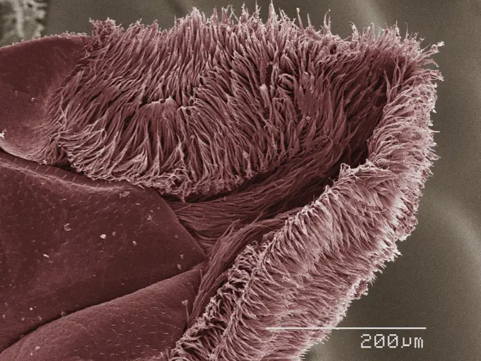
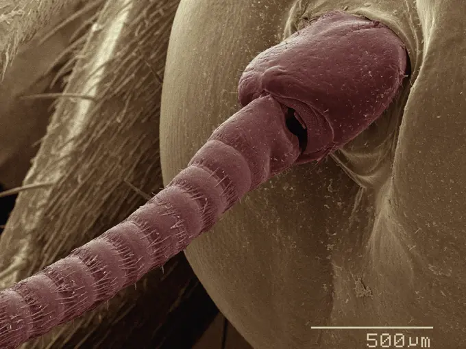
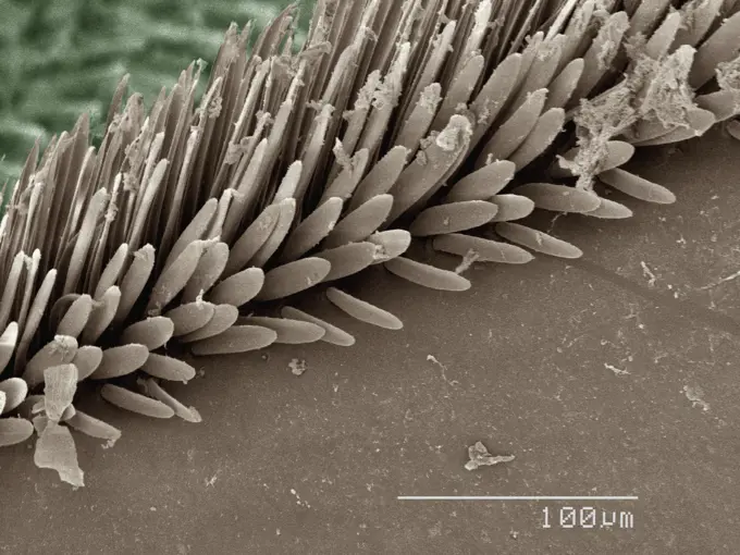
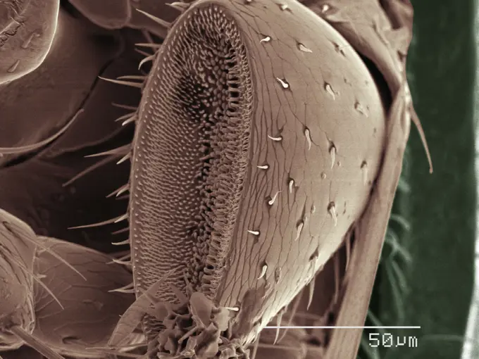
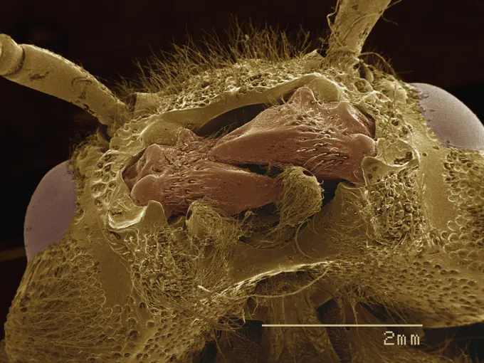
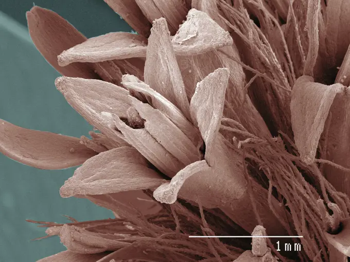
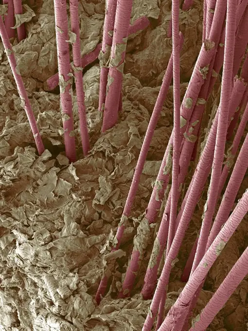
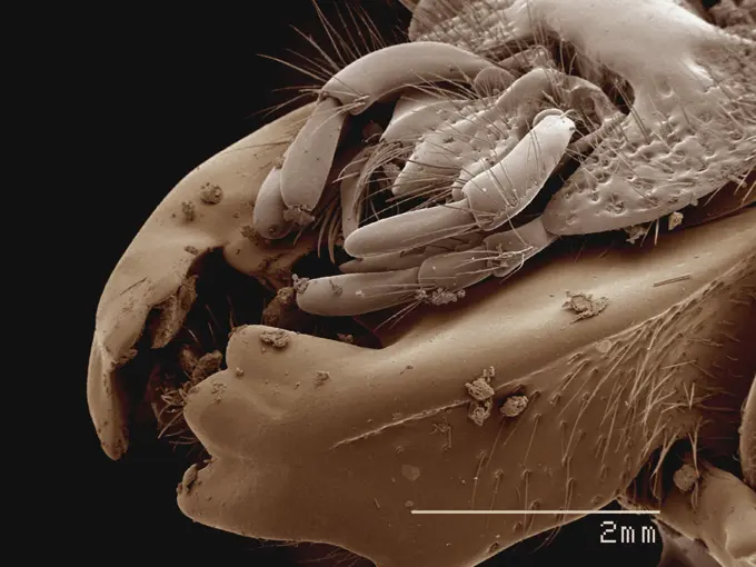
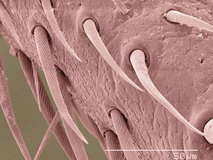
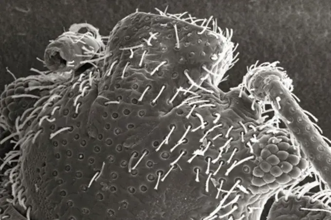
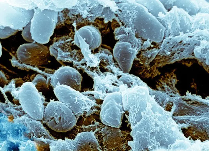
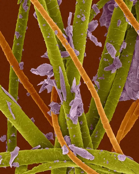
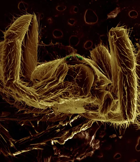
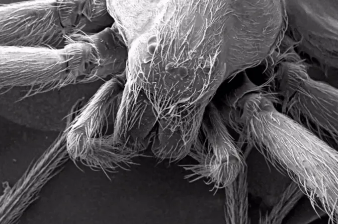
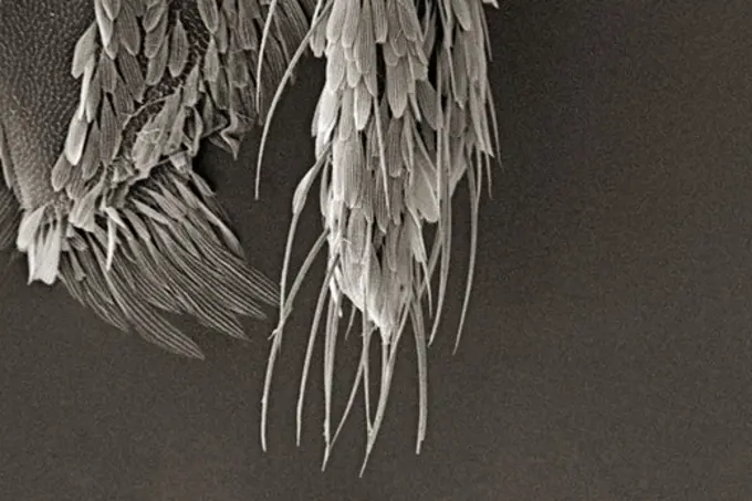
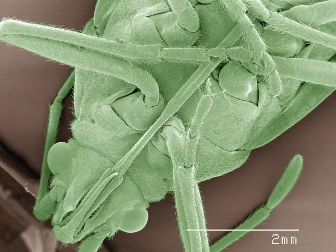
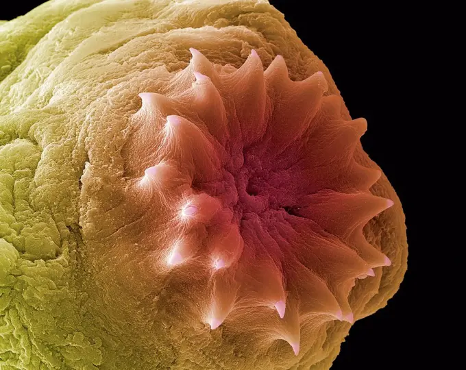
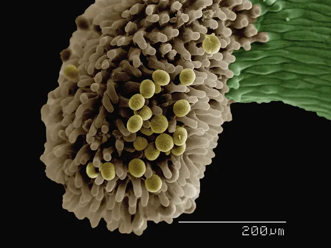
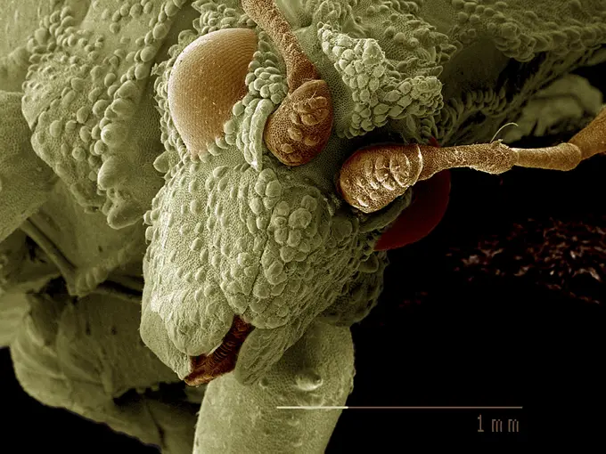
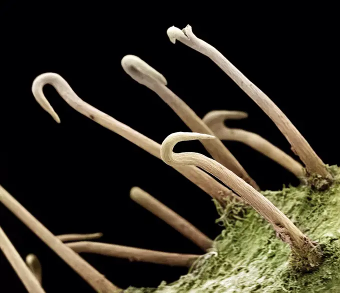
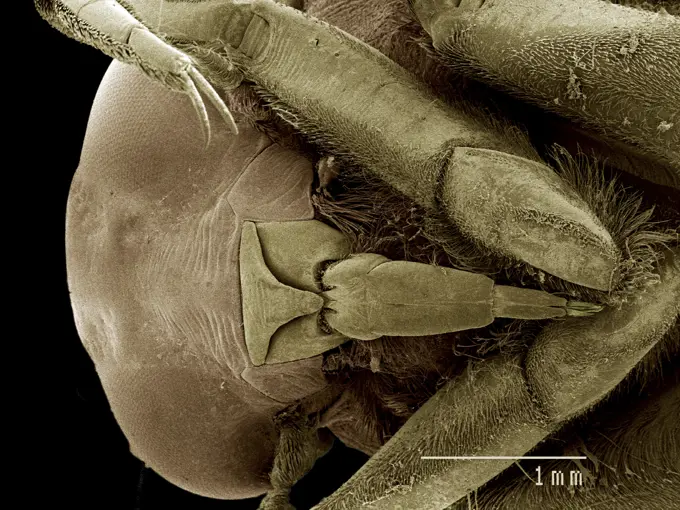
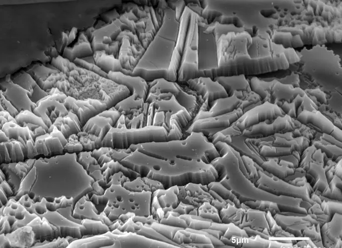
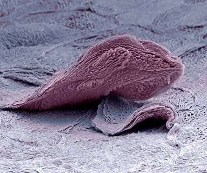
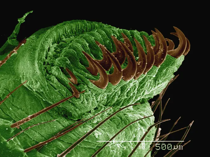
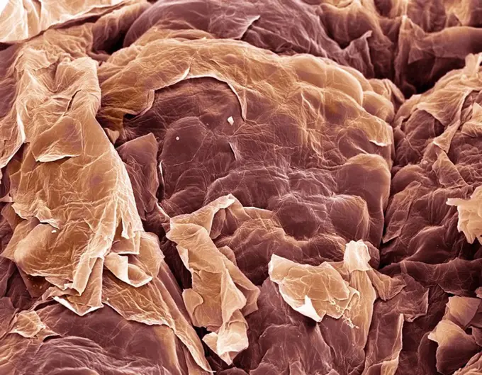
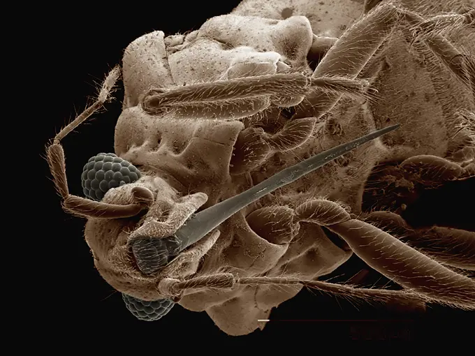
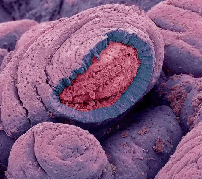
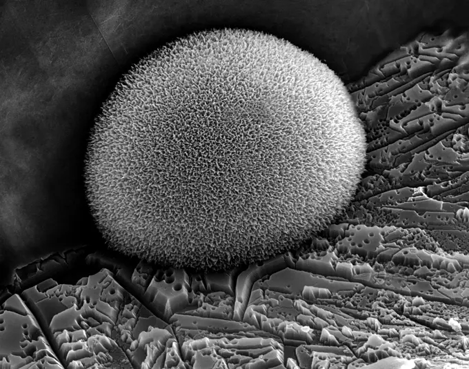
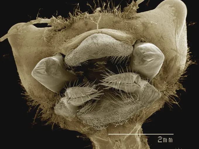
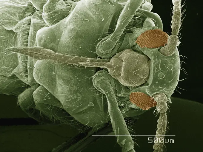
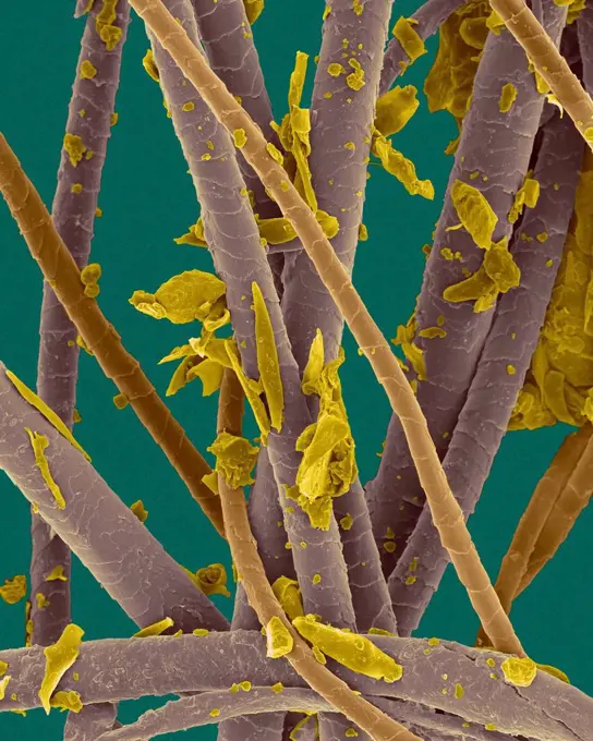
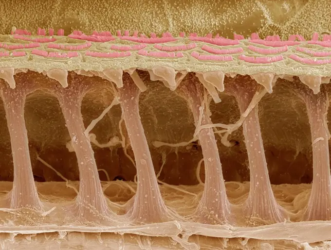
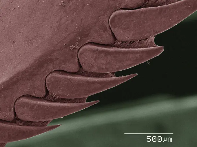
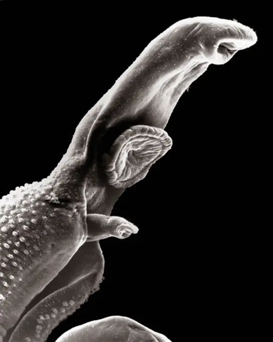
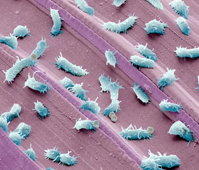
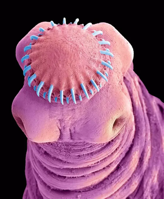
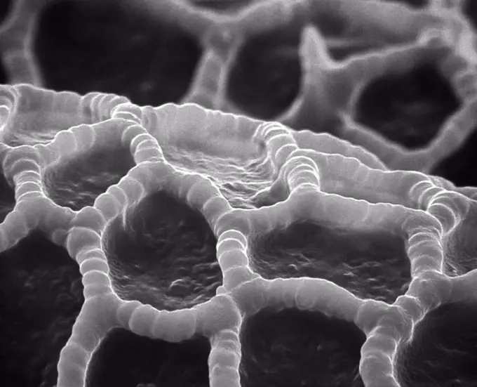
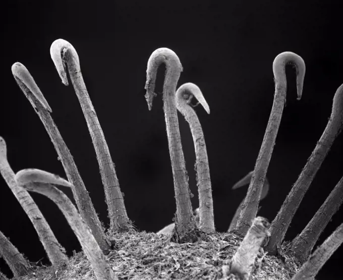
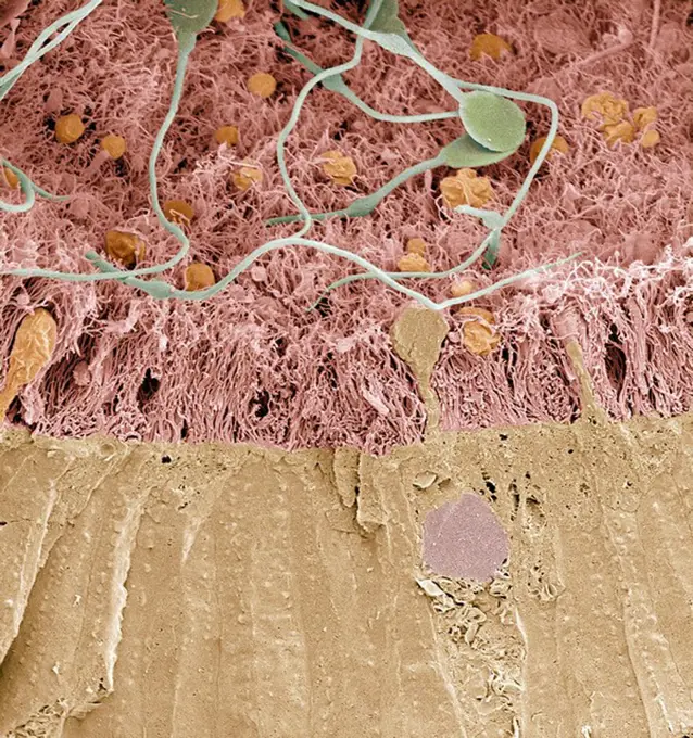
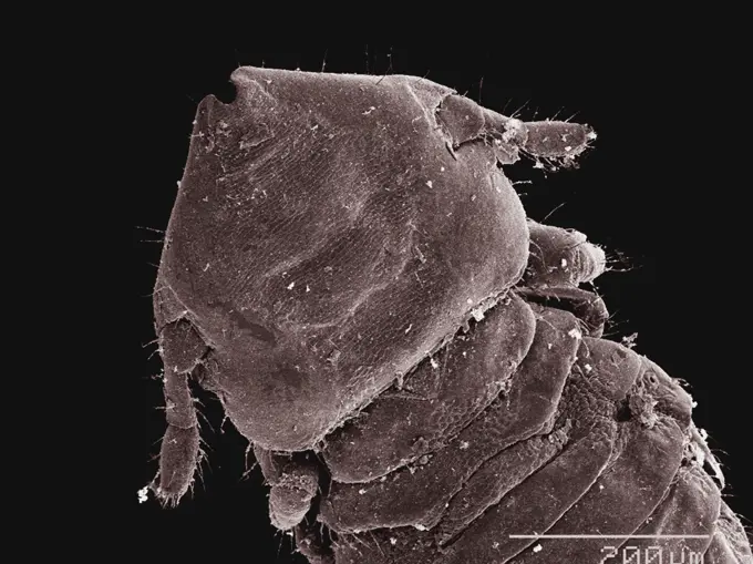
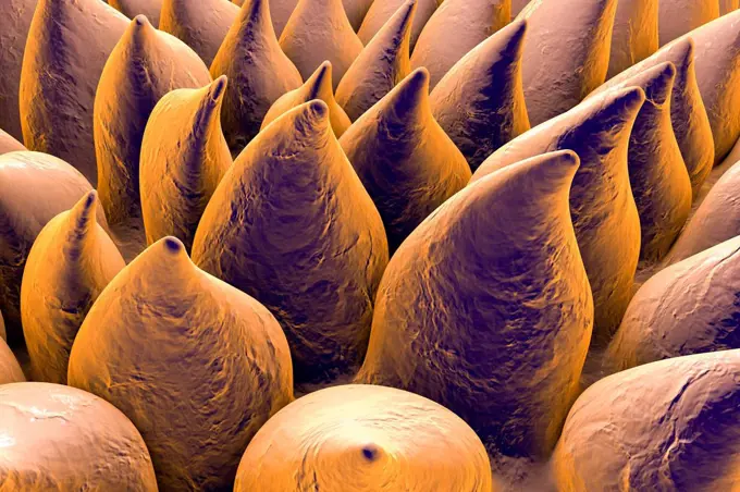
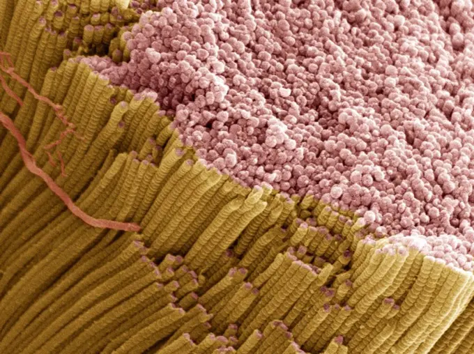
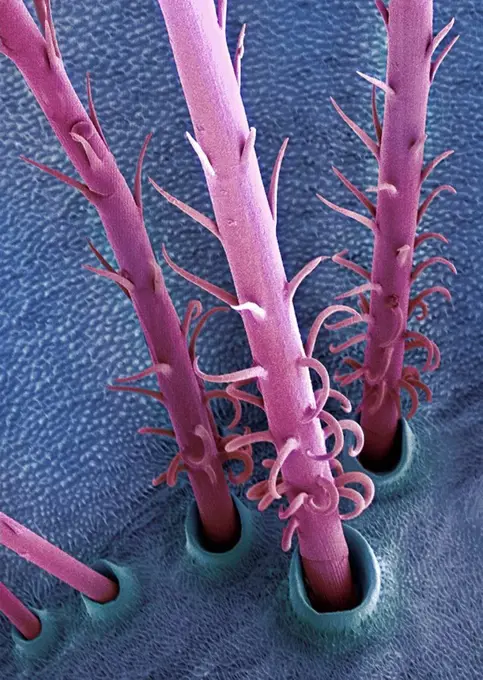
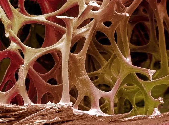
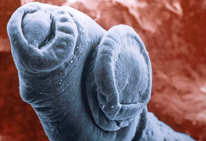
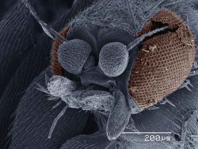
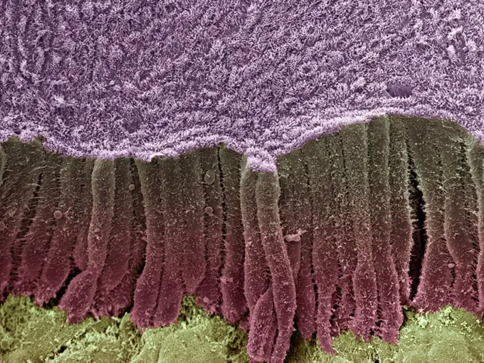
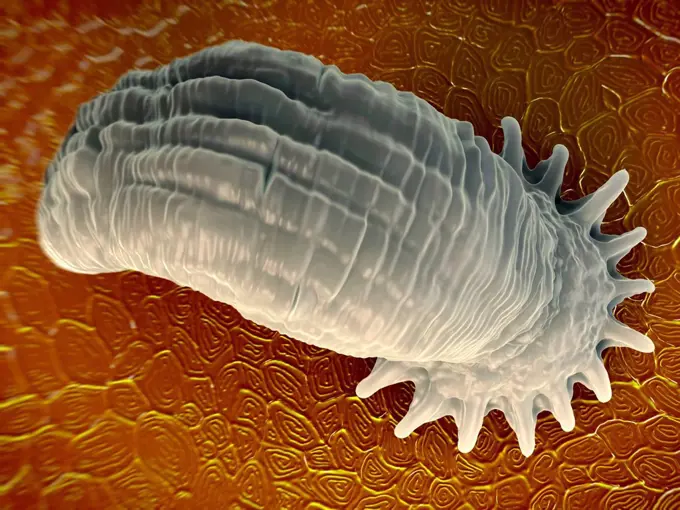
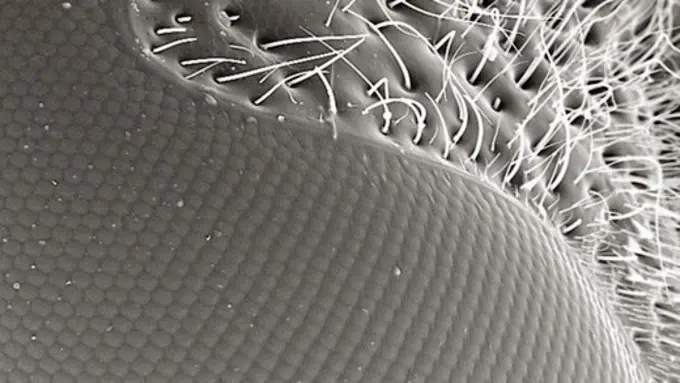
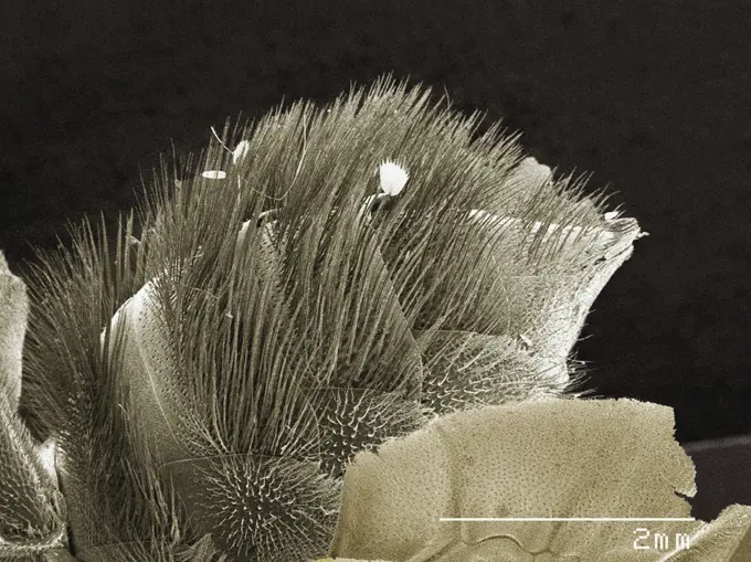
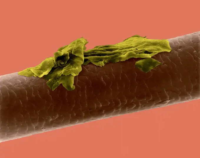
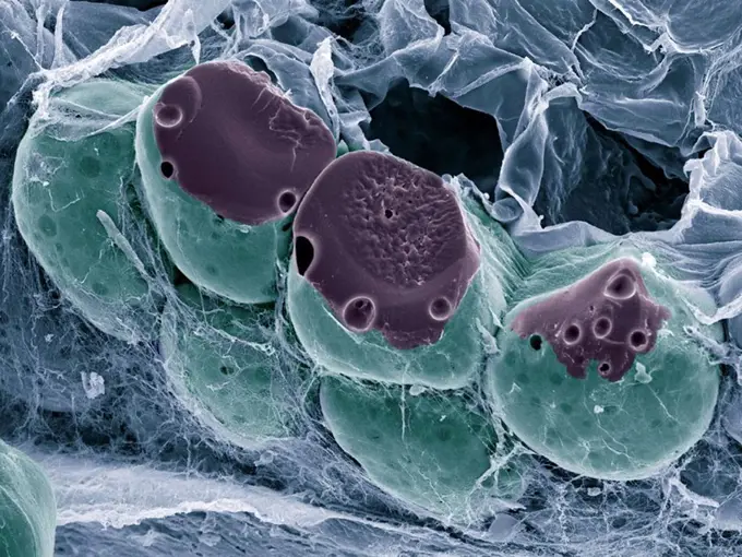
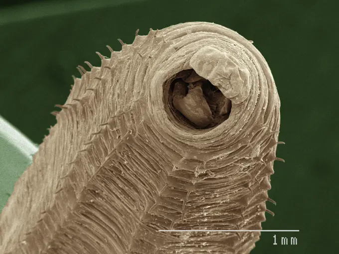
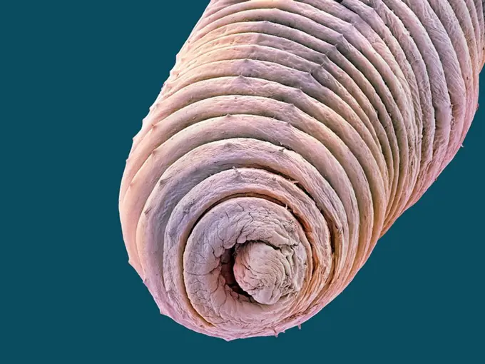
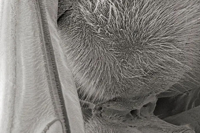
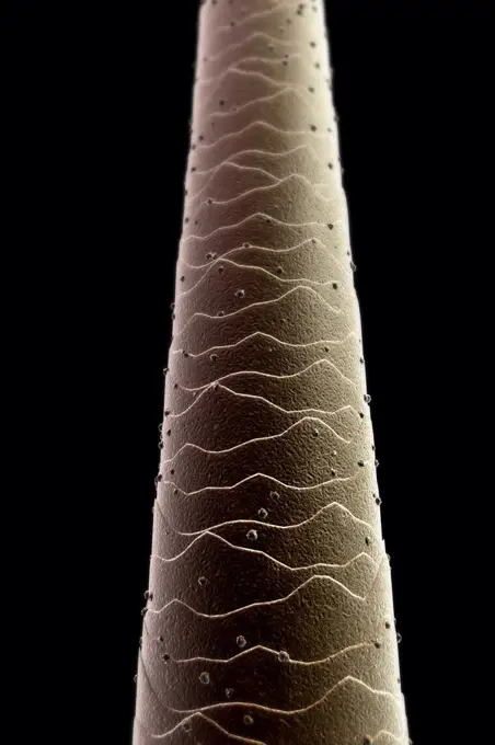
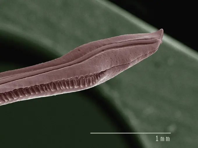
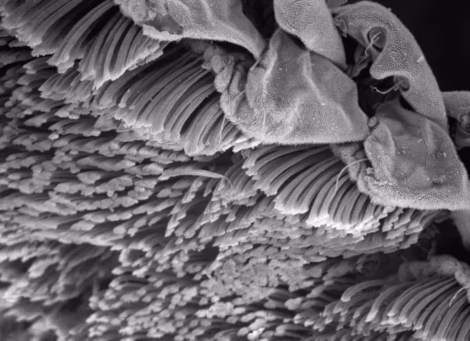
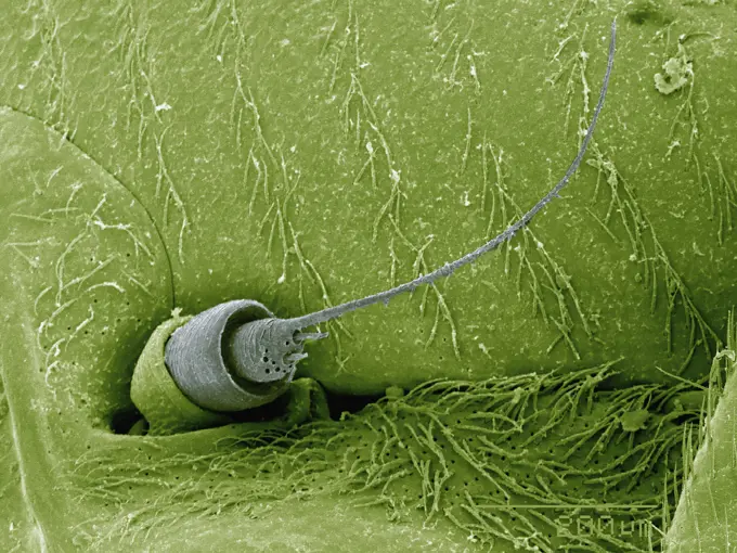
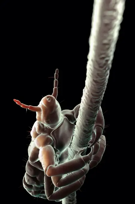
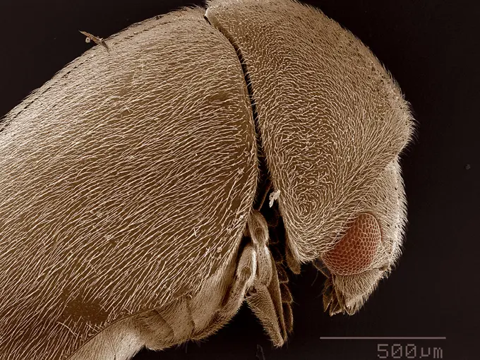
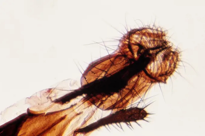
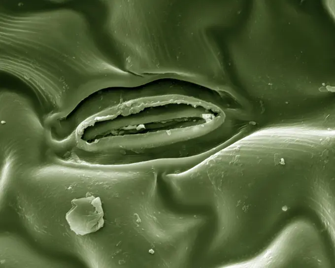
Fine ArtVintageFootageSubscriptionBoardsFree ResearchPricing
  • Fine Art
  • Vintage
  • Footage
  • Subscription
  • Boards
  • Free Research
  • Pricing

Microscopic Life Forms

Detailed close-ups of microscopic textures and insect features captured under an electron microscope, revealing the complexity of natural patterns.

Closeup view of a microscopic surface revealing intricate textures
190 assets in this story
1439-57940144
Closeup view of a microscopic surface revealing intricate textures
1439-57940215
Extreme closeup of an insect eye and antennae under a scanning electron microscope
1439-57940161
A closeup view of microscopic structures resembling a textured landscape
1439-57947639
Intricate patterns of nature seen through a microscope.
1439-57940141
A closeup look at an insect eye through an electron microscope
4269-24645
Muscle. Muscle fibre with 2 red blood cells SEM (Scanning Electron Microscope)
4384-423
Microscopic Detail of a Carpenter Bee's Antenna
1439-57940188
Up close with the miniature landscape of a moth's antennae
1439-57949047
Intricate patterns of a microscopic landscape
1439-57940000
Closeup view of microscopic organisms in a vivid scientific image.
1439-57948992
Intricate closeup of a spider under a microscope.
1439-57940113
Closeup of a delicate microstructure under electron microscopy
4128R-6851
Human hair, SEM
1439-57940186
Closeup of a mosquito proboscis under the microscope.
4384-150
This digitally-colorized scanning electron micrograph (SEM) of an untreated water specimen extracted from a wild stream mainly used to control flooding during inclement weather revealed the presence of unidentified organisms, which included bacteria, protozoa, and algae. In this particular view, it appears that a diatom had been fractured in the processing of this specimen. Surrounding this diatomic microorganism, was a large biofilm mass within, and around which were numerous protozoan amoeboid
1439-57949020
Closeup of an insect claw under a microscope
1439-57940213
Up close with microworld wonders, the intricate landscape of a gecko's skin and its fine structures in sharp focus.
1439-57949058
Closeup view of an insect head under a microscope
4128R-13586654
Nanostructures on titanium. Coloured scanning electron micrograph (SEM) of nanostructures formed on a titanium surface by a laser beam. This research ...
4384-257
Scanning Electron Micrograph of Bed Bug
4297-1800
Colorized SEM image of Yersinia pestis bacteria
4128R-13620211
Coloured scanning electron micrograph (SEM) Pet dander. dog hair, cat hair and dead skin (cat hair. orange, dog hair. green, dander. purple). Dog and ...
4220-21334718
Scanning Electron Micrograph (SEM): Spectacle Moth.     Date:
1439-57949081
A closeup view of microscopic structures resembling a dense forest under magnification.
1439-57940168
Closeup of a pointed object under magnification
1439-57940199
Intricate microscopic landscape in shades of pink.
1439-57949073
A closeup view of a microworld, resembling an alien landscape.
1773-100036
SEM image of spider
4384-104
Under a low magnification of only 27x, this scanning electron micrograph (SEM) depicted the dorsal cephalothorax, i.e., a combination of its head and thoracic regions, of a venomous brown recluse spider, Loxosceles recluse,  found inhabiting a Kentucky farm. Most spiders possess eight eyes (4 pairs), however, as is evidenced in this image, recluse spiders only possess six.   L. reclusa is sometimes referred to as the violin or fiddle spider, for on its cephalothorax one will see what appears
4384-410
Microscopic Detail of Mosquito's Palps
1439-57940177
Ultradetailed micrograph of an insect
4128R-12576706
Dog tapeworm head. Coloured scanning electron micrograph (SEM) of the head (scolex) of a dog tapeworm (Taenia pisiformis). This tapeworm has dogs and ...
1439-57952415
Closeup view of a pollencovered stamen under the microscope
1439-57943307
Closeup view of a bee under the microscope
4201-21590100
Spiny Cocklebur (Xanthium spinosum) hooks on fruit
1439-57943343
Closeup view of an insect under a microscope
1439-57941086
Intricate microscopic landscape in grayscale
4128R-6322
Skin cell. Coloured scanning electron micrograph SEM of a squamous cell on the surface of the skin. This is a flat, keratinised, dead cell. Squamous c...
1439-57949011
Closeup of a caterpillar's proleg with hooklike structures on a leaf surface
4128R-4241
Skin, SEM
1439-57952413
A closeup scientific image of a tiny insect viewed under electron microscopy
4128-16171802
Small intestine. Coloured scanning electron micrograph (SEM) of a freeze-fractured of the small intestine. The surface consists of deep folds, called ...
1439-57941089
A highly detailed micrograph revealing the intricate textures of a surface and spherical structure.
4128R-8234
Albumen crystals, SEM
1439-57940036
A closeup of a leaf under the microscope revealing intricate textures and patterns.
1899-61460835
This scanning electron microscopic (SEM) image of an untreated water specimen extracted from a wild stream mainly used to control flooding during inclement weather, revealed the presence of unidentified organisms, which included bacteria, protozoa, and algae. In this particular view, a mass of gelatinous biofilm had enmeshed numbers of microorganisms, including amoebae and bacteria. For a colorized version of this image, see PHIL 11707. CDC/ Janice Haney Carr 2009
4128R-4549
Trachea mucous membrane, SEM
1439-57943322
A detailed micrograph of an insect's anatomy
1439-57949023
Closeup of a spider under a microscope.
1439-57949072
Closeup of an insect under the microscope provides a detailed look at the tiny world often unnoticed.
1439-57949048
Closeup of microscopic landscape with textured surface and hairlike structures
4128R-13620210
Coloured scanning electron micrograph (SEM) Pet dander. dog hair, cat hair and dead skin (cat hair. brown, dog hair. purple, dander. yellow). Dog and ...
4201-66234
Carpenter Bee (Xylocopa violacea) SEM close-up of mandibles of at 14x magnification
1439-57940020
A closeup view of a spherical object through an electron microscope.
4128R-14147
Inner ear hair cells. Coloured scanning electron micrograph SEM of sensory hair cells in the cochlea of the inner ear. The crescent_shaped arrangement...
824-63225899
Colorized scanning electron micrograph of a cell infected with SARS-CoV-2 virus particles (pink), isolated from a patient sample. Image captured at the NIAID Integrated Research Facility (IRF) in Fort Detrick, Maryland.
4201-66273
Peruvian Lily (Alstroemeria sp) SEM close-up view of pollen grains of at 700x magnification
1439-57940067
Microscopic view of velcro hooks closeup.
4391-334
The schistosome parasite enters the body through the skin of persons coming in contact with infested waters, causing schistosomiasis. The adult worm lives in the veins of its host. The parasite is magnified x256 in this photograph. Photo by Bruce Wetzel and Harry Schaefer.
4128R-9054
Stagnant water bacteria, SEM
4201-21264687
Carrot (Daucus carota) hooked spines on seed enable animal dispersal, magnification 3000x
4128R-15545697
Tapeworm cysticercus, SEM
4201-66280
Trumpet Lily (Lilium longiflorum) SEM close-up view of pollen at 2800x magnification
4201-66267
Spiny Cocklebur (Xanthium spinosum) SEM close-up of curved spines on the fruit which adhere to the pelage of animals or clothes of humans and thus assist in dispersal away from the parent plant
4128R-10559
Epididymis, SEM
1439-57949062
Upclose and personal with an insect under the microscope.
1439-57952409
Intricate Details of a Microscopic World
1439-57948996
Extreme closeup of an insect under the microscope
1439-57947638
Closeup of an intriguing microscopic landscape.
1439-57940063
Closeup view of an insect under the microscope
6145-29297479
Human bronchus, excess mucin. SEM
4128R-8677
Asparagus beetle, SEM
4128R-13376745
Tongue surface, illustration
4128R-2850
Tendon, SEM
4128R-3042
Caterpillar hairs. Coloured scanning electron micrograph SEM of hairs from the vapourer moth Orgyia antiqua caterpillar. Magnification: x250 when prin...
4128R-7174
Bird bone tissue, SEM
4269-24582
Schistosoma. SEM (Scanning Electron Microscope)
1439-57949021
Extreme closeup of an insect head under the microscope
4128R-4654
Bile duct, SEM
1439-57940180
Closeup of a creature under the microscope
4128R-12923039
Gyrodactylus aquatic parasite
4384-314
Electron Micrograph of Wasp's Head
1439-57949025
Closeup of a textured surface under magnification
4128R-13619953
Human hair with dandruff, coloured scanning electron micrograph (SEM). The outer layer of hair (the cuticle) has overlapping scales of keratin. These ...
4128R-458
Fat tissue, SEM
1439-57940047
Closeup view of an intriguing microscopic texture
4128R-4483
Earthworm, SEM
4384-222
Scanning Electron Micrograph of Hornet
4128R-8807
Sutured wound, SEM
4378-1092
Scanning electromicrograph styled close-up of human hair.
1439-57940209
A closeup view of an earthworm under the microscope
4201-66266
Moorish Wall Gecko (Tarentola mauritanica) SEM close-up view of the adhesive filaments on the bottom of the foot at 16.5x magnification
6145-29294766
embryo wound healing SEM
1439-57952640
Microscopic view of a plant stoma with guard cells
4201-21258383
Carrot (Daucus carota) hooked spines on seed enable animal dispersal, magnification 1500x
4378-2644
Parasitic head louse clinging to a hair follicle.
1439-57943289
Closeup of an ant under the microscope in great detail
1788-111177020
Mouthparts, housefly (Musca domestica), Muscidae.
4384-358
Microscopic Detail of Eggshell
4220-21334720
Scanning Electron Micrograph (SEM) Leaf Stomata.     Date:
PREVIOUS
of 2
NEXT
2401 S. Ervay, Suite 206
Dallas, Texas 75215
United States
Browse
CategoriesGalleriesFree Research
For Creators
License Your ContentSuperStock For StudentsFrame of Mind BlogContributor PortalNew Image SpotlightLicense AI Imagery
Resources
About UsLicensingTerms & ConditionsAPI AccessPrivacyPricing
Contact
+1 866 236 0087help@superstock.com Contact form
©2026 SuperStock. All Rights Reserved. -A
This site is protected by reCAPTCHA and Google Privacy Policy and Terms of Service apply.