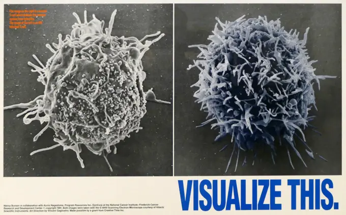
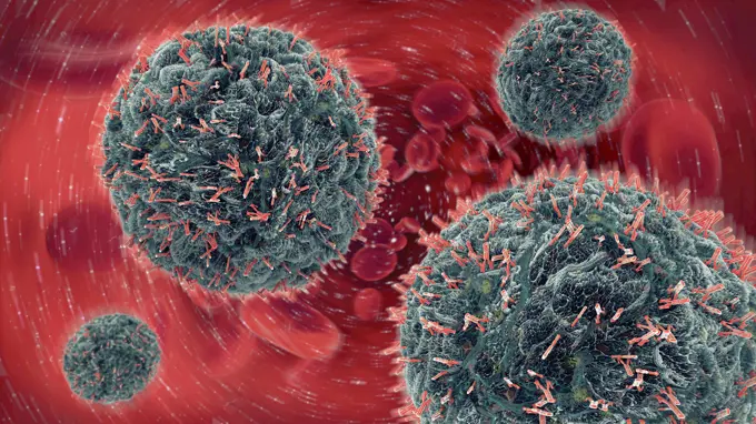
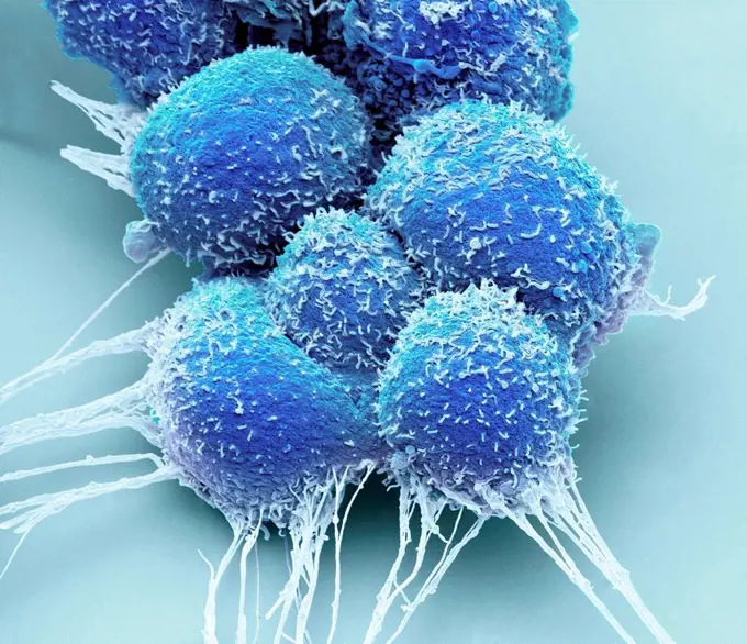
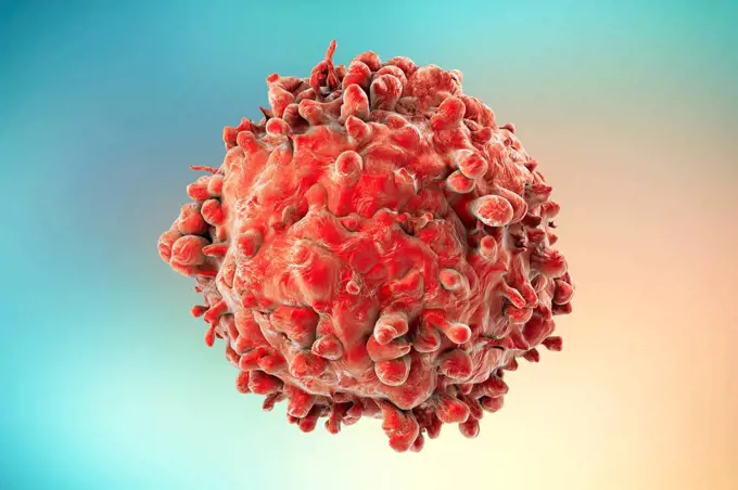
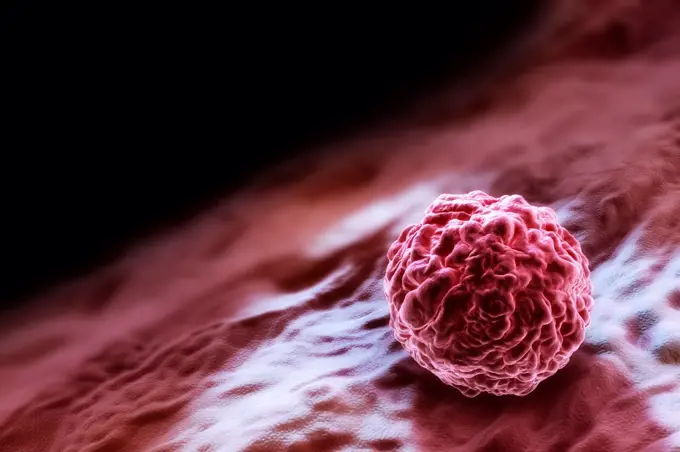
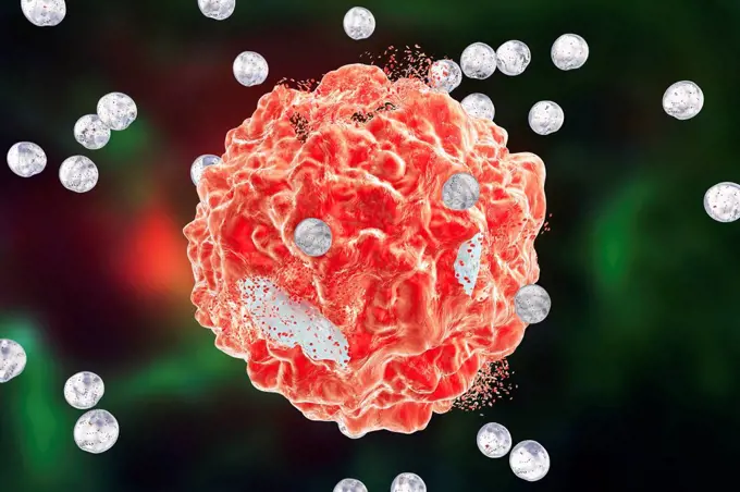
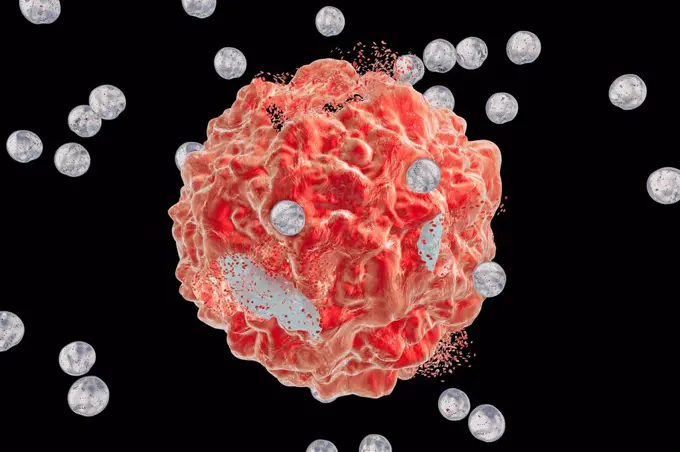
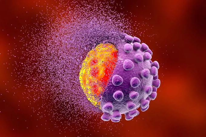
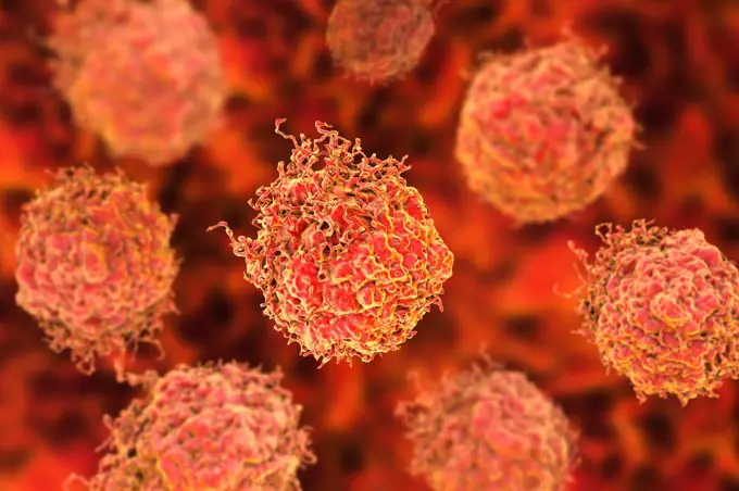
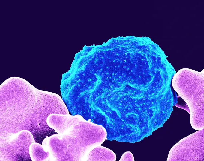
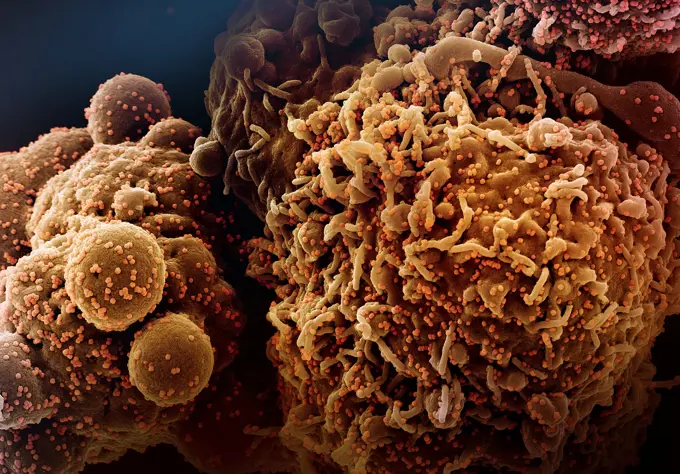
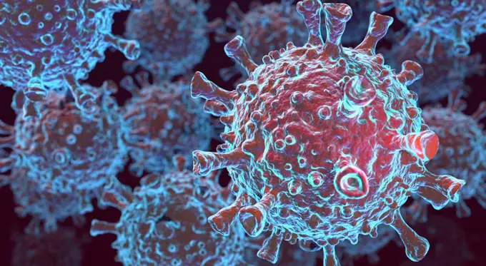
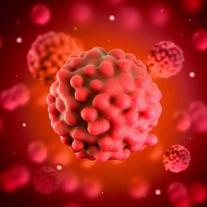
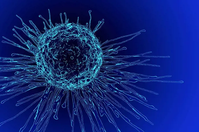
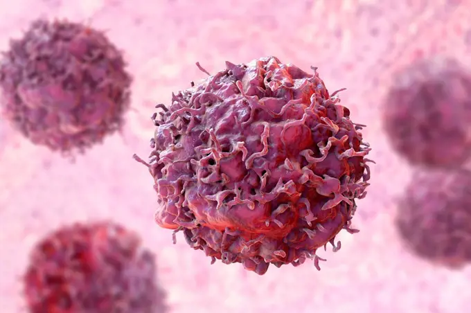
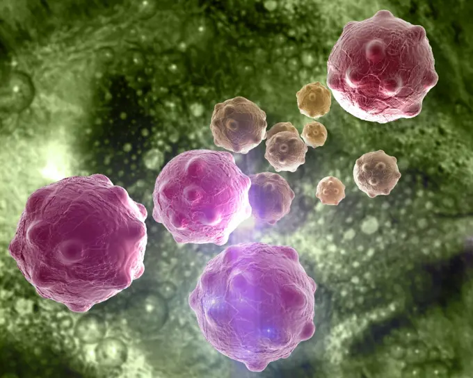
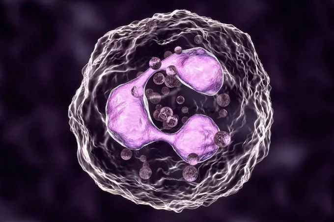
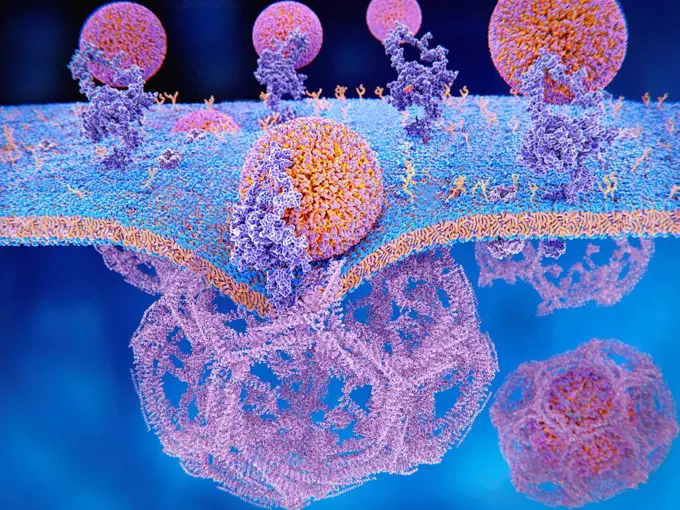
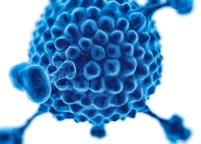
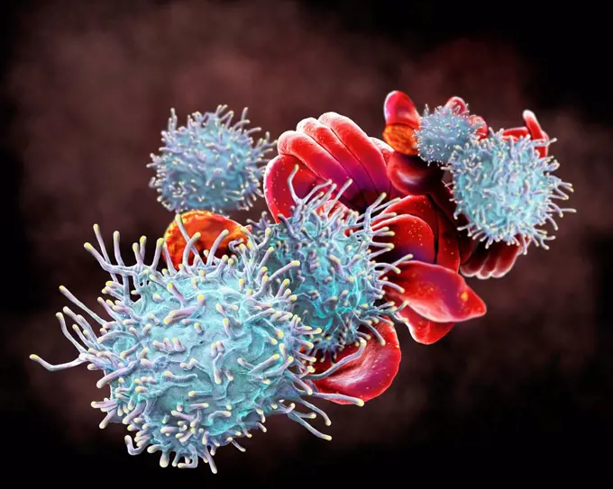
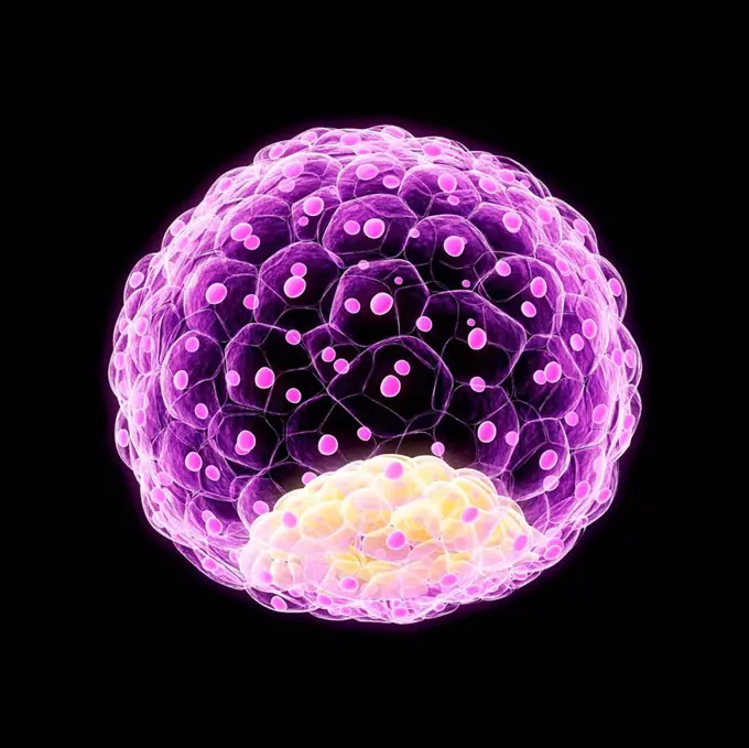
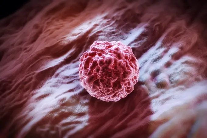
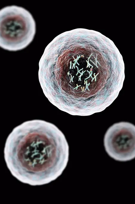
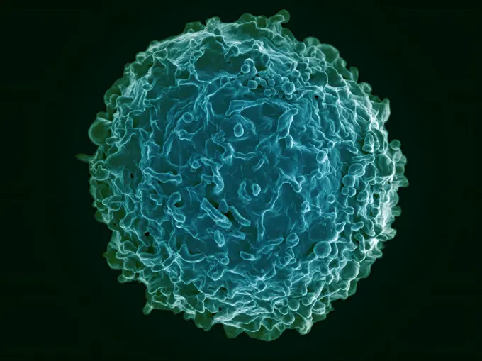
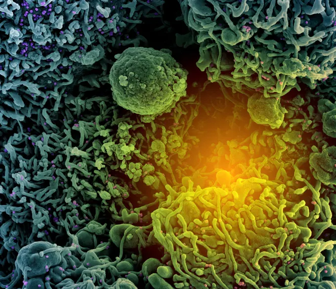
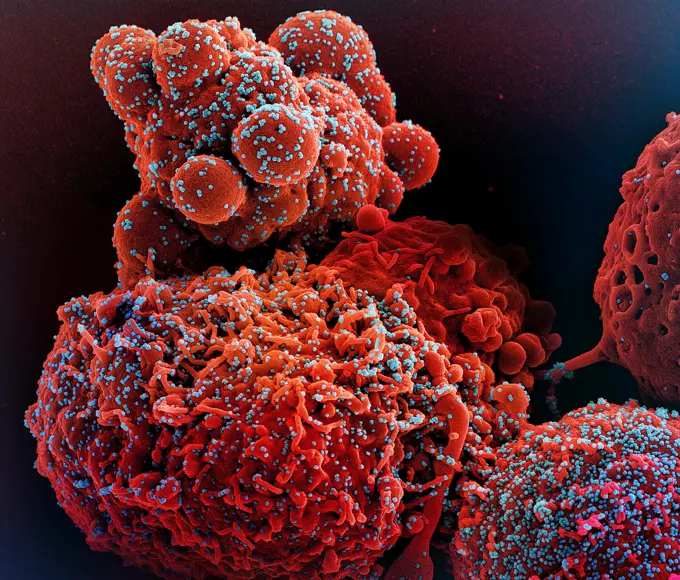
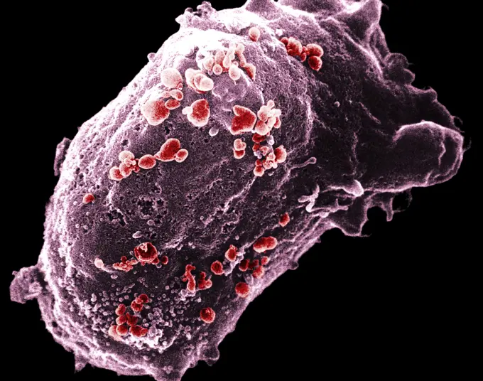
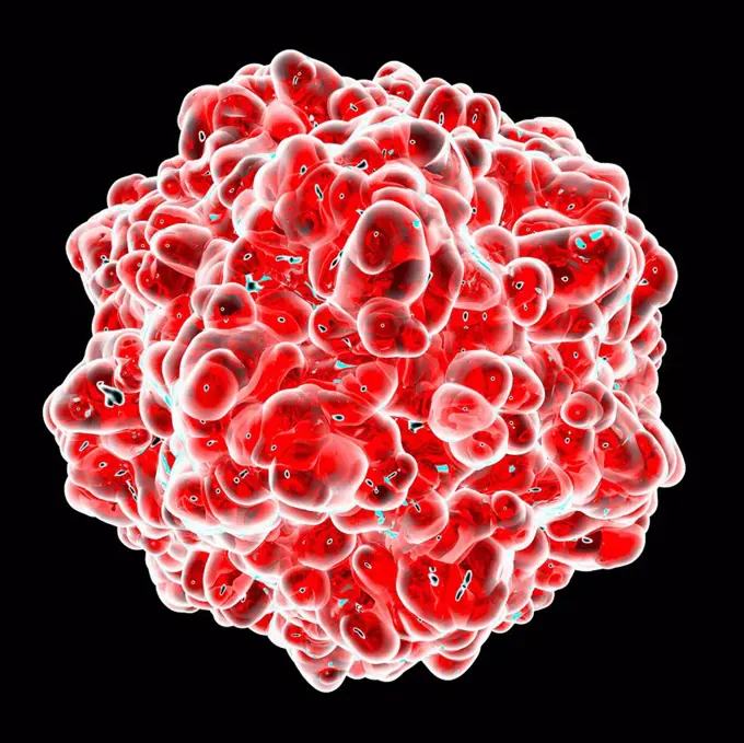
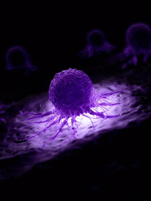
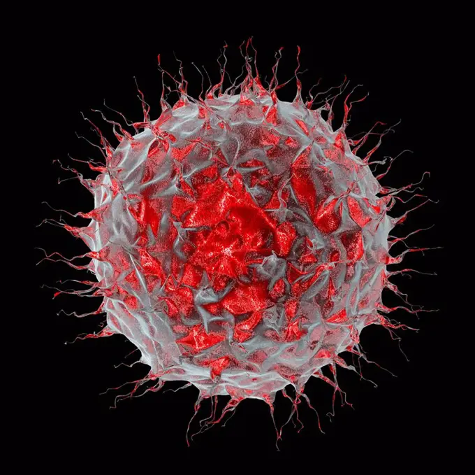
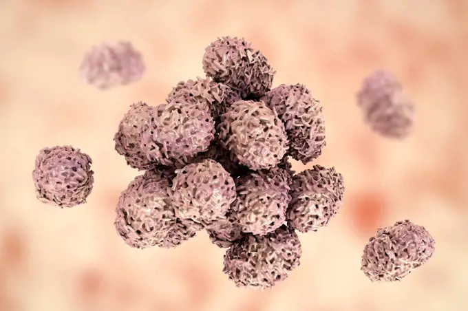
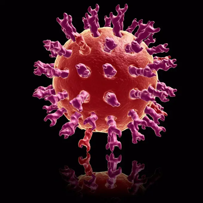
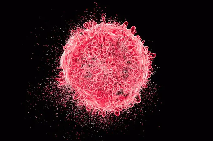
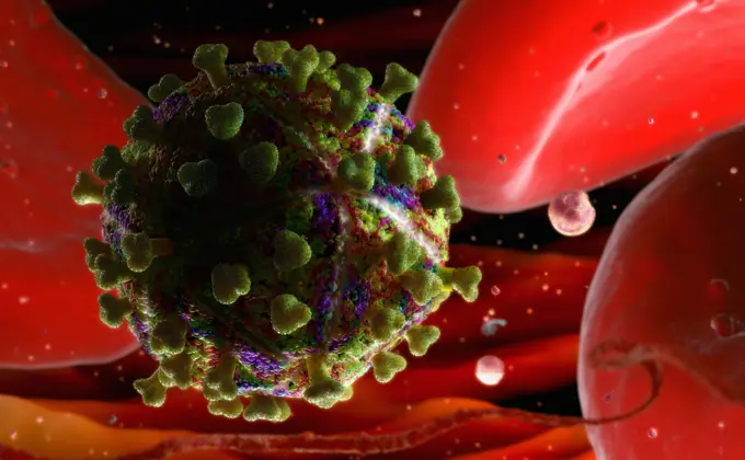
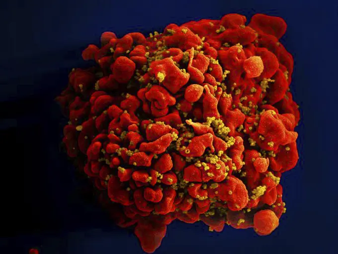
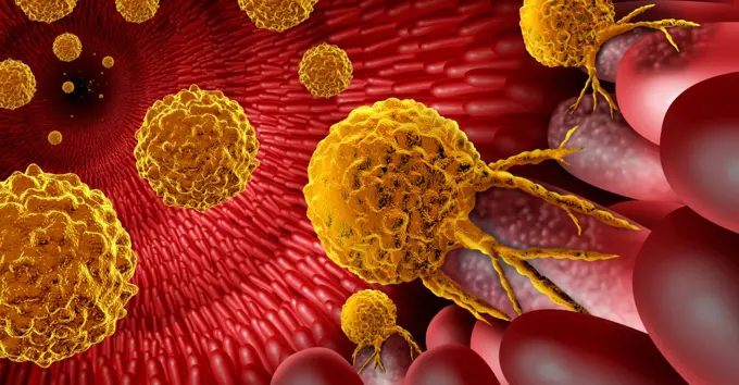
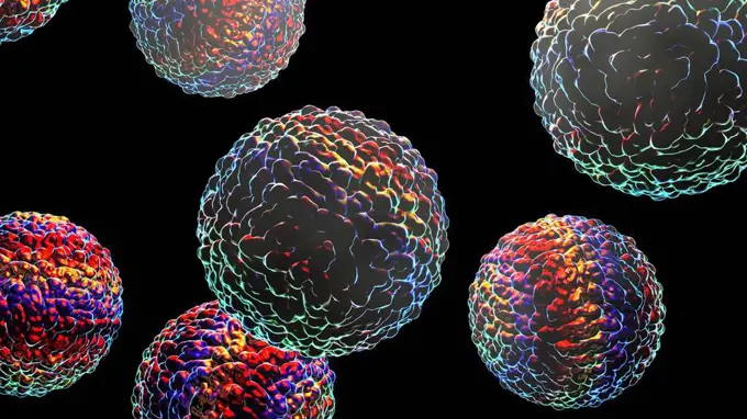
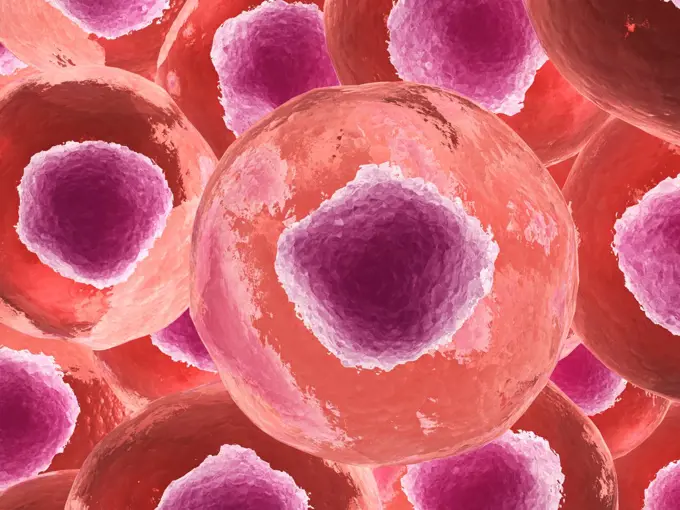
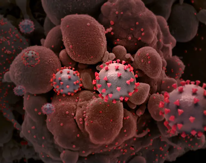
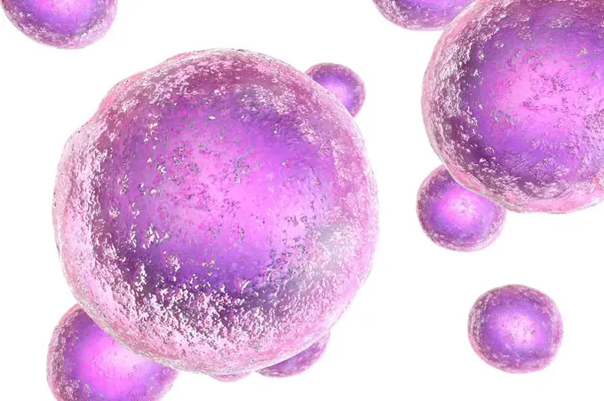
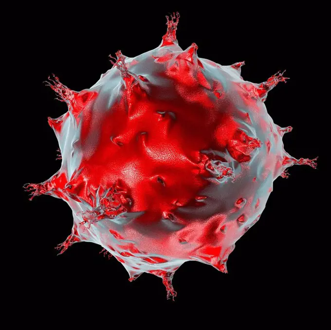
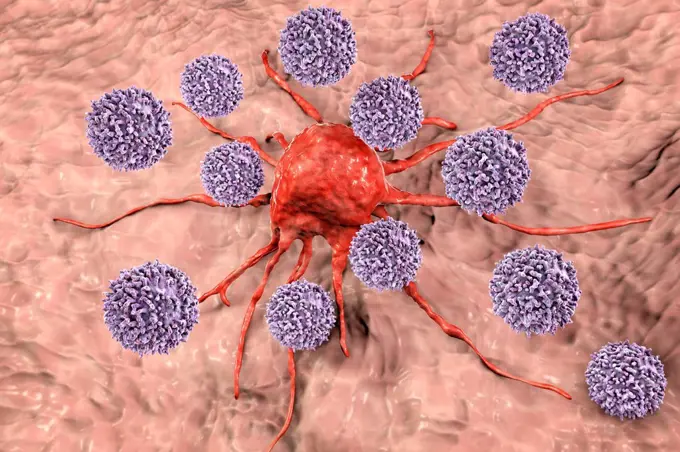
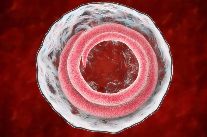
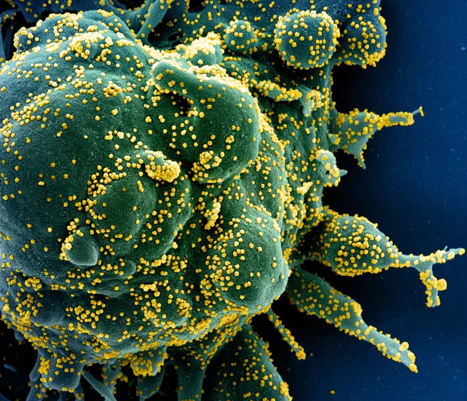
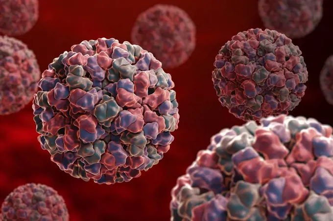
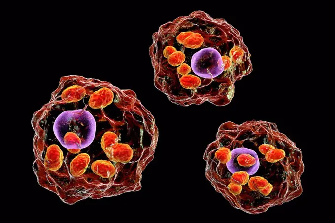
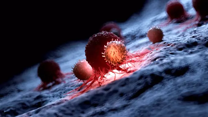
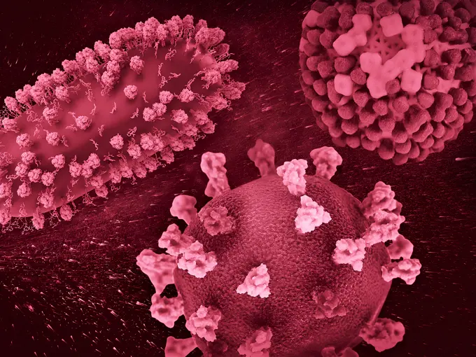
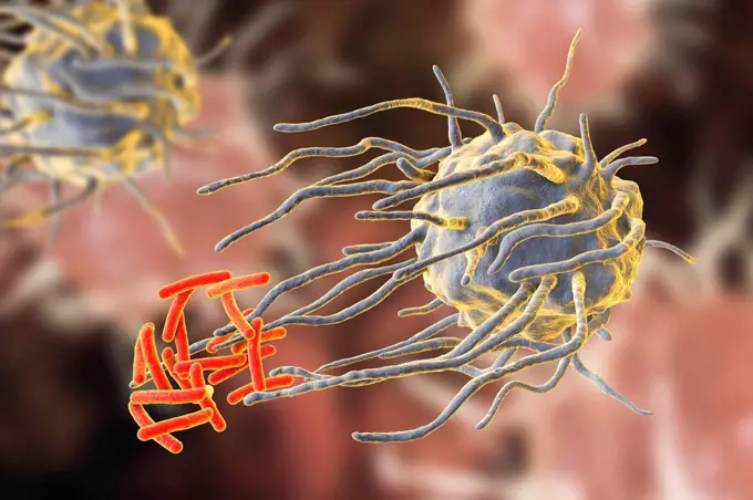
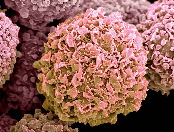
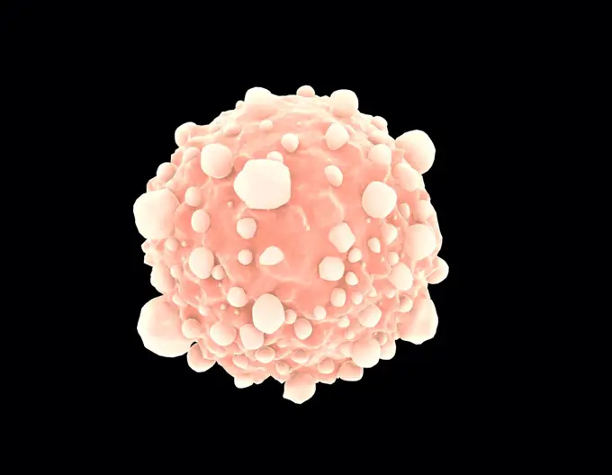
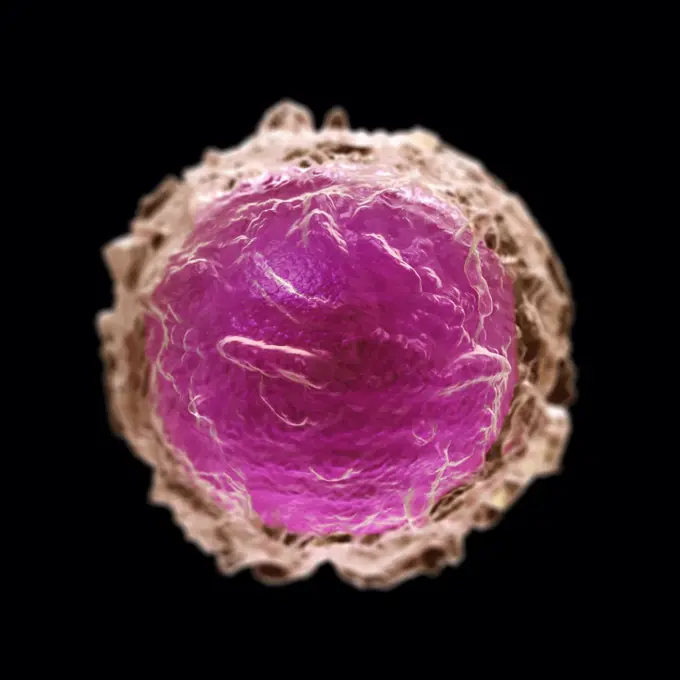
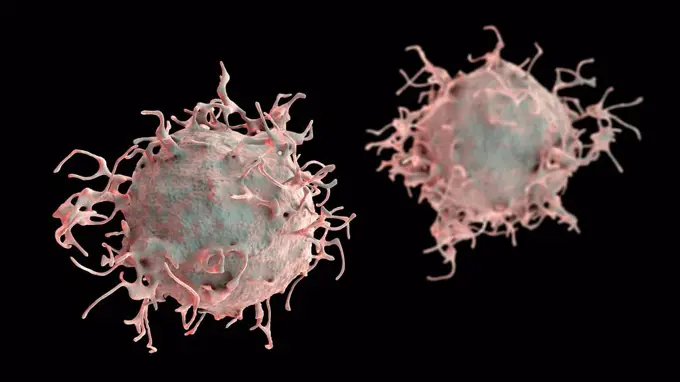
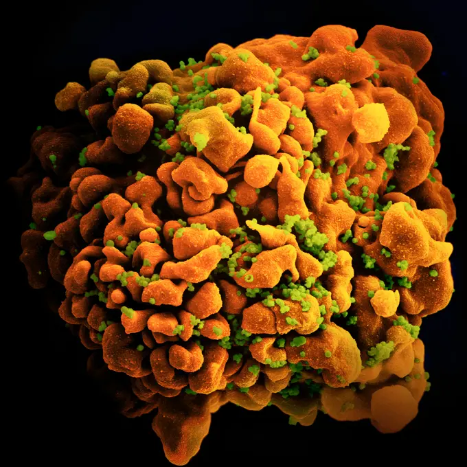
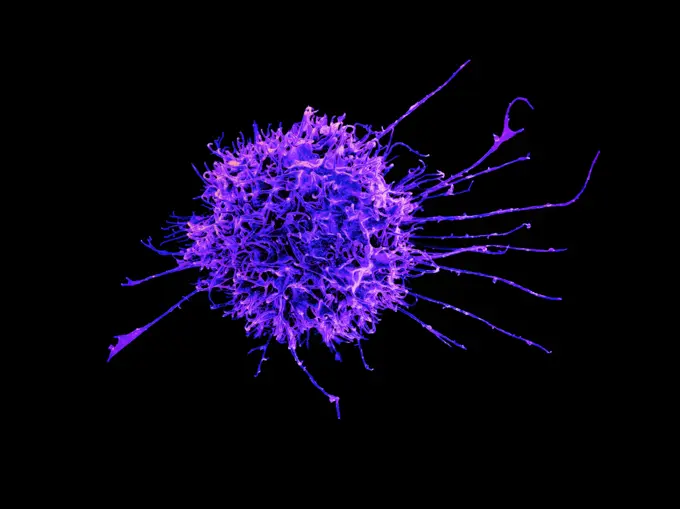
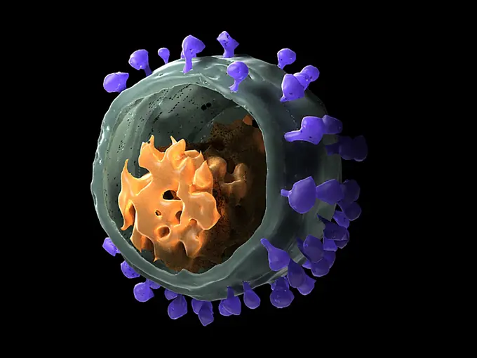
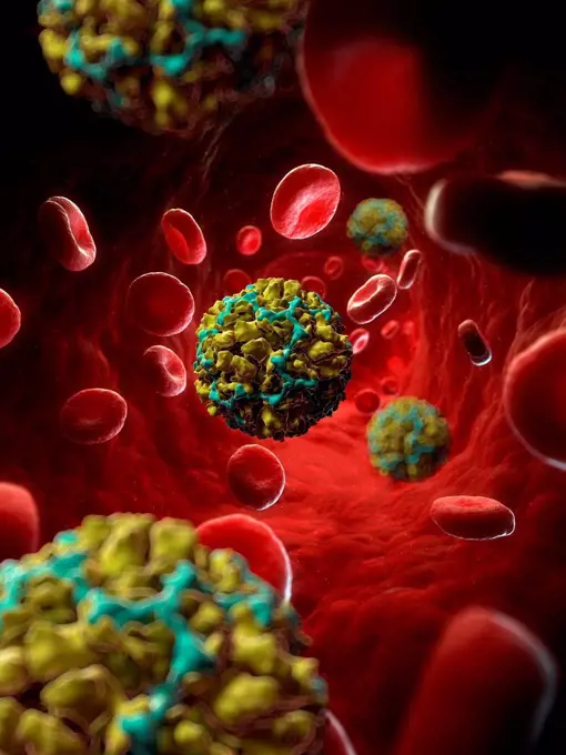
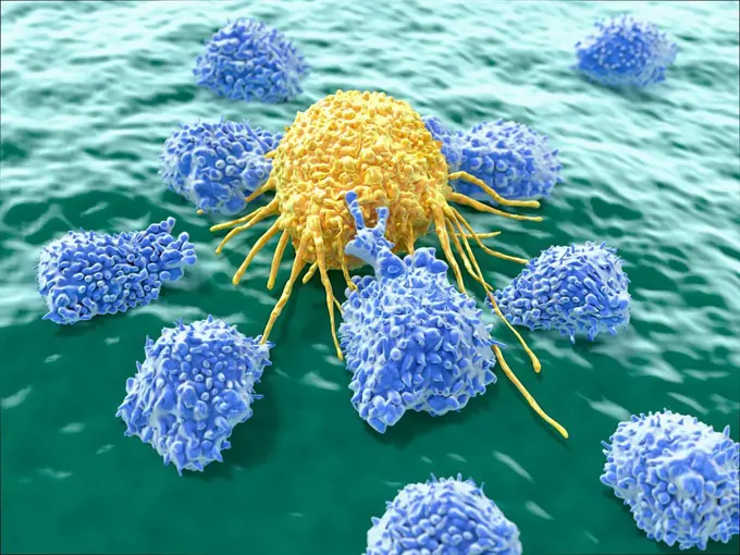
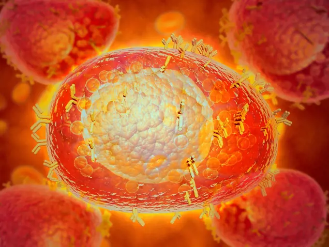
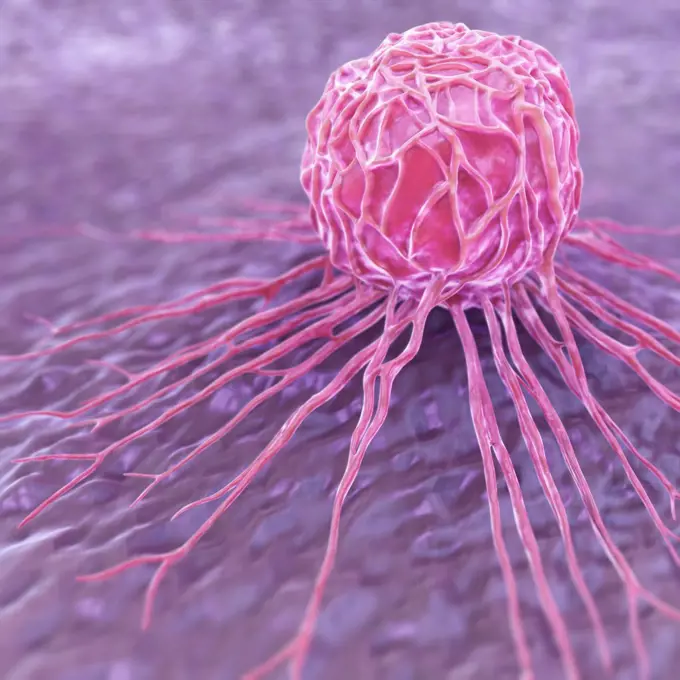
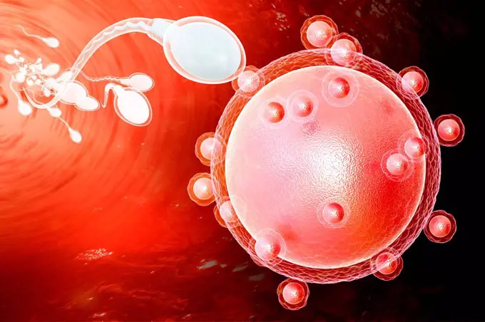
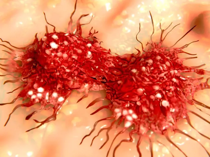
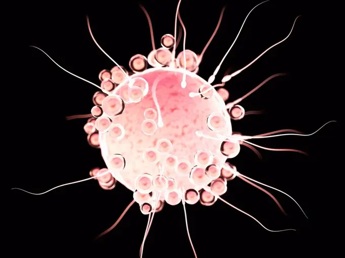
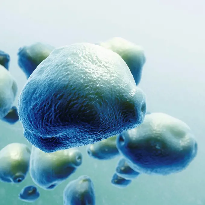
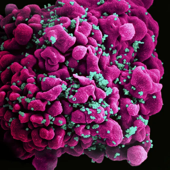
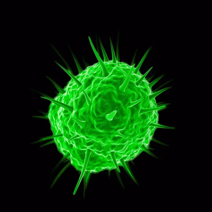
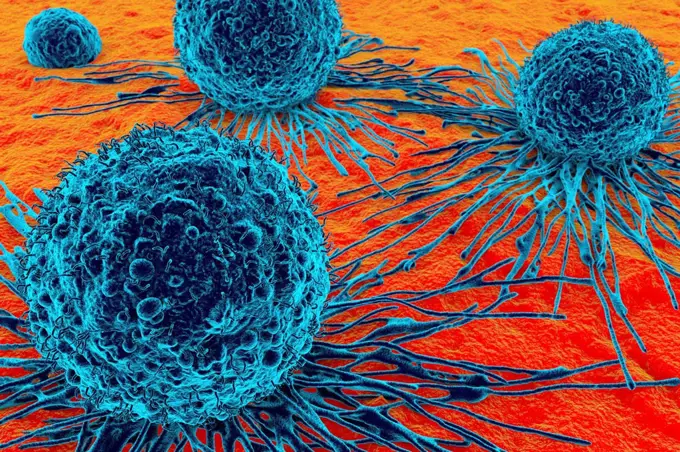
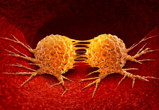
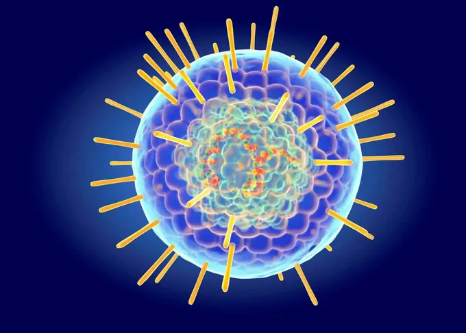
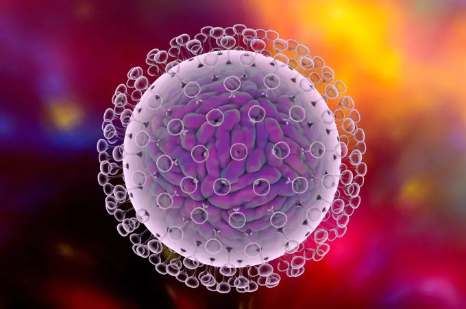
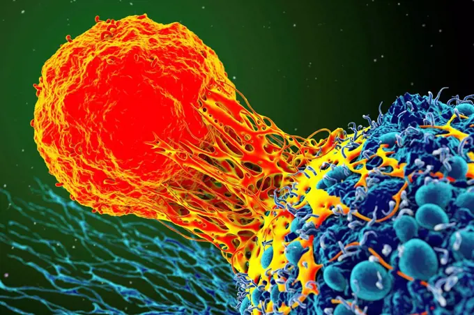
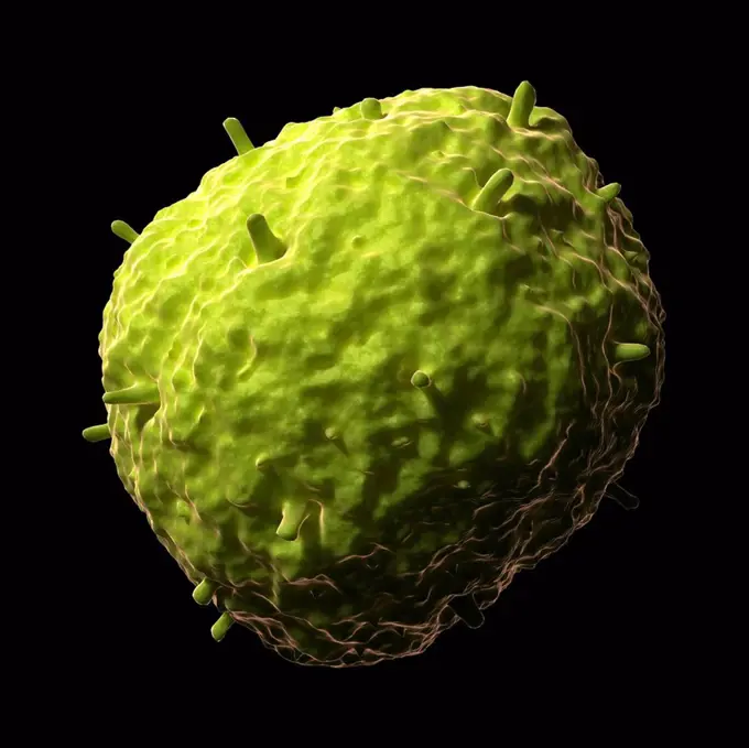
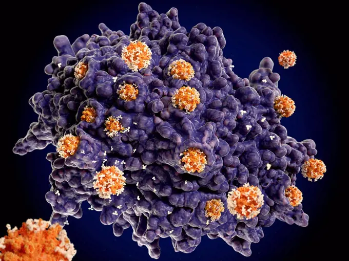
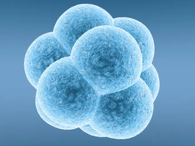
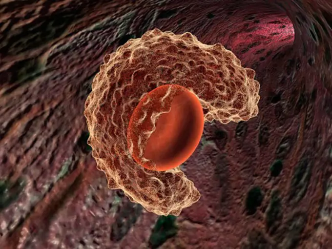
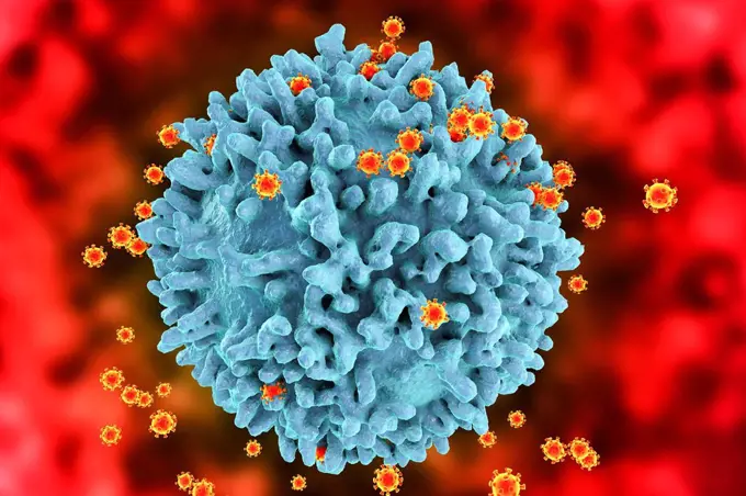
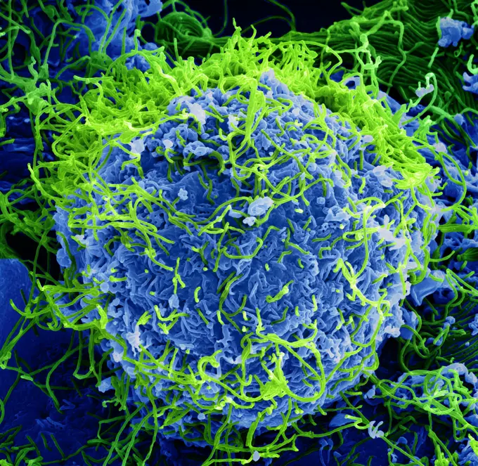
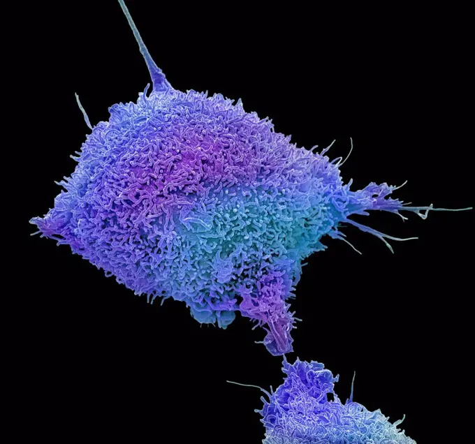
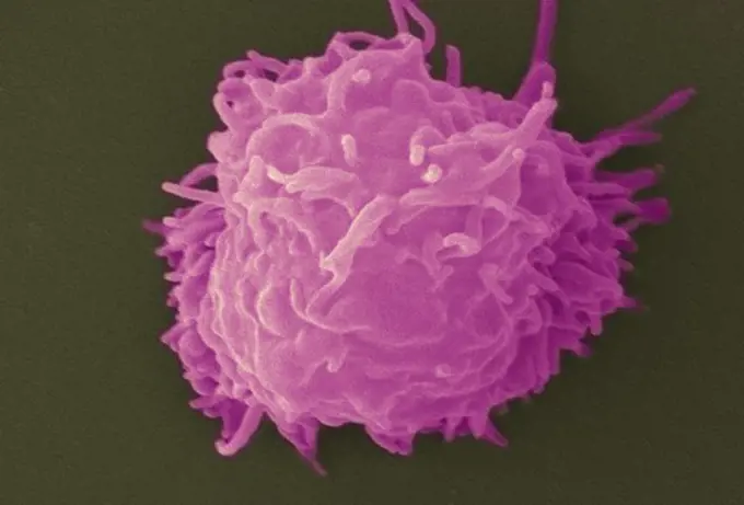
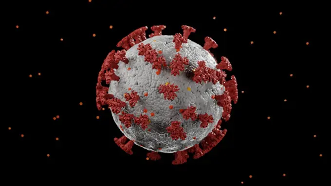
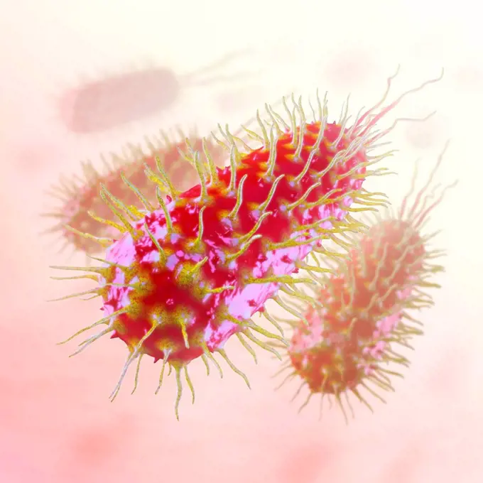
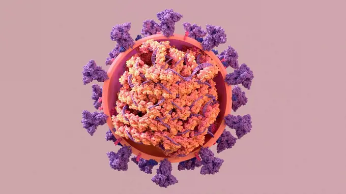
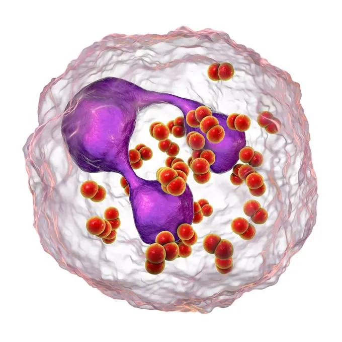
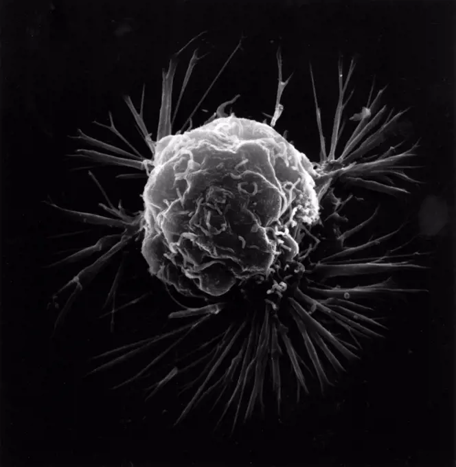
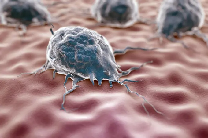
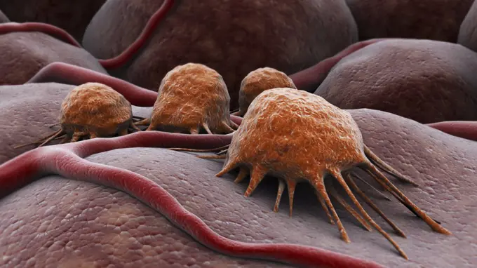
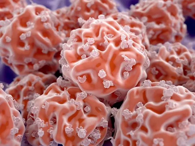
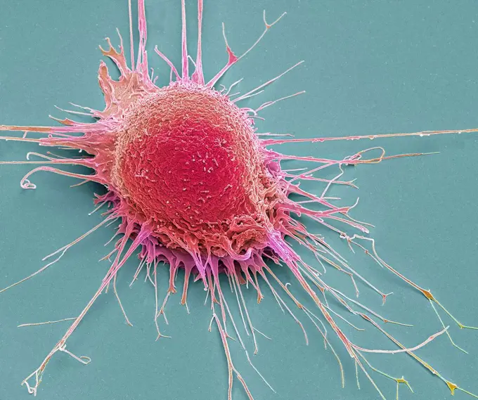
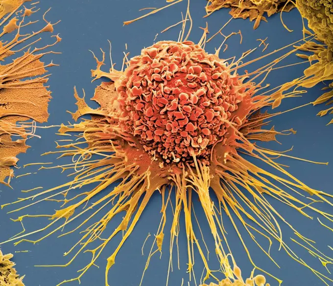
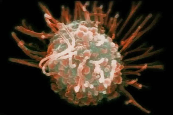
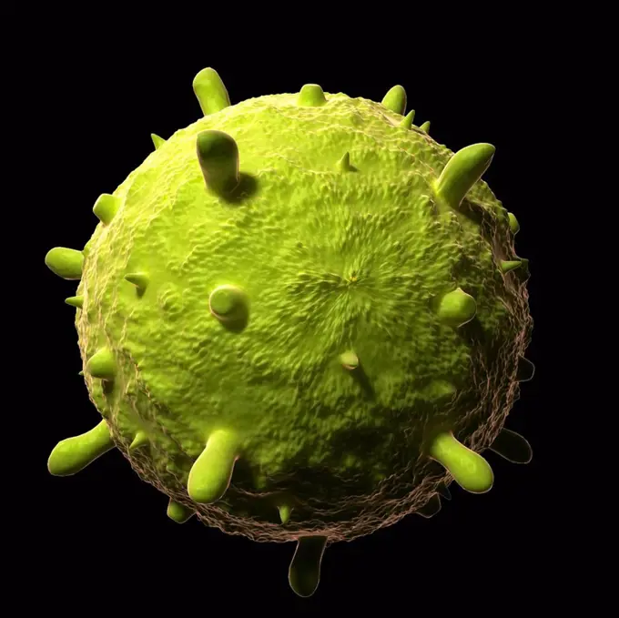
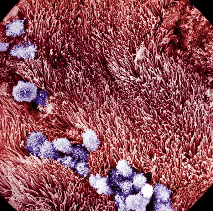
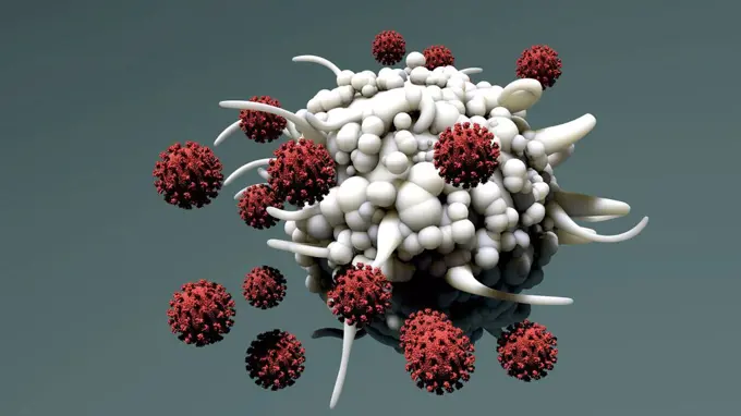
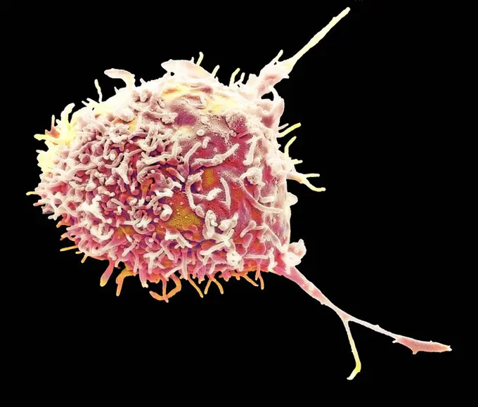
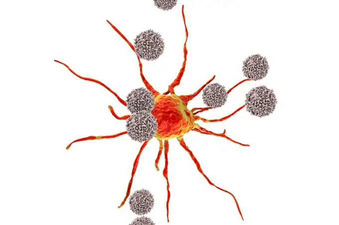
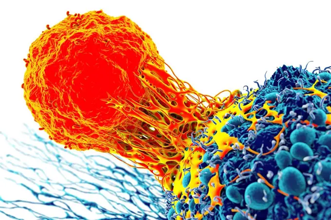
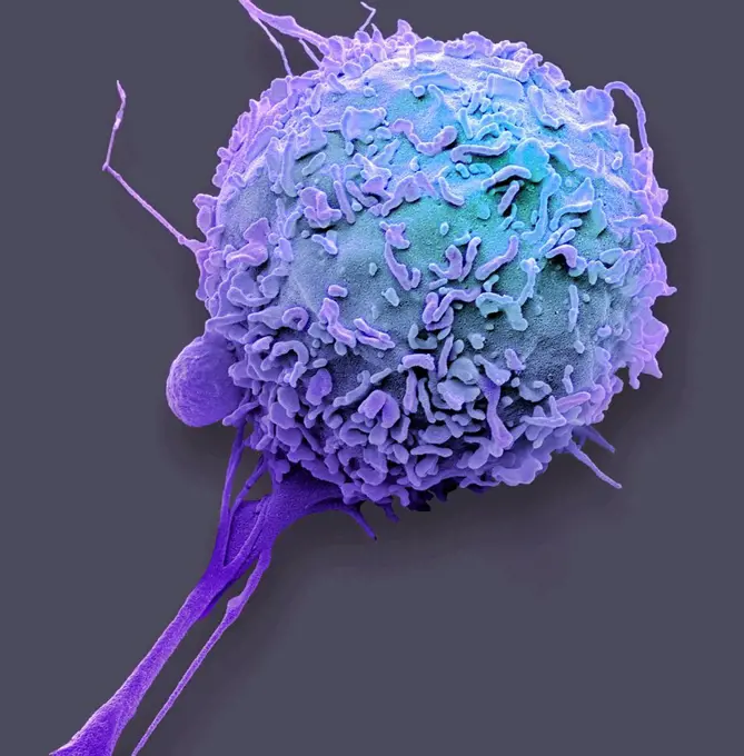
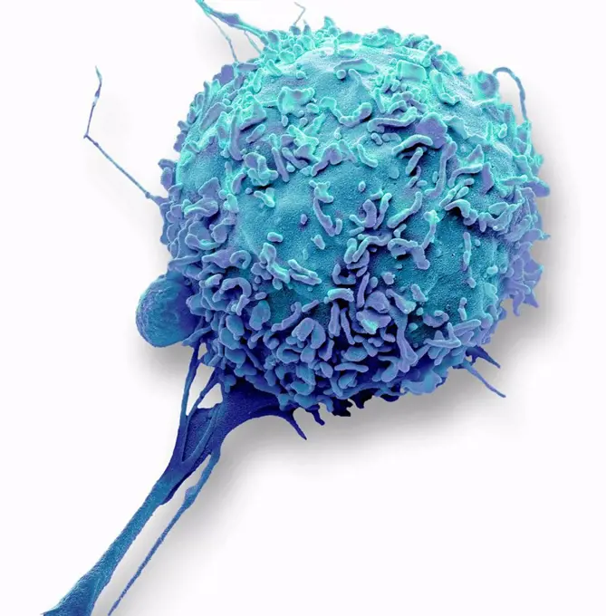
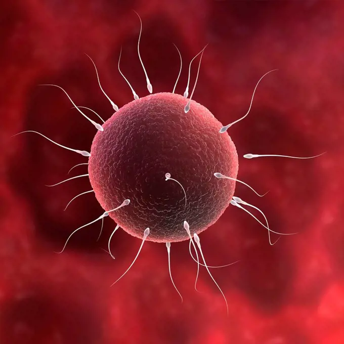
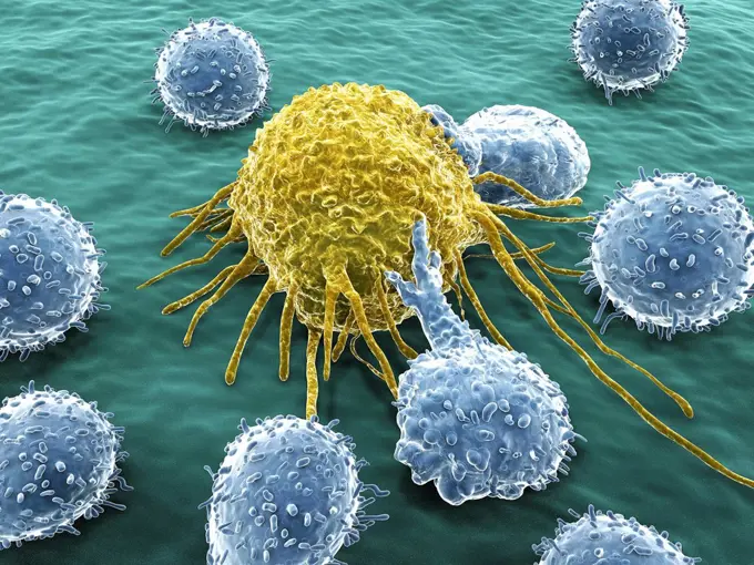
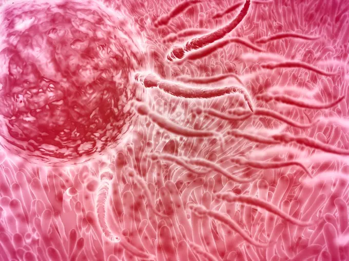
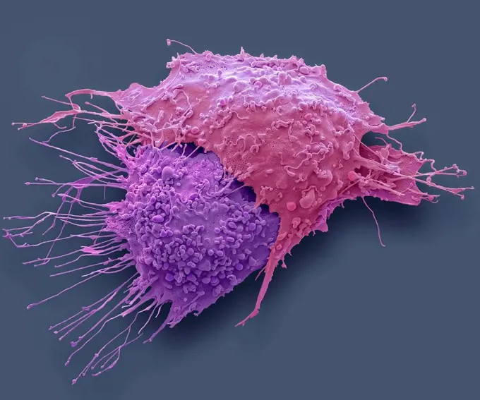
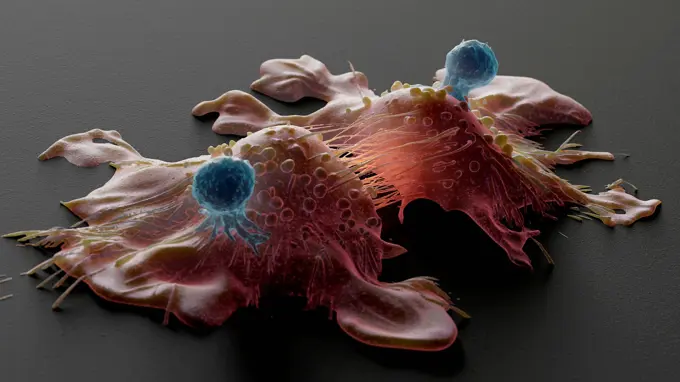
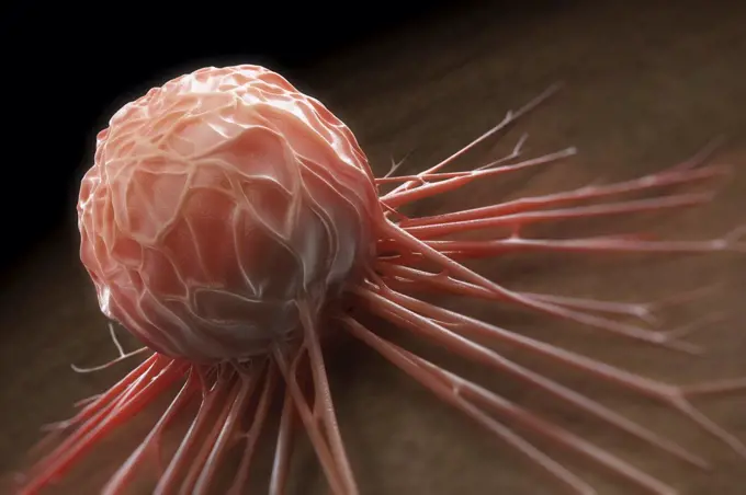
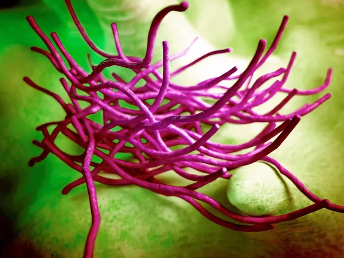
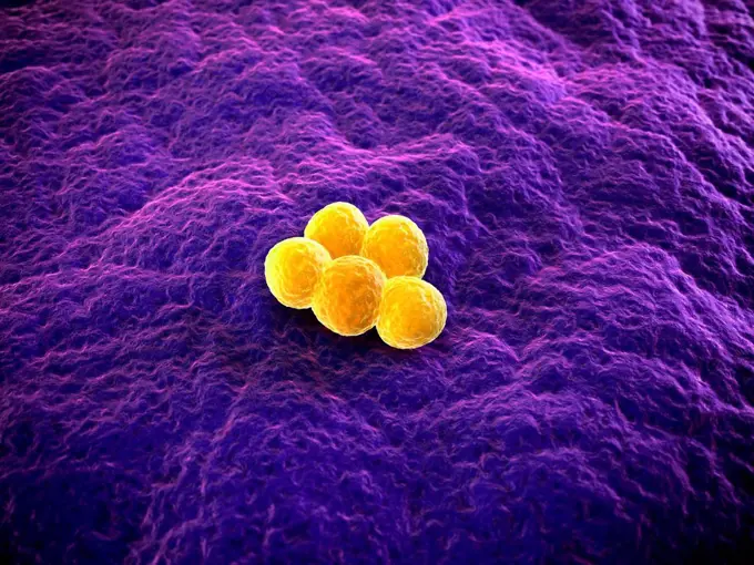
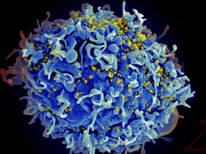
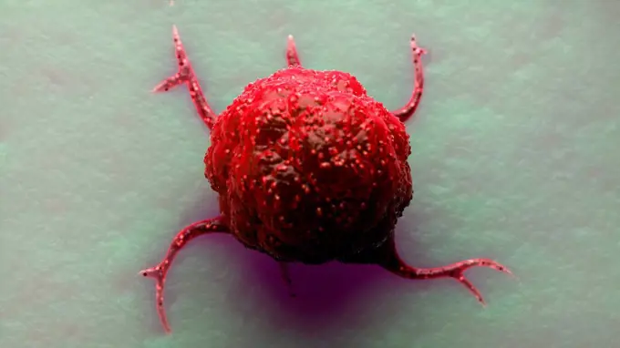
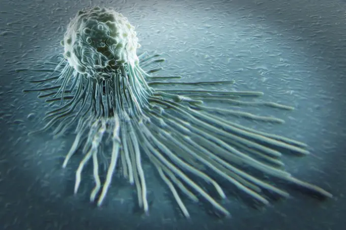
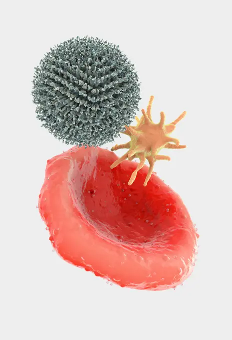
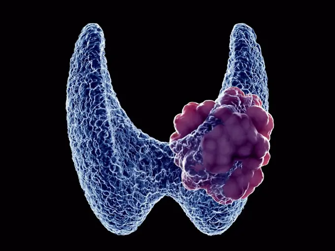
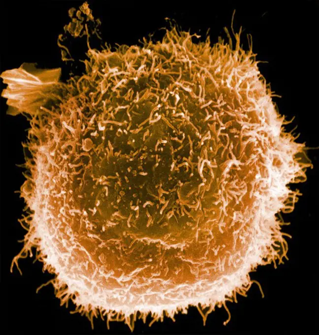
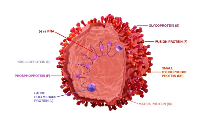
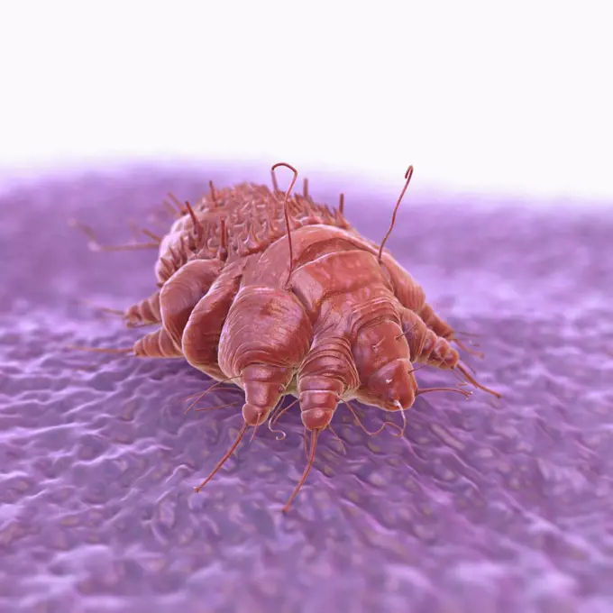
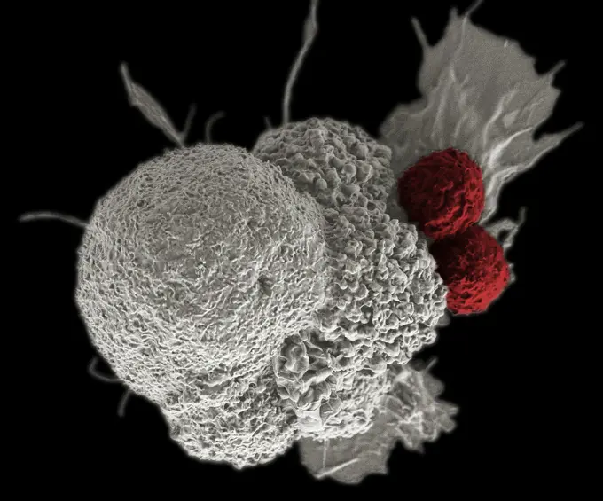
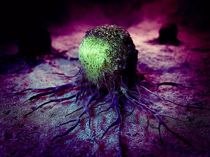
Microscopic Cells and Immunology

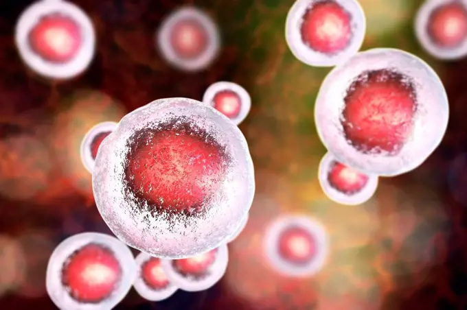
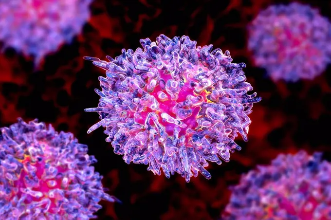
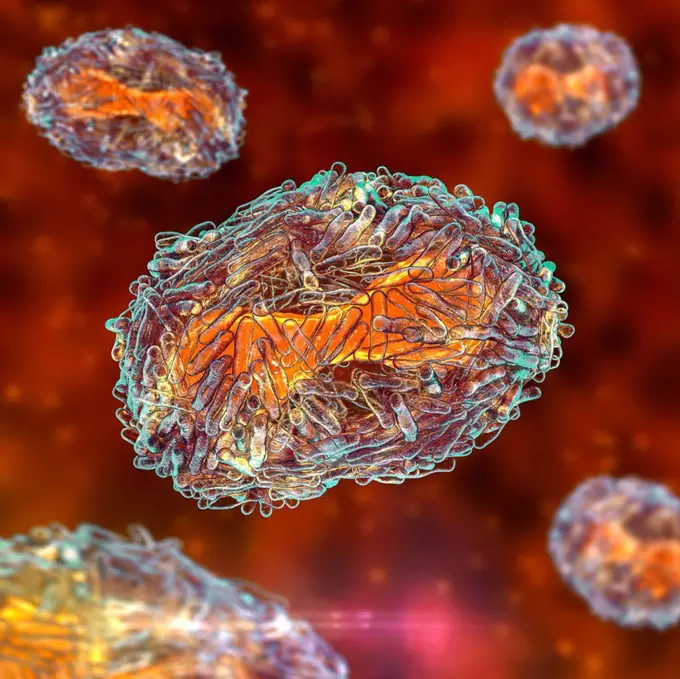
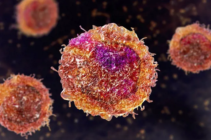
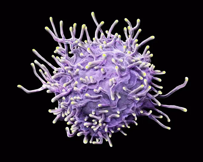
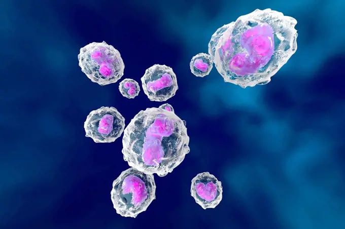
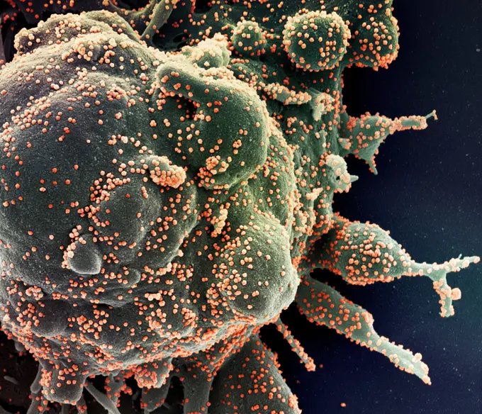
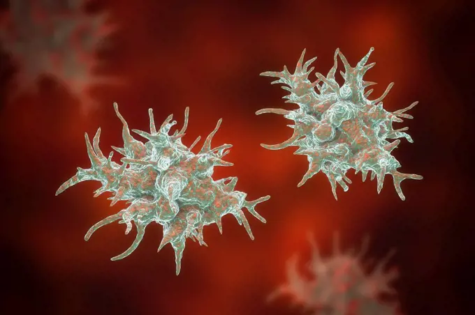
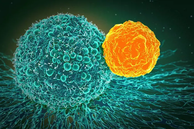
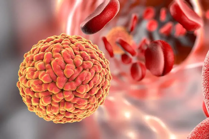
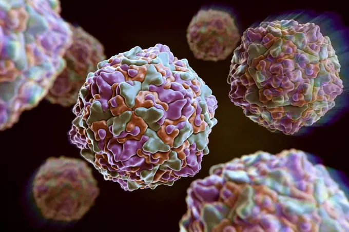
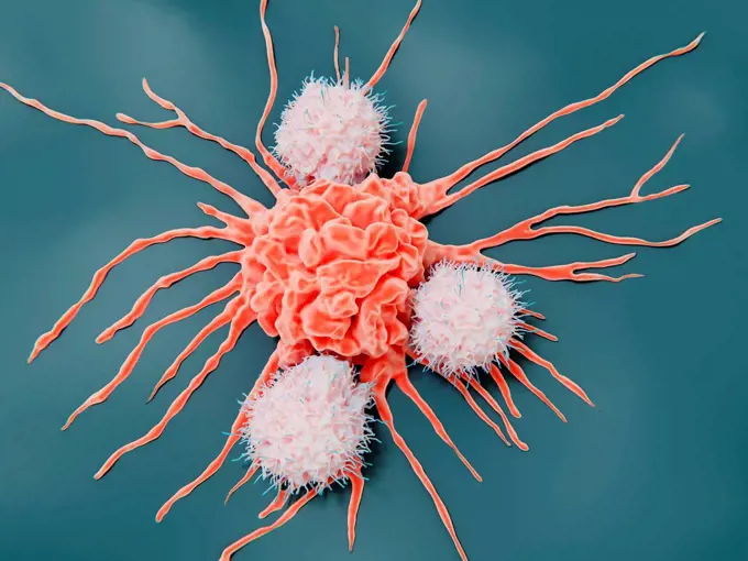
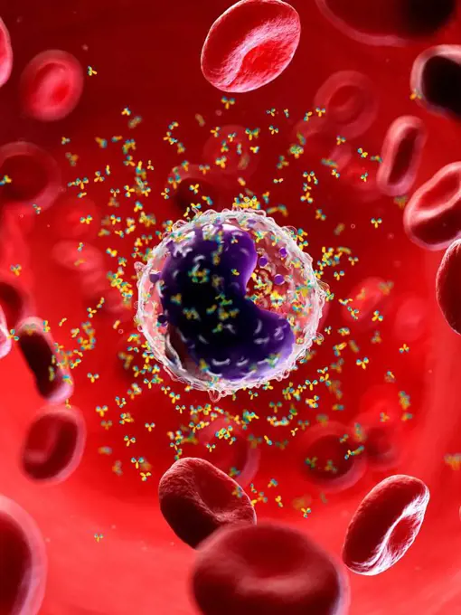
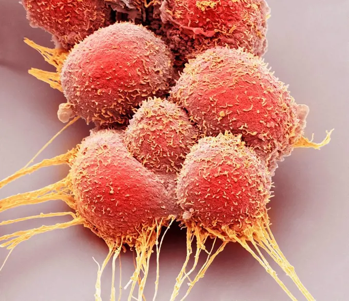
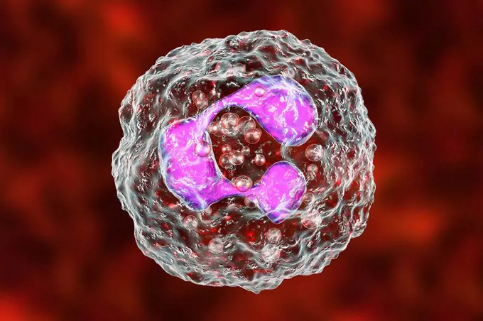
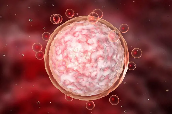
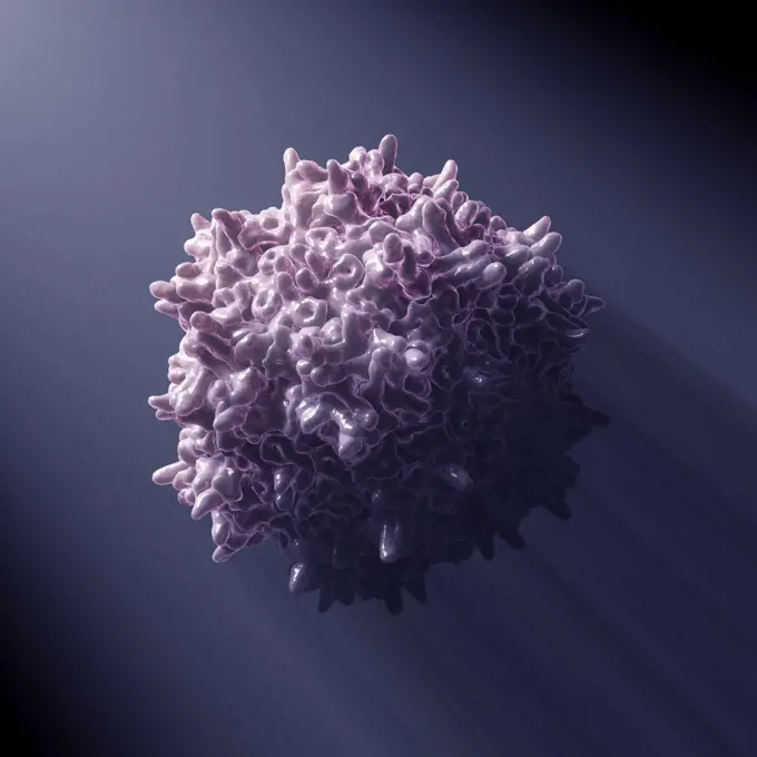
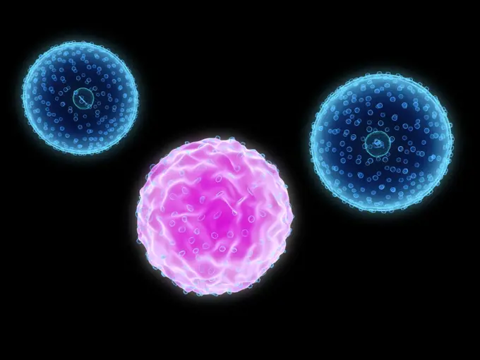
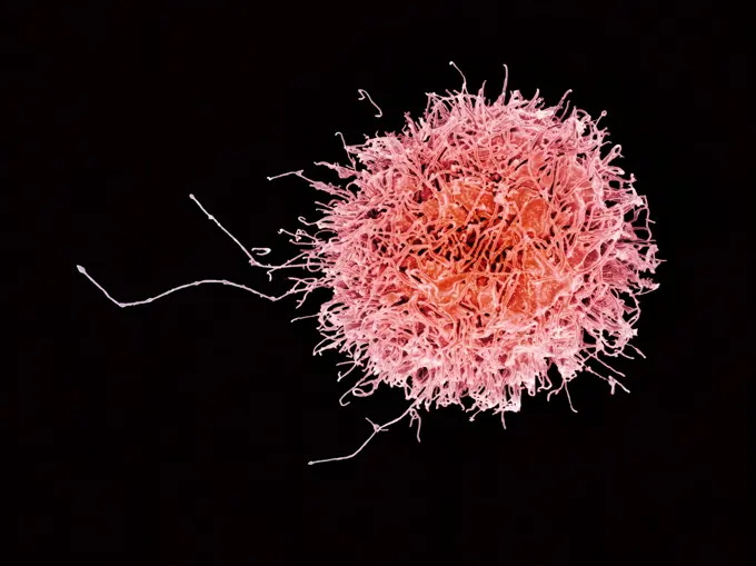
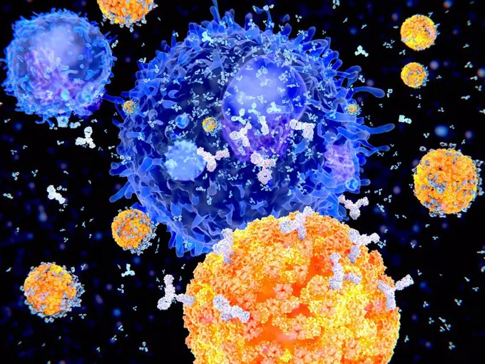
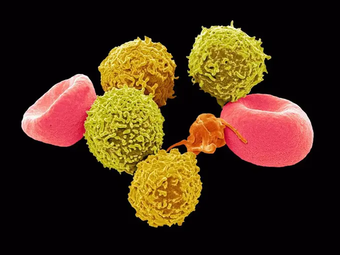
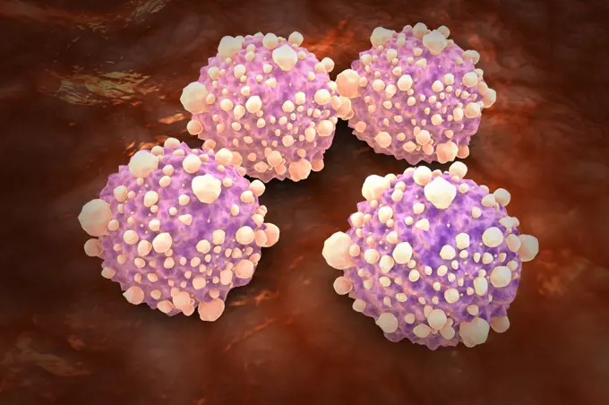
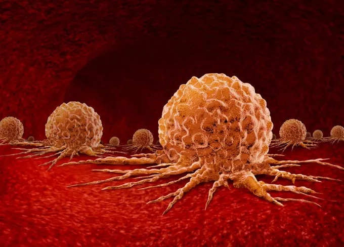
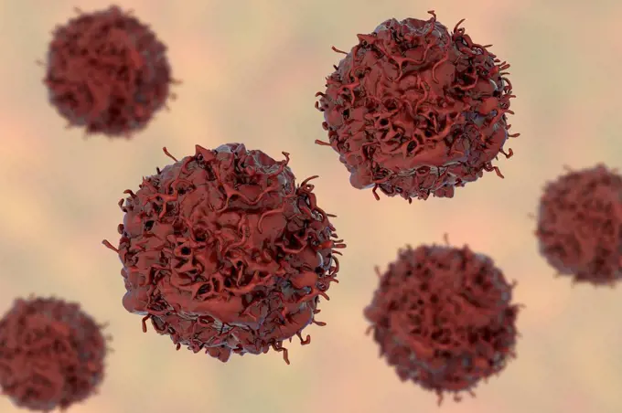
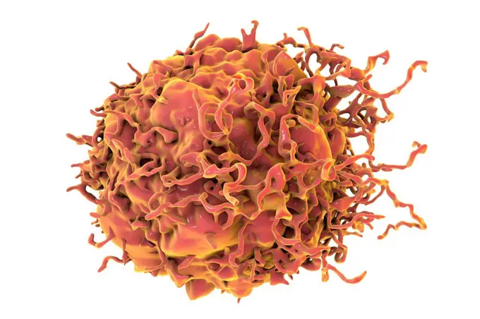
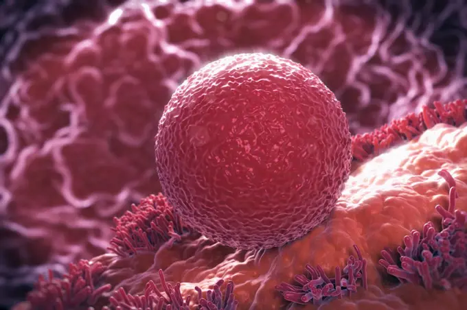
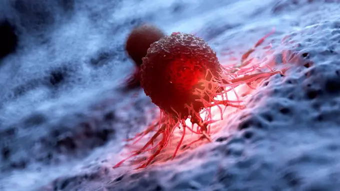
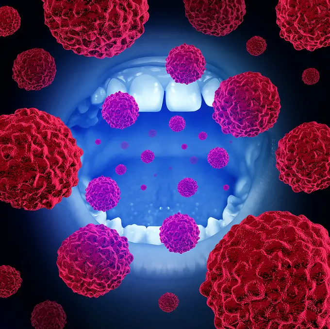
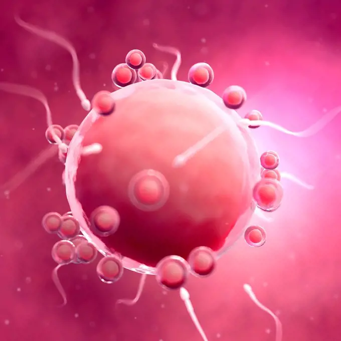
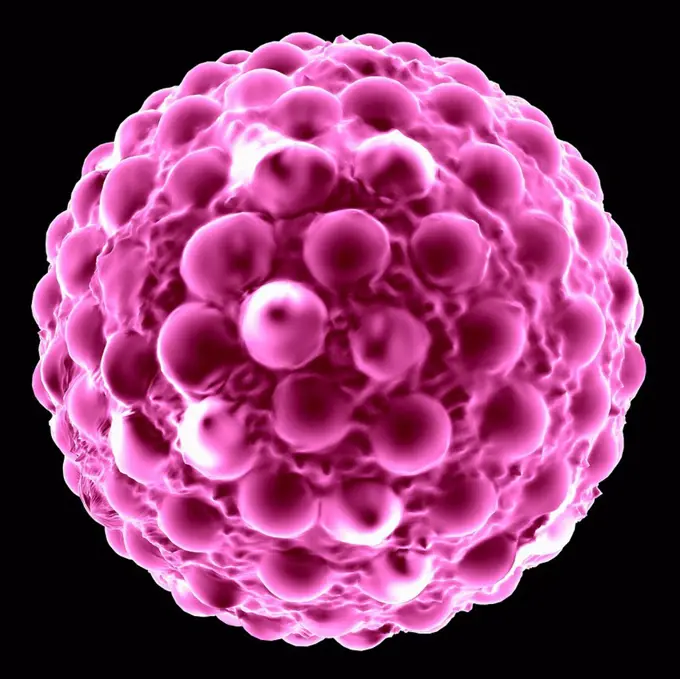
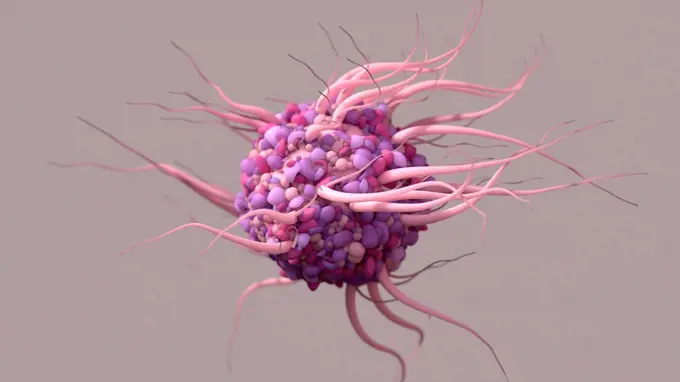
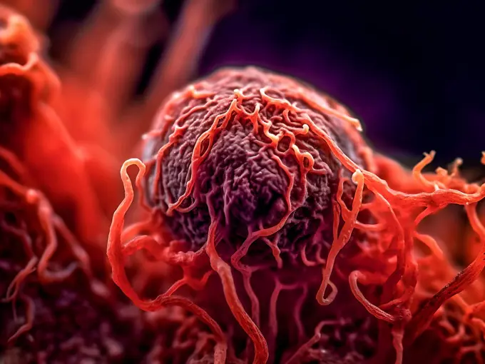
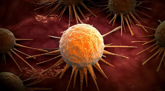
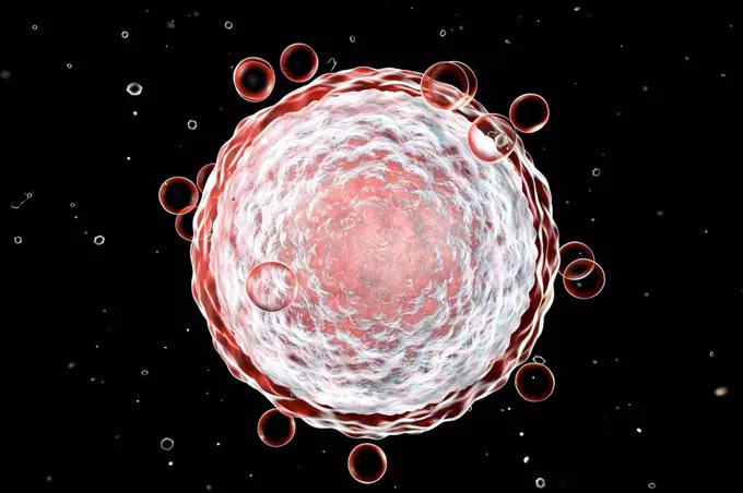
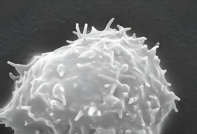
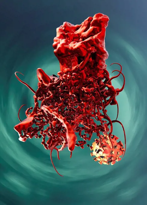
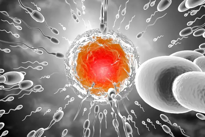
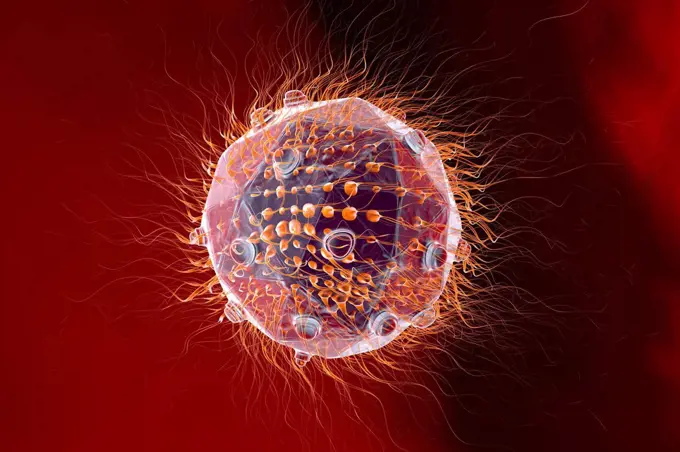
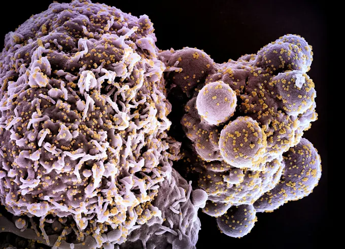
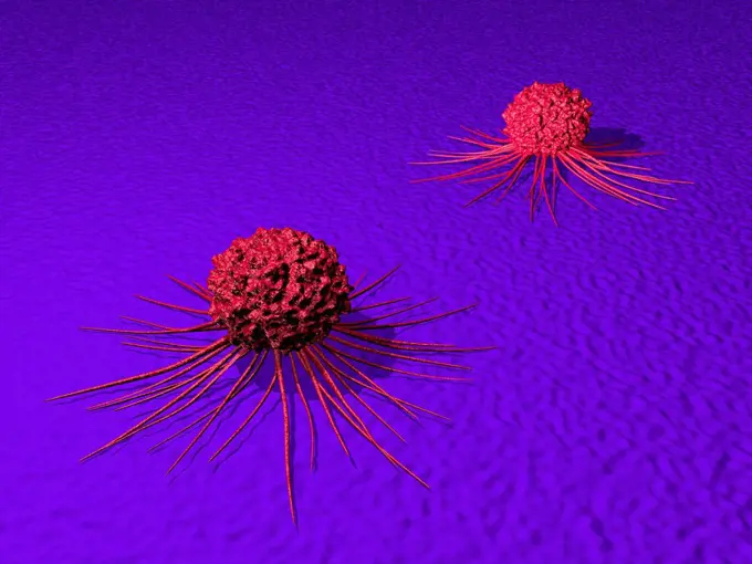
3D illustrations of various cells, including antibodies, cancerous cells, sperm and ovum, showcasing intricate details and vibrant colors in a scientific context.

Sperm and Ovum
Sperm and Ovum

Assets in this Story

6188-65582611
3D illustration of antibodies that attack a virus cell in the blood 3D illustration of antibodies that attack a virus cell in the blood
4128-111453068
Prostate cancer cells, SEM
4378-5408
Sperm and Ovum
4128R-13682117
Computer illustration of a cancerous white blood cell in leukaemia.
4378-5396
Embryonic Stem Cell
1815R-13386195
Virus similar to HI virus, 3D Rendering
4128R-13586614
Destruction of a cancer cell by nanoparticles, computer illustration. Conceptual image which illustrates the potential of nanoparticles in the treatme...
4378-20013834
Sperm and Ovum
4128R-13586613
Destruction of a cancer cell by nanoparticles, computer illustration. Conceptual image which illustrates the potential of nanoparticles in the treatme...
4128-16073625
Eradicating covid-19 coronavirus, conceptual illustration
4128R-14328996
Prostate cancer cells, illustration
824-65830627
Colorized scanning electron micrograph of red blood cell infected with malaria parasites (blue). The small bumps on the infected cell show how the parasite remodels its host cell by forming protrusions called 'knobs' on the surface, enabling it to avoid destruction and cause inflammation. Uninfected cells (pink) have smoother surfaces.
1899-65662499
Colorized scanning electron micrograph of a cell (brown) infected with the Omicron strain of SARS-CoV-2 virus particles (orange), isolated from a patient sample.
4417-16039635
Illustration of the Corona Virus DOF On Blue
4128R-13941203
Rectal cancer cell, illustration.
4128R-14057771
Cancer cell, illustration
4128-18799748
Cancer cells, illustration
1525-19757770
cancer cell made in 3d software
4239-V53647176
4128R-15366576
Neutrophil white blood cells, illustration
4128R-15046065
LDL receptors on cell membrane, illustration
4128R-10758
Adenovirus
4128R-15046998
RBCs and activated T lymphocytes, SEM
4128R-15366499
Human embryonic stem cells, illustration
4128R-15366541
Lymphocytes in hairy cell leukaemia, illustration
4128-19489543
Monkeypox virus particles, illustration
4128-18799723
Cancer cells, illustration
4128R-25364
Blastocyst. Computer artwork of a 100-cell blastocyst embryo.
4378-20013841
Embryonic Stem Cell
4128R-14181716
Human cell with chromosomes, illustration
824-63227221
Colorized scanning electron micrograph of a B cell from a human donor.
824-63227337
Colorized scanning electron micrograph of a cell (teal and green) infected with a variant strain of SARS-CoV-2 virus particles (UK B.1.1.7- purple and pink), isolated from a patient sample . Image requested from the NIAID Integrated Research Facility (IRF) in Fort Detrick, Maryland.
824-65831025
Colorized scanning electron micrograph of a cell (red) infected with the Omicron strain of SARS-CoV-2 virus particles (blue), isolated from a patient sample.
4269-27691
HIV. Scanning electron micrograph (SEM) of a lymphocyte with HIV cluster.
4128R-11473793
Canine panleukopenia, computer artwork.
4128R-15047003
Activated T lymphocyte, SEM
1815R-13380200
Monocytes, immune system, defense cells, 3D rendered illustration
824-63223227
Colorized scanning electron micrograph of an apoptotic cell (green) heavily infected with SARS-COV-2 virus particles (orange), isolated from a patient sample. Image captured at the NIAID Integrated Research Facility (IRF) in Fort Detrick, Maryland. Credit: NIAID.
4128R-14587958
Acanthamoeba amoeba, illustration
4128R-14057776
Cancer cell and T cell, illustration
4128R-13022167
Zika virus, computer illustration. This is an RNA (ribonucleic acid) virus from the Flaviviridae family. It is transmitted to humans via the bite of a...
4378-3379
Crystal structure of Coxsackievirus (PDB 1COV).
4128-18631823
Natural killer cells attacking a cancer cell, illustration
4128-V58569136
4128R-15516277
Illustration of a cancer cell
4378-1808
Fertilization between egg and sperm.
4128R-11543403
Virus, conceptual illustration.
4128-111612430
Toxic mould spores (Stachybotrys chartarum), computer illustration. Several strains of this fungus (S. atra, S. chartarum and S. alternans) are synony...
4128R-13586489
Illustration of Rotavirus.
4128R-12901
Puffball fungus spore, coloured scanning electron micrograph SEM. This is the reproductive cell of the fungus. Magnification: x3000 when printed at 10...
4128-15981012
B cell and antibodies, computer illustration.
4128-19357717
Illustration of a cancer cell.
4239-18641368
Conceptual image of hepatitis virus. Viral hepatitis is liver inflammation due to a viral infection. The most common causes of viral hepatitis are the five unrelated hepatotropic viruses
4128-111453066
Prostate cancer cells, SEM
4128R-15366572
Neutrophil white blood cells, illustration
4128R-13575538
Human egg cell, computer illustration.
1525R-178728
4128-28970688
Cancer cells dividing, illustration
4239-18641386
Microscopic view of Sindbis virus (SINV). SINV is a mosquito-borne virus that causes rash and arthritis, has been causing outbreaks in humans
4128R-14057764
Cancer cells, illustration
4128R-13619874
Destruction of leukaemia blood cell, computer illustration. Conceptual image for leukaemia treatment.
1849-66221385
Close up of HIV virus in bloodstream
4269-20408825
H9-T cell infected by numerous, spheroid-shaped, human immunodeficiency virus (HIV) particles, which can be seen sttached to the cell's surface membrane, (SEM).
1525-24404837
Inner intestine cancer anatomy concept as the malignant tumor growth inside of a digestion organ with intestinal villi and crypts in a 3D illustration style.
1899-65662496
Colorized scanning electron micrograph of a cell (purple) infected with the Omicron strain of SARS-CoV-2 virus particles (green), isolated from a patient sample.
4378-4603
Blood Cells
4128-18799714
Cancer cells, illustration
4128R-11474824
Swine influenza virus, computer artwork.
4128-15980254
Zika viruses, computer illustration. This is an RNA (ribonucleic acid) virus from the Flaviviridae family. It is transmitted to humans via the bite of...
4239R-20483554
Microscopic view of animal cell.
824-63223251
Creative rendition of SARS-CoV-2 virus particles. Note: not to scale. Credit: NIAID.
4128R-15366502
Human embryonic stem cells, illustration
4128R-11543412
Virus, conceptual illustration.
4128R-11543487
Virus, conceptual illustration.
4128R-11542043
Virus, computer illustration.
4128R-15405935
T-lymphocytes attacking cancer cell, illustration
4128R-13282744
Cyst with Trichina spiralis roundworm (formerly called Trichinella spiralis), computer illustration. These parasites infect mammals, including humans,...
824-63223228
Colorized scanning electron micrograph of an apoptotic cell (green) heavily infected with SARS-COV-2 virus particles (yellow), isolated from a patient sample. Image captured at the NIAID Integrated Research Facility (IRF) in Fort Detrick, Maryland. Credit: NIAID.
4378-3978
Structure of the Norwalk virus (1HIM), an RNA virus from the family Caliciviridae and the major cause of epidemic gastroenteritis in humans.
4128R-14057508
Leishmania parasites, illustration
4128R-15516261
Illustration of white blood cells attacking a cancer cell
4128-V58557899
824-65831070
Creative work featuring 3D renditions of three dangerous respiratory viruses: RSV (top left), SARS-CoV-2 (bottom center), and flu (top right).
1428R-340
Close-up of a macrophage cell
4378-3830
Atomic structure of Adeno-Associated Virus (PDB 1LP3), a vector for human gene therapy.
1525-22197389
leukemia
824-63227231
Colorized scanning electron micrograph of a natural killer cell from a human donor.
4128-15701630
Immune response to a virus, illustration
4128R-15545707
Blood cells, SEM
4128-19357718
Illustration of a T helper cell, also known as a CD4+ (cluster of differentiation 4) cell.
4239R-20483588
Microscipic view of pancreatic cancer cells.
1525-56205030
Malignant Cancer Growth and Metastasis anatomy concept as a growing tumor cell and Malignancy disease growth on an organ inside the human body as a 3D illustration.
4128R-15290458
Lung cancer cell, illustration
4128R-14324122
Macrophage engulfing tuberculosis bacteria, illustration
4128-15982702
Breast cancer cells. Coloured scanning electron micrograph (SEM) of breast cancer cells. The cells shows numerous processes and microplicae (fine surf...
4239R-20483499
Microscopic view of pancreatic cancer cell.
4378-20013829
Lymphocyte Cell
4128R-13575639
Colon cancer cell, computer illustration.
4378-3202
An ovum surrounded by hair like structures called cilia which form part of the lining of the fallopian tubes.
4378-20014123
Sperm and Ovum
4128R-15516249
Illustration of a human cancer cell
1525-20986989
Oral cancer medical concept as a mouth with malignant disease cells with 3D illustration elements.
4128-16506724
Skin cancer cell, computer illustration.
824-63227039
Scanning electron micrograph of an HIV-infected H9 T cell.
824-63227040
Scanning electron micrograph of a human natural killer cell. Credit: NIAID
824-63191270
Rendering of 3D structure of a single simian immunodeficiency virus obtained using cryo-electron tomography.
4128R-13371814
Fertilisation of human egg, illustration.
4128R-11474565
Hepatitis a, computer artwork.
4128-18498245
Dendritic cell, illustration
4128-48285955
Blood vessel formation in tumour, illustration
4128R-11312916
Human rhinovirus 14, computer artwork.
4128R-13022947
Illustration of lymphocytes attacking a cancer cell. Natural killer cells are a type of lymphocyte that destroys cancer cells and other altered cells ...
4239R-8337
Microscopic view of a mast cell found in connective tissue that contains numerous basophilic granules and releases substances such as heparin and histamine in response to injury.
4378-3925
Close up view of a single breast cancer cell on a surface.
4128R-14060108
Human conception, illustration
4128R-12576630
Dividing cancer cell, computer illustration.
4128-111581935
Fertilisation, computer illustration.
4378-4579
Birch Tree Pollen
824-63191249
Colorized image of HIV-infected H9 T-cell.
4128R-11474110
Mimivirus, computer artwork.
4128R-14057766
Cancer cells, illustration
1525-25739610
Dividing cancer cell metastasis division as a disease anatomy concept as a growing malignant tumor on an organ inside the human body as a 3D illustration.. Dividing Cancer Cell
4128-111493687
Herpes Simplex virus structure, illustration. Herpes simplex is one of the most common viral parasites in humans. They cause small, fluid-filled blist...
4378-1060
Conceptual visualization of the gonorrhea bacterium that causes the sexually transmitted disease gonorrhea.
4128R-13023068
Zika virus. Computer illustration showing the protein spikes in the outer viral envelope. This is an RNA (ribonucleic acid) virus from the Flavivirida...
4128R-14057789
Cancer cell and T cell, illustration
4128R-11313715
H1N1 influenza virus, computer artwork.
4128-16102446
Macrophage engulfing coronaviruses, illustration
4128R-22400
Fertilisation, computer artwork.
1525-19806055
Embryo
1574R-018975
Macrophage Cell
4128-V58562335
4128R-13372949
HIV viruses infecting T-lymphocytes, computer illustration. The surface of the T-cell has a lumpy appearance with large irregular surface protrusions....
4128R-11543433
Virus, conceptual illustration.
4239R-20483464
Conceptual image of cancer virus.
1525-27361598
Digital Illustration of the human Ovum
824-63225901
Colorized scanning electron micrograph of a cell infected with a variant strain of SARS-CoV-2 virus particles (UK B.1.1.7 green), isolated from a patient sample. Image captured at the NIAID Integrated Research Facility (IRF) in Fort Detrick, Maryland.
4128R-13575541
Human egg cell, computer illustration.
1899-61460841
This scanning electron microscope (SEM) image revealed some of the ultrastructural features on the surface of a protozoan Acanthamoeba polyphaga. Note the numerous pseudopodia, protruding from the surface of the organisms. These allow the amoeba to move and grasp objects in its environment. CDC/Catherine Armbruster Margaret Williams 2009
4128R-13682013
Cell destruction, illustration.
1815-111782703
3D Rendered Illustration, visualisation of sperm cells racing to a egg to fertilise
1815R-14189247
Insemination, 3D Rendering
1815R-13377832
Virus, 3D Rendering
1899-65662500
Colorized scanning electron micrograph of a cell (pink) infected with the Omicron strain of SARS-CoV-2 virus particles (yellow), isolated from a patient sample.
4128R-14649629
Cancer cells, illustration
824-63223154
Colorized scanning electron micrograph of Ebola virus particles (green) both budding and attached to the surface of infected VERO E6 cells (blue). Image captured and color-enhanced at the NIAID Integrated Research Facility in Fort Detrick, Maryland. Credit: NIAID.
4128R-15465091
Cervical cancer cell, SEM
4297-1728
SEM of an Acanthamoeba polyphaga protozoa
1525-24096304
3D Render Coronavirus cells or bacteria molecule. Virus Covid-19. Close-up of Flu, view of a virus under a microscope, infectious disease. Coronavirus influenza background,viral disease epidemic. Health care concept. Isolation period. Precautionary measures. 3D Render Coronavirus cells or bacteria molecule. Virus Covid-19. Close-up of Flu, view of a virus under a microscope, infectious disease
4128R-30489
Bacteria, computer artwork.
4128-V58562083
4128-18301091
Covid-19 coronavirus particle, illustration
4128R-15366566
Gonorrhoeae bacteria inside neutrophils, illustration
1899-85561
Breast Cancer SEM
4128R-11474552
Influenza virus H1N1, computer artwork.
824-63227036
Scanning electron micrograph of Ebola virus particles (green) budding and attached to the surface of infected VERO E6 cells (orange). Focused image at the NIAID Integrated Research Center in Fort Detrick, Maryland. Credit: NIAID
4128-18631819
Natural killer cells attacking a cancer cell, illustration
1525-56190062
Stem cells in the bone marrow are responsible for producing white blood cells. 3d illustration. Stem cells in the bone marrow are responsible for producing white blood cells.
4128R-2704
Diatom alga, SEM
824-63227177
Colorized scanning electron micrograph of Ebola virus filamentous particles (blue) budding from a chronically infected VERO E6 cell (yellow-green).
4378-1173
Stylized macrophage cells on a cellular surface.
1815-111459846
Insemination, 3D Rendering
4239-18641710
Conceptual image of cancer virus.
4128R-13192835
Stem cells, illustration. Stem cells are undifferentiated cells that can produce other types of cell when they divide. There are three main stem cell ...
4128-111576787
Prostate cancer cell, Coloured scanning electron micrograph (SEM). Such cells can clump to form tumours, which often invade and destroy surrounding ti...
4128R-30460
Activated human macrophage, coloured scanning electron micrograph (SEM). Magnification: x2,700 when printed at 10 centimetres wide.
4297-1798
SEM image of a human white blood cell
4128R-11312883
Influenza virus, computer artwork.
4269-6800
Scanning electron micrograph (SEM) of the cilia lining up the Fallopian tubes.
4128-16171701
Dendritic cell and virus particles, illustration. Dendritic cells are a component of the body's immune system. The cell gets it name from the long mem...
4128-20041801
Mesenchymal stem cell, SEM
4128R-15405929
T-lymphocytes attacking cancer cell, illustration
4128R-14057791
Cancer cell and T cell, illustration
4128R-15465085
Macrophage, SEM
4128R-15465078
Macrophage, SEM
4128-V58557841
4128R-14490
Fertilisation. Computer artwork of sperm fertilising an egg.
4128R-11291558
Artwork of lymphocytes attacking a cancer cell.
4239R-8403
Sperm traveling towards egg with cellia.
4128R-15465075
Ovarian cancer cells, sem
824-63227132
Shown here is a pseudo-colored scanning electron micrograph of an oral squamous cancer cell (white) attacked by two cytotoxic T cells (red), part of a natural immune response. Nanomedicine researchers create personalized cancer vaccines by loading neo-antigens identified from patient and tumor
4128-28970696
CAR-T cell therapy, illustration
4378-2927
Close up view of a single breast cancer cell.
4128R-13587285
Anthrax bacteria, illustration.
4128R-26274
MRSA bacteria. Computer artwork of methicillin-resistant Staphylococcus aureus (MRSA) bacteria.
4128R-13410450
HIV virus infecting a cell, illustration
824-63191251
Scanning electron micrograph of HIV particles infecting a human T cell.
4128R-15516293
Illustration of a cancer cell
4378-3369
A close up of a macrophage cell. Macrophages are phagocytes and their function is involved in both innate immunity as well as adaptive immunity.
6188-66486097
3d-Illustration der sehr genau beobachteten roten, Erythrozyten genannten, weißen Blutkörperchen, Leukozyten genannt, und Blutplättchen, Thrombozyten genannt, im menschlichen Körper 3d illustration of very closely observed red, called erythrocytes, white blood cells, called leukocytes and platelets, called thrombocytes in the human body Copyright: xZoonar.com/ChristophxBurgstedtx 22601134
4128-15665288
Thyroid tumour, illustration
4297-1825
SEM image of a human macrophage
4128-30419946
Spherical respiratory syncytial virus, illustration
4378-3223
A single Sarcoptes scabiei mite which is the cause of the contagious skin infection Scabies. The mite burrows under the host's skin, causing intense allergic itching.
4269-21219147
T lymphocytes and cancer cell, Coloured scanning electron micrograph (SEM) .
4128R-13371444
Cancer cell, illustration.
1848-54783061
Tumour in an early stage, illustration
4298-1055
Bean root with nitrogen nodule  Magnification 40x
6188-67458302
3D Isometric Flat Illustration of Cancer Cells, Tumor Developmentbr3D Isometric Flat Illustration of Cancer Cells, Tumor Development 3D Isometric Flat Illustration of Cancer Cells, Tumor Developmentbr3D Isometric Flat Illustration of Cancer Cells, Tumor Development Copyright: xZoonar.com/AnastasiiaxTorianykx 23013229
1746-30005089
Nancy Burson's 'Visualize This'. Nancy Burson (1948-) an American artist