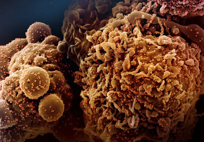
Scanning electron micrograph of Ebola virus particles (green) budding and attached to the surface of infected VERO E6 cells (orange). Focused image at the NIAID Integrated Research Center in Fort Detrick, Maryland. Credit: NIAID

SuperStock offers millions of photos, videos, and stock assets to creatives around the world. This image of Scanning electron micrograph of Ebola virus particles (green) budding and attached to the surface of infected VERO E6 cells (orange). Focused image at the NIAID Integrated Research Center in Fort Detrick, Maryland. Credit: NIAID by NIH-NIAID/IMAGE POINT FR/BSIP is available for licensing today.
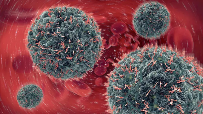
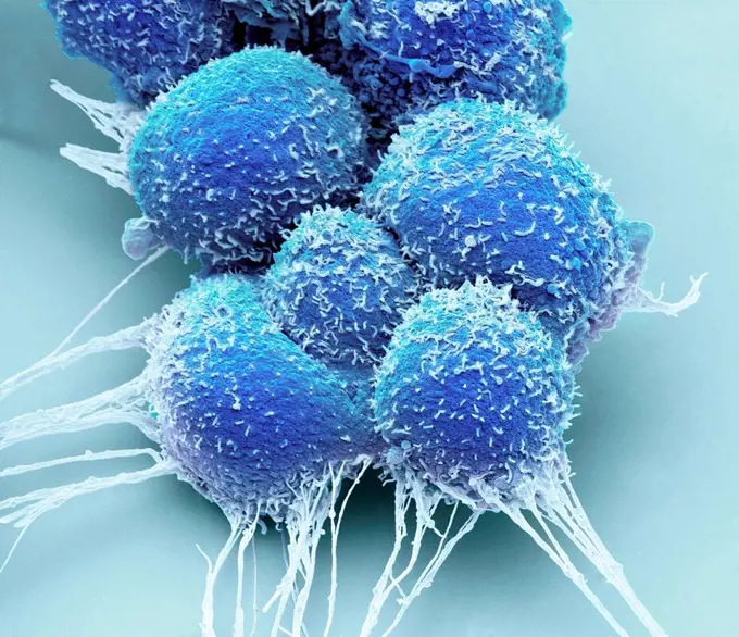
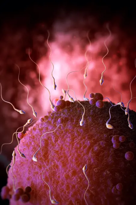
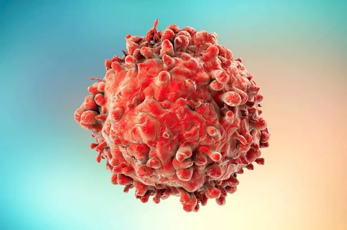
Visually Similar More from Microscopic Cells and Immunology story

Looking for a license?
Click here, and we'll help you find it! Questions? Just ask!
Click here, and we'll help you find it! Questions? Just ask!
DETAILS
Image Number: 824-63227036Rights ManagedCredit Line:NIH-NIAID/IMAGE POINT FR/BSIP/SuperStockCollection:BSIP Story:Microscopic Cells and ImmunologyContributor:NIH-NIAID / IMAGE POINT FR / BSIP Model Release:NoProperty Release:NoResolution:4200×4100