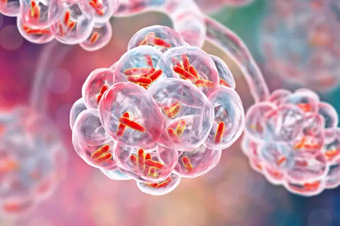
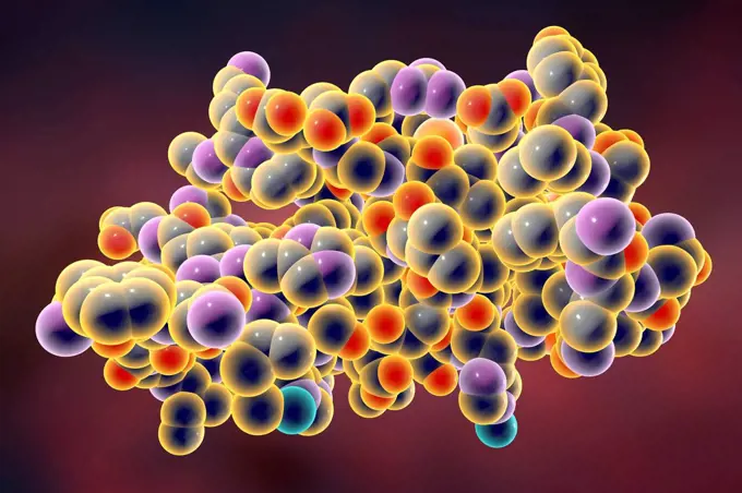
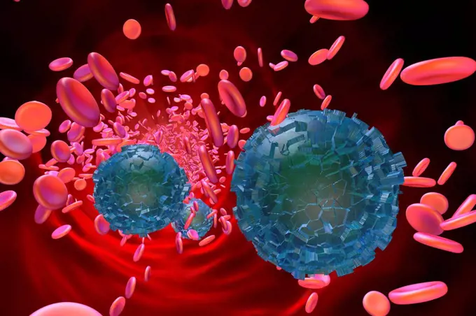
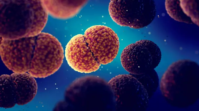
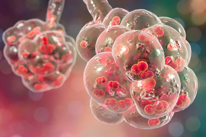
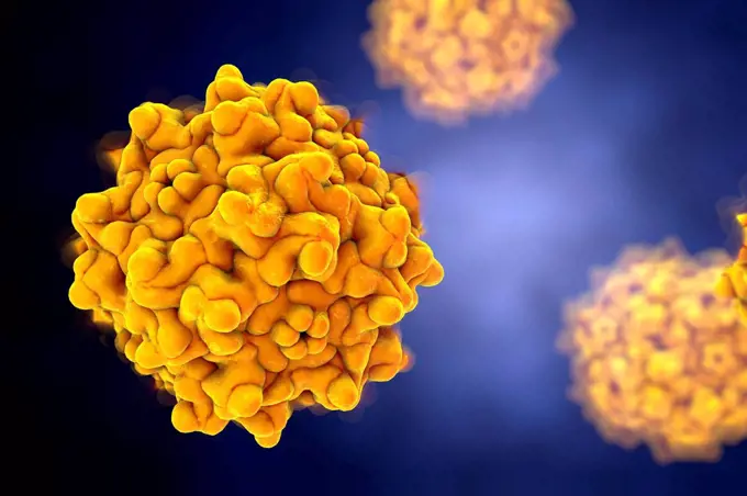
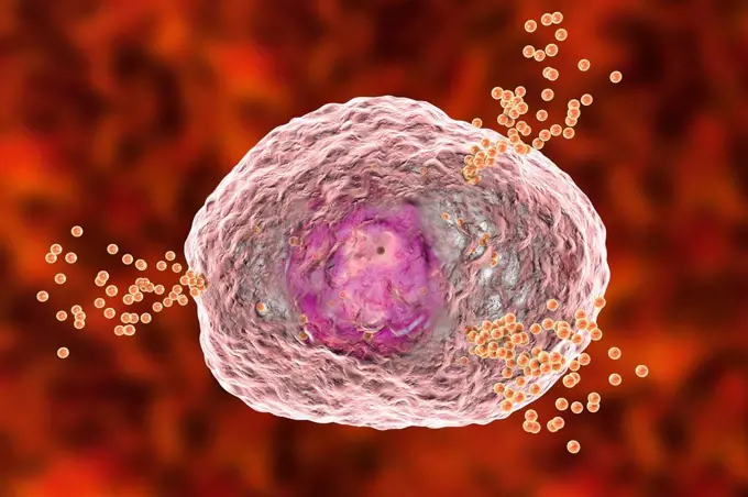
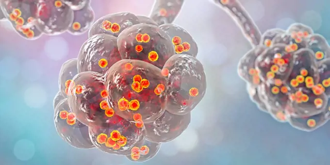
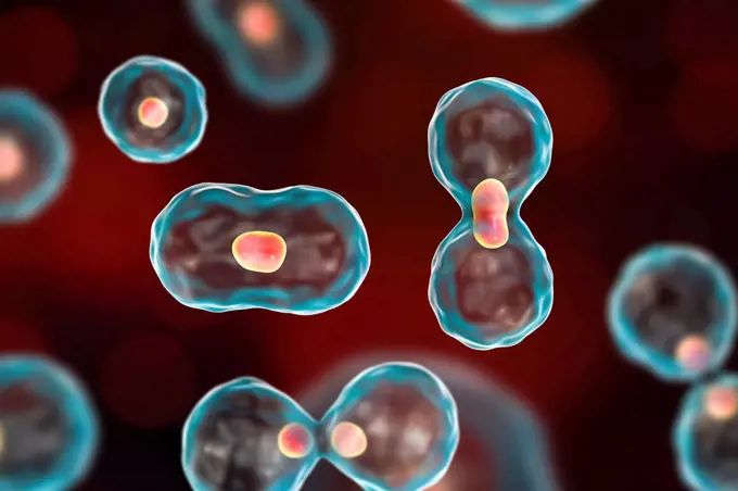
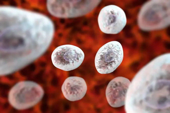
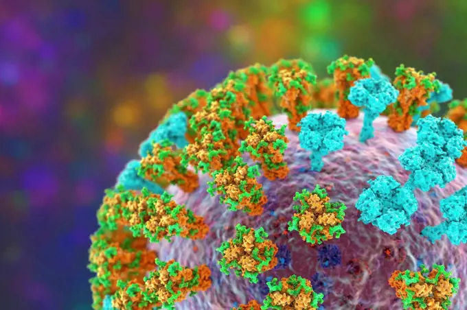
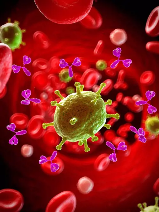
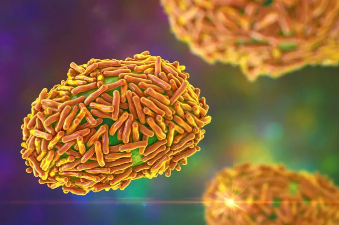
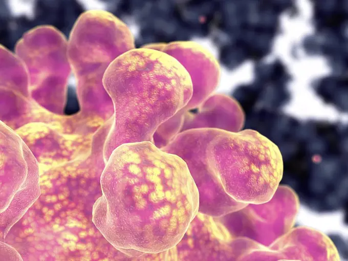
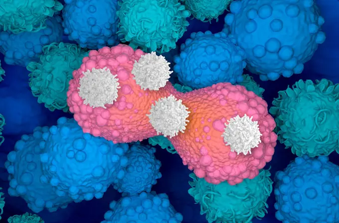
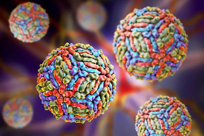
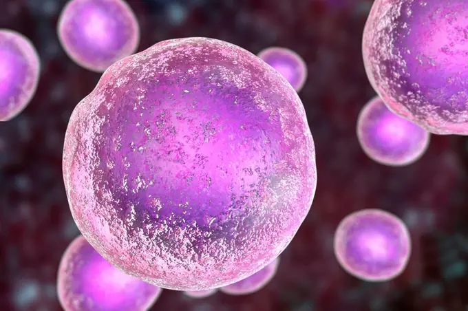
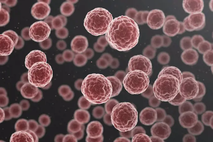
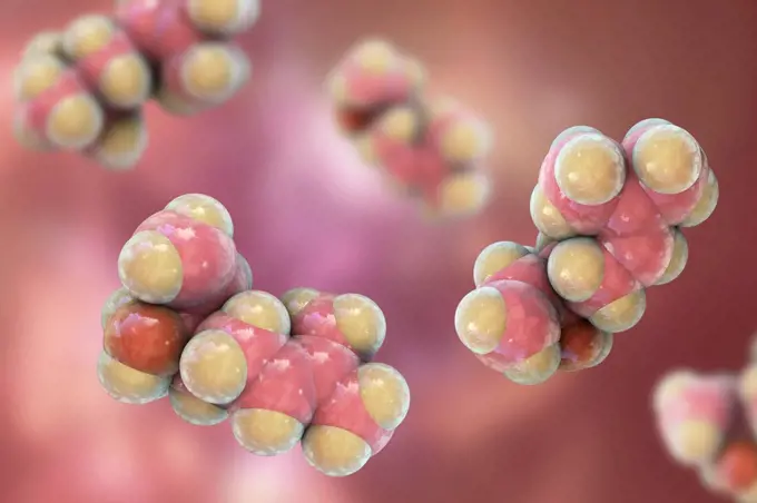
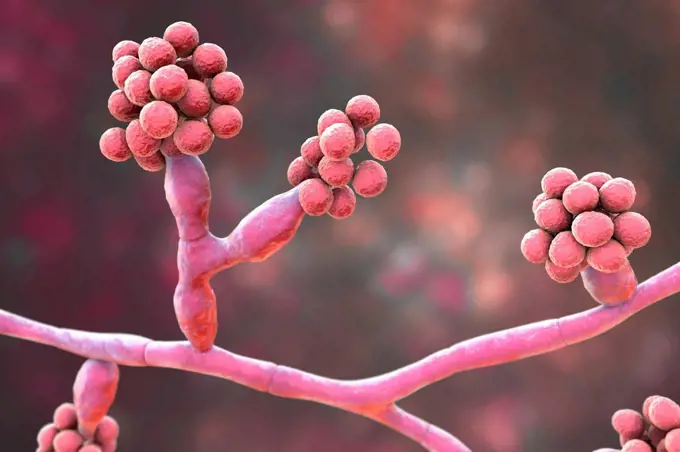
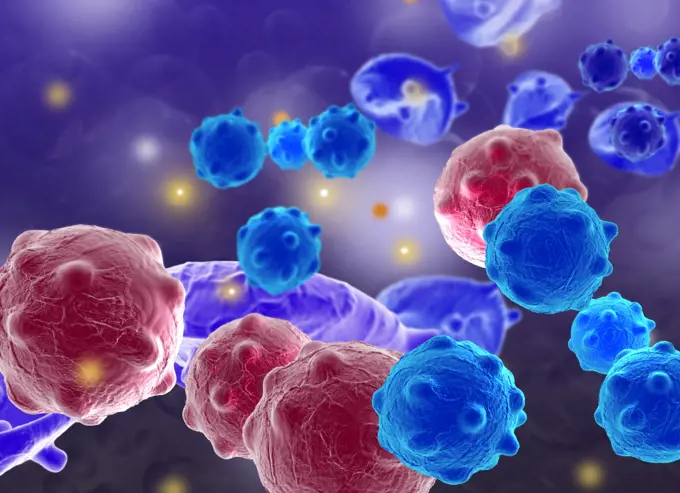
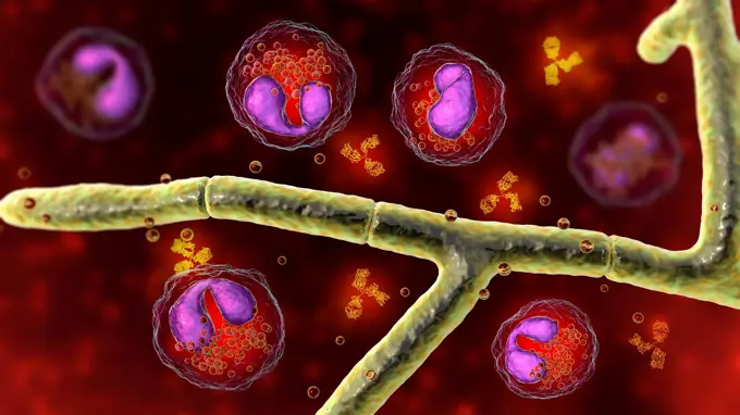
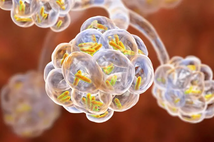
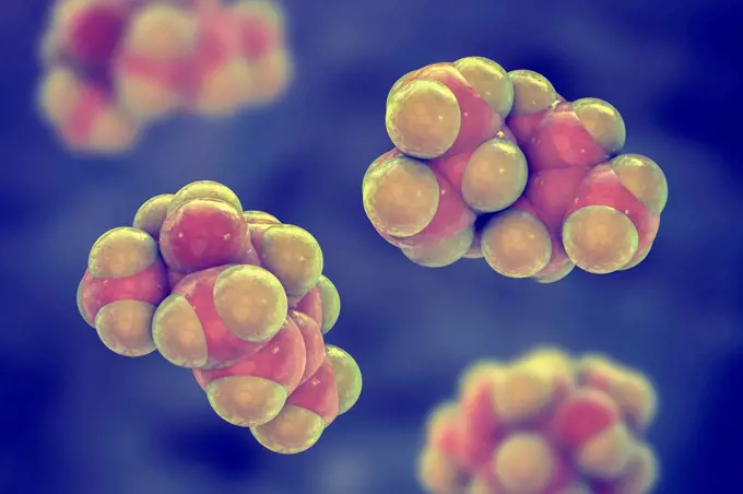
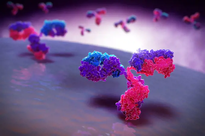
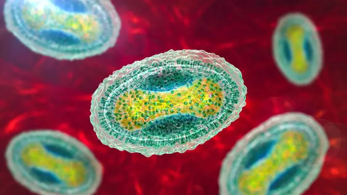
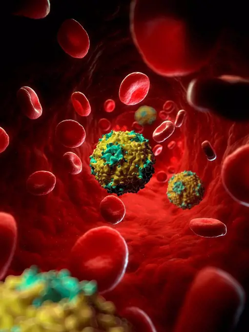
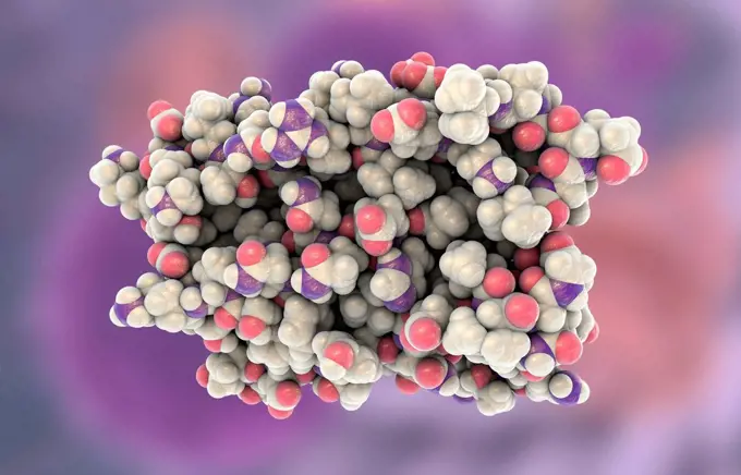
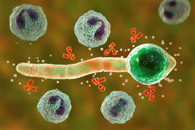
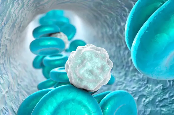
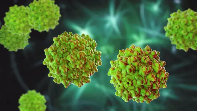
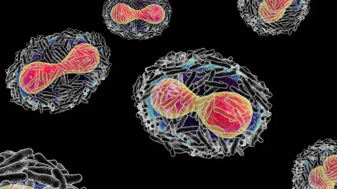
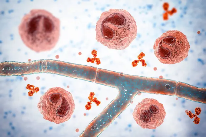
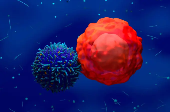
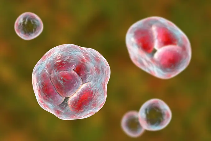
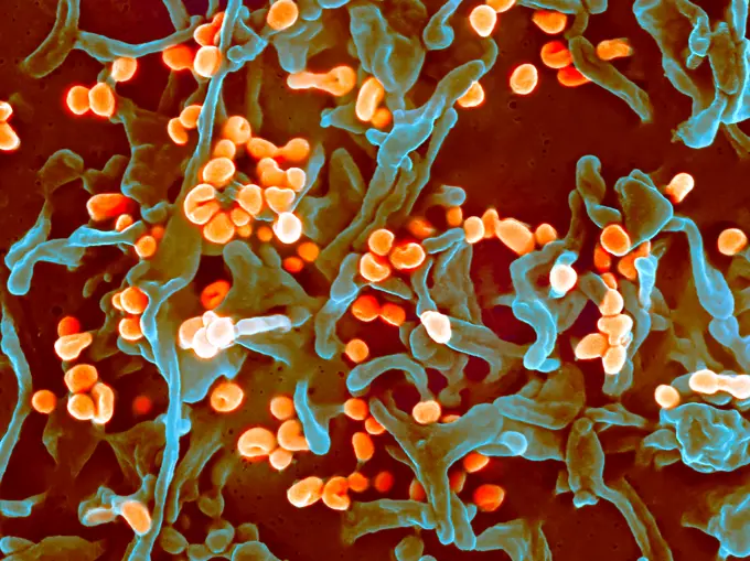
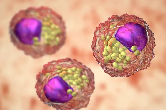
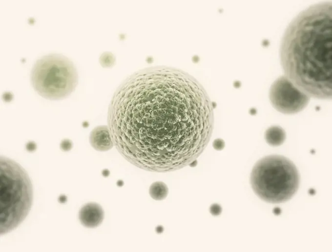
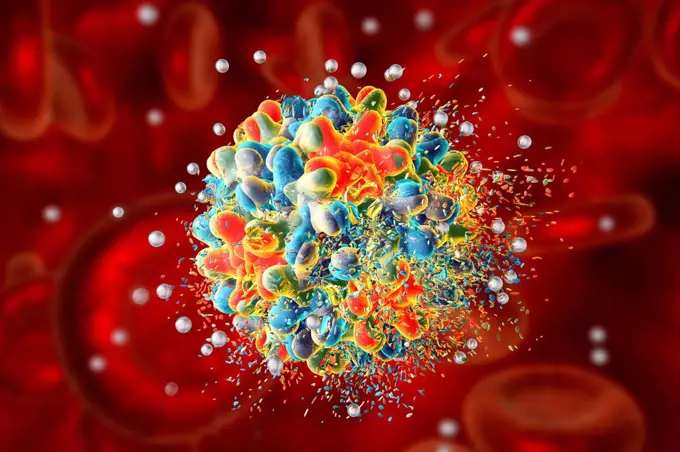
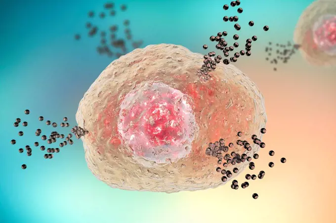
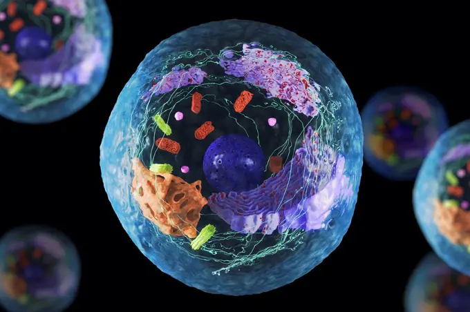
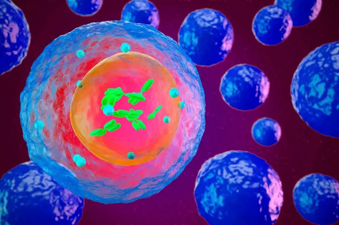
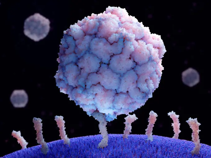
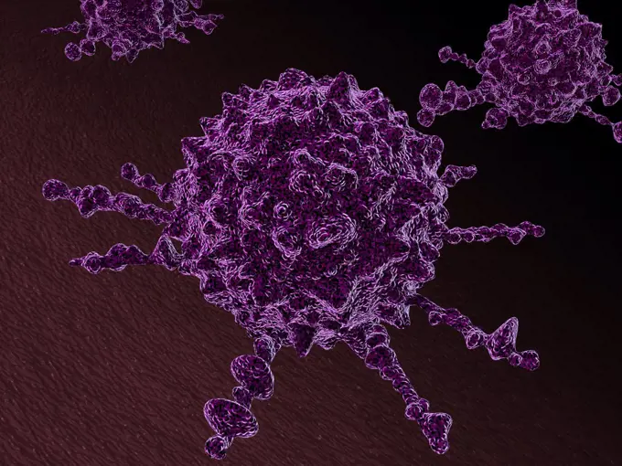
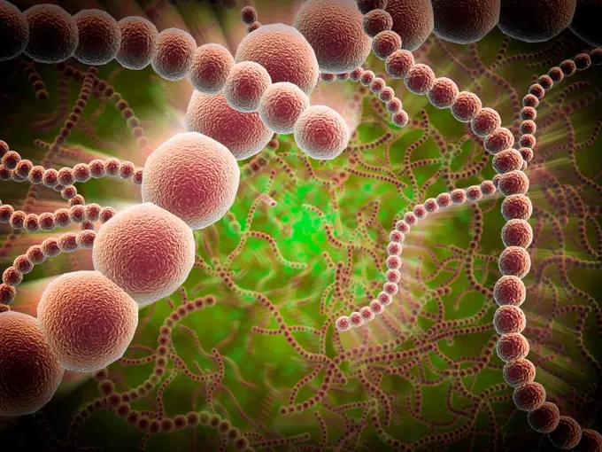
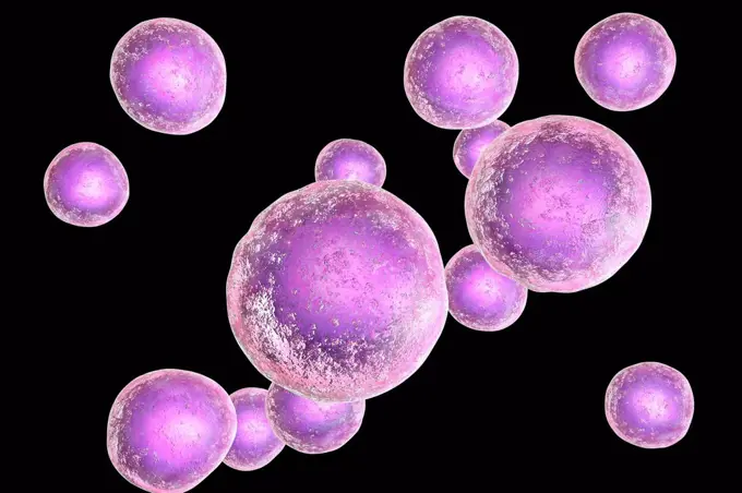
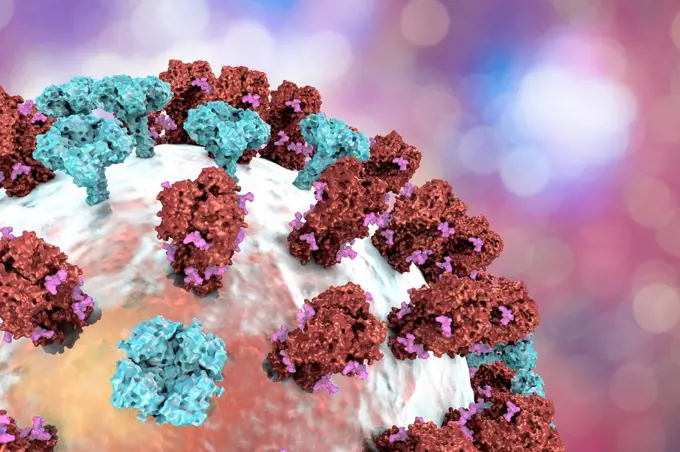
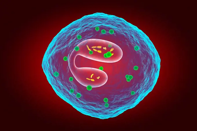
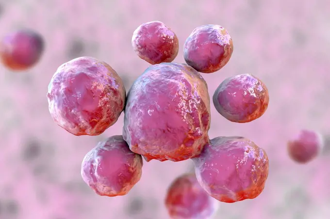
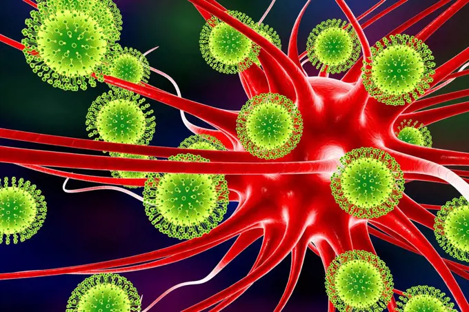
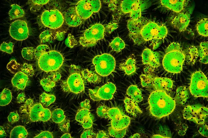
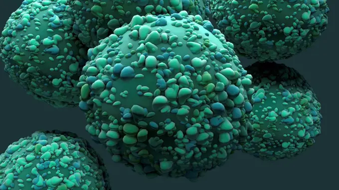
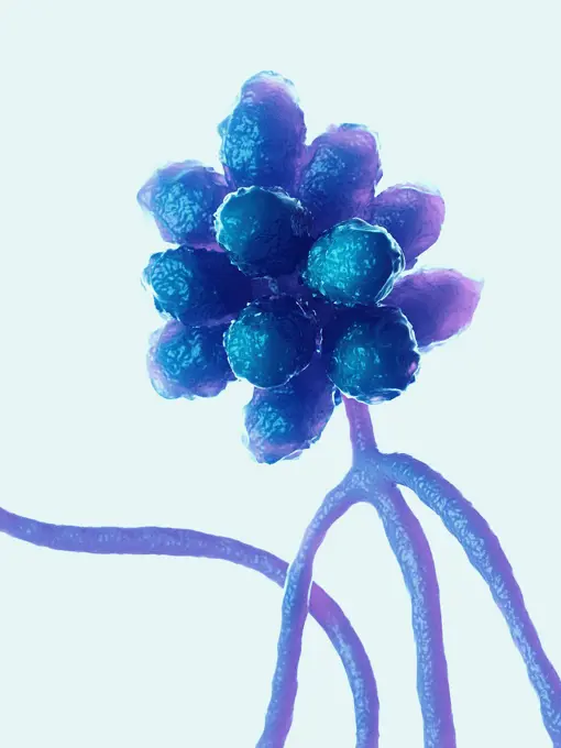
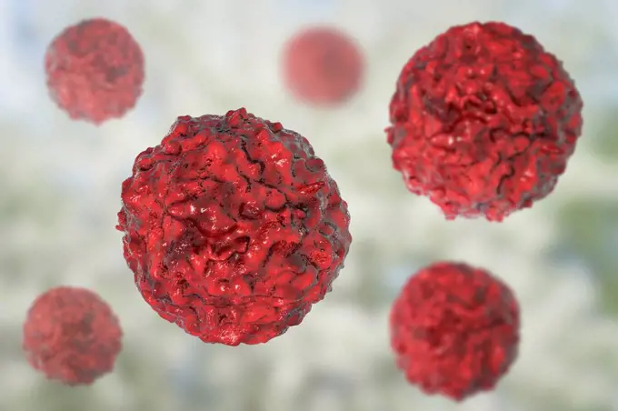
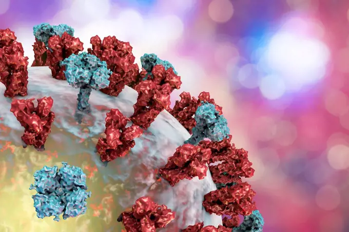
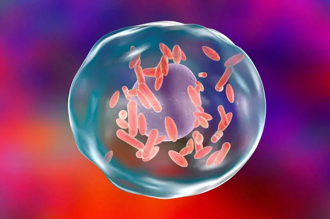
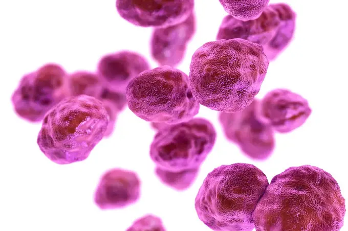
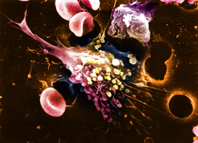
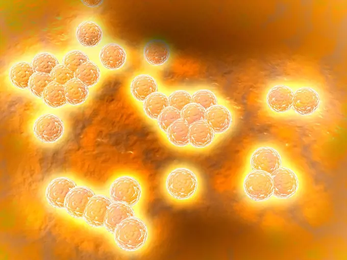
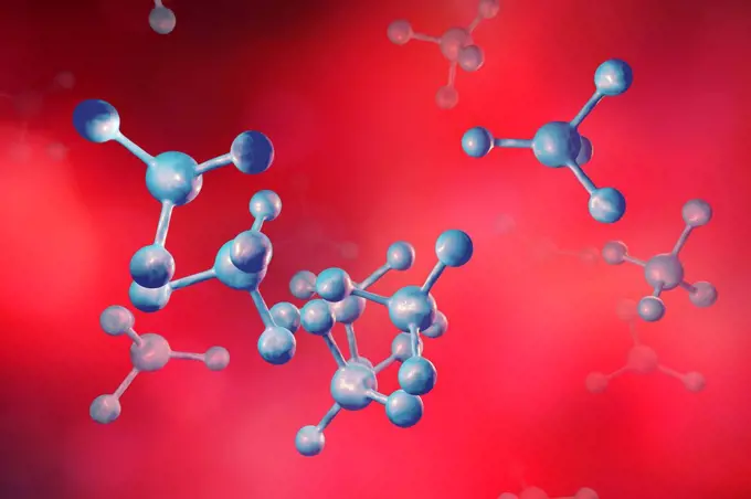
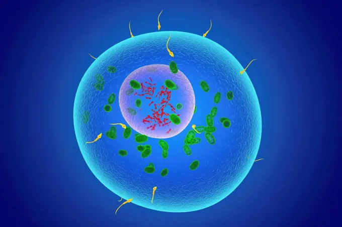
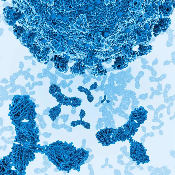
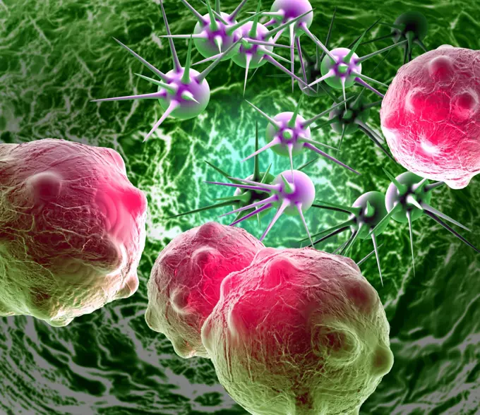
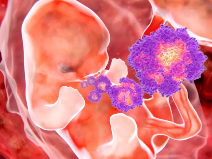
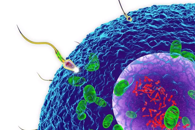
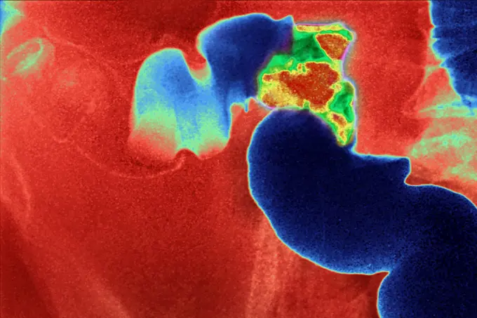
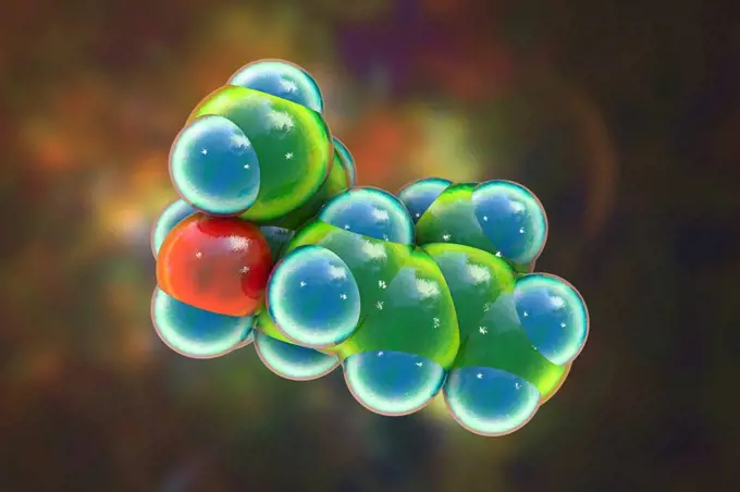
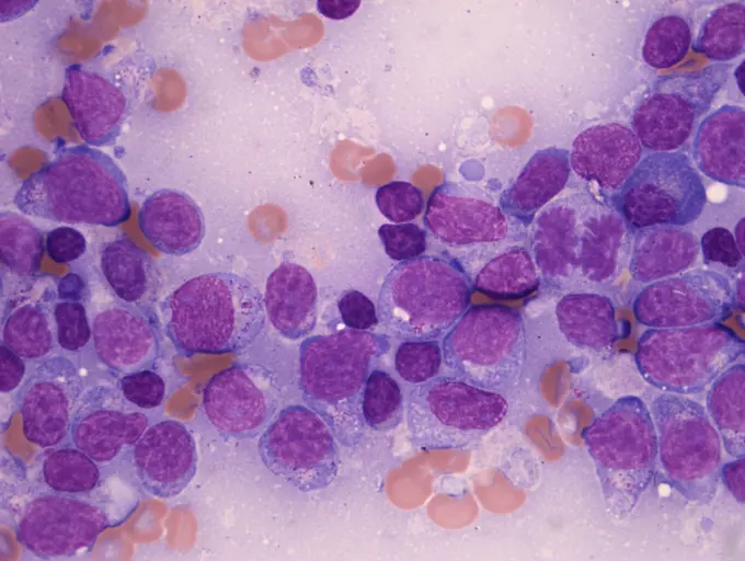
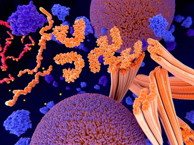
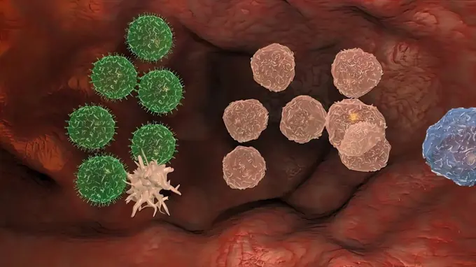
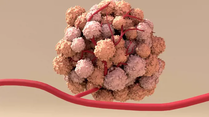
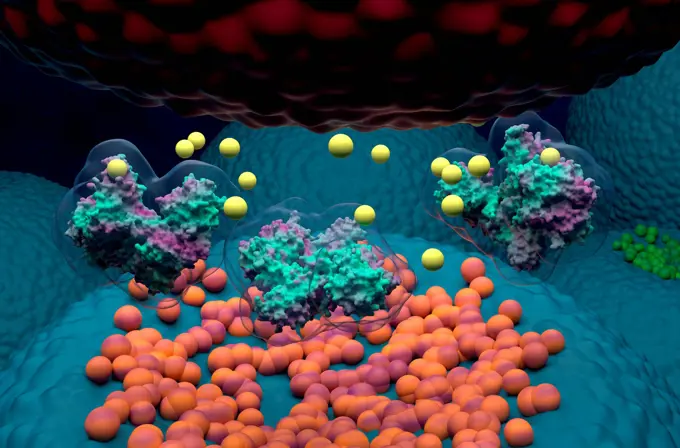
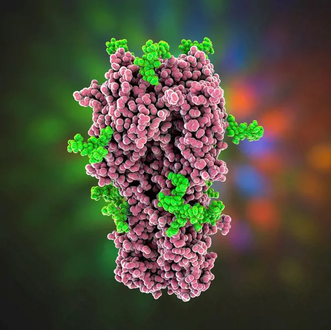
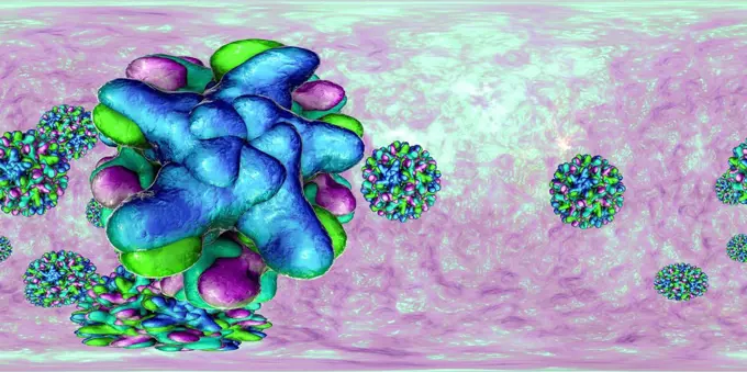
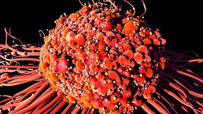
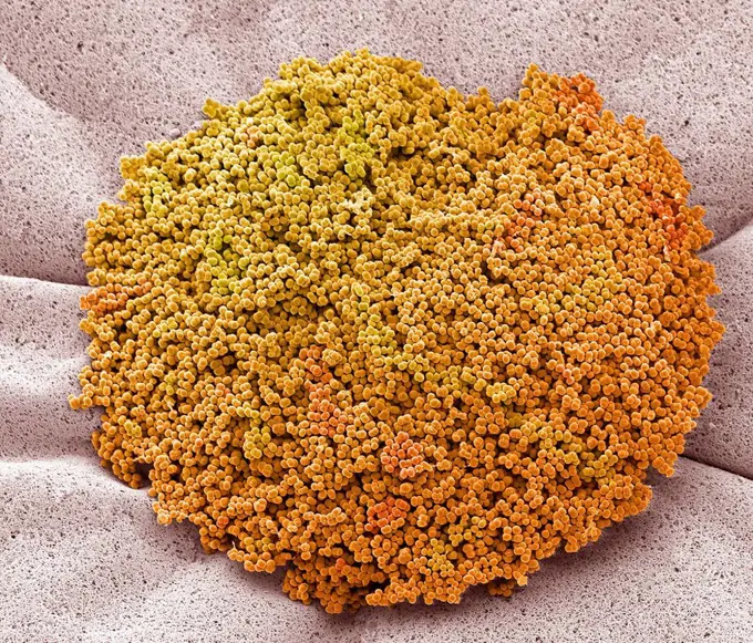
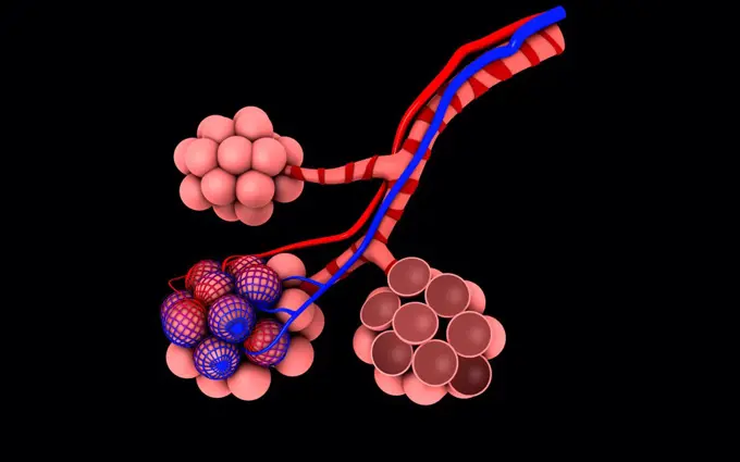
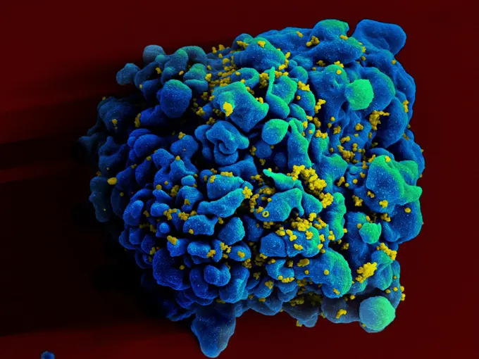
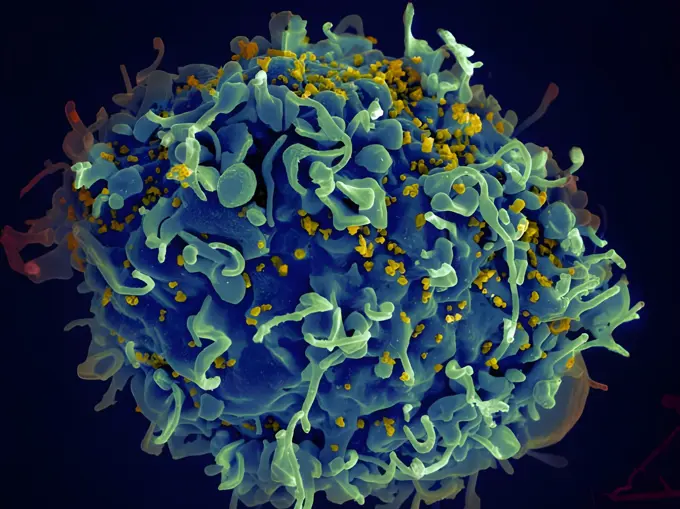
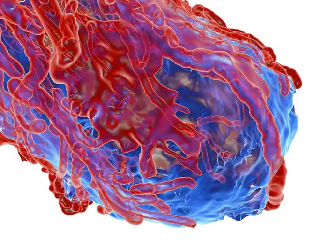
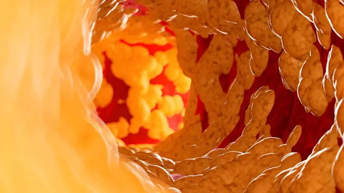
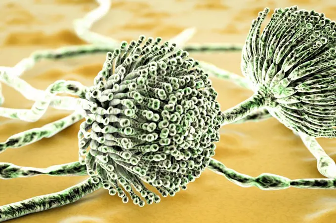
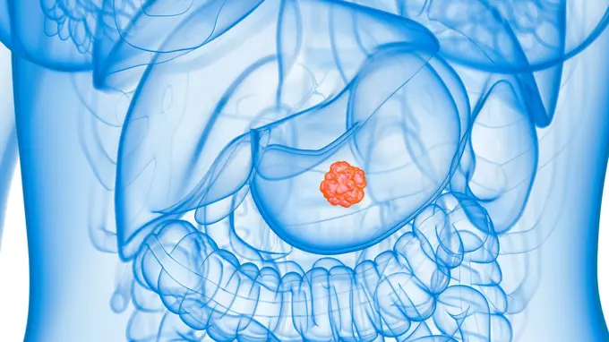
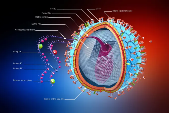
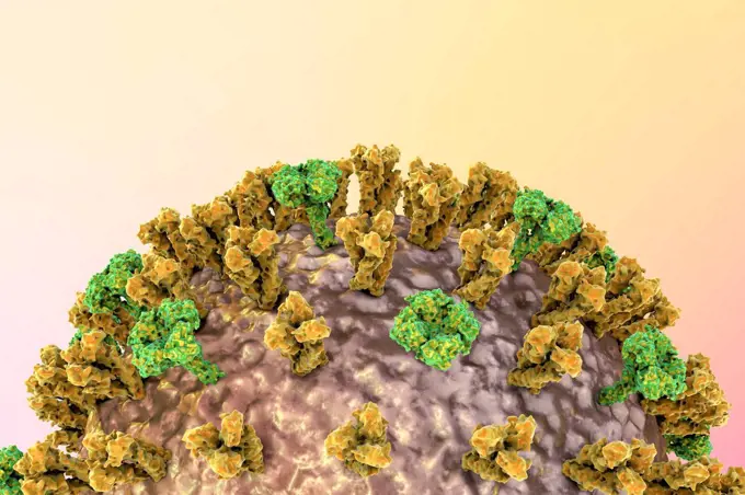
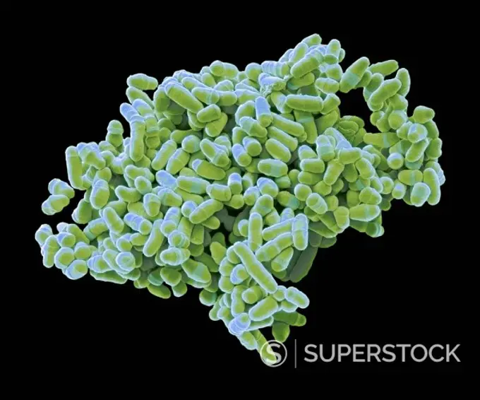
Microscopic Biological Structures

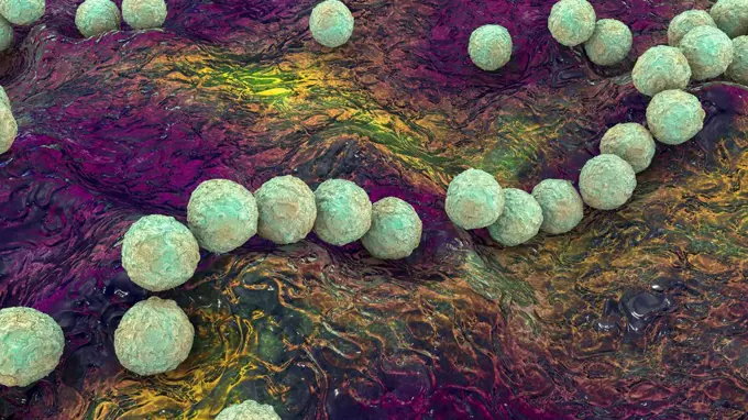
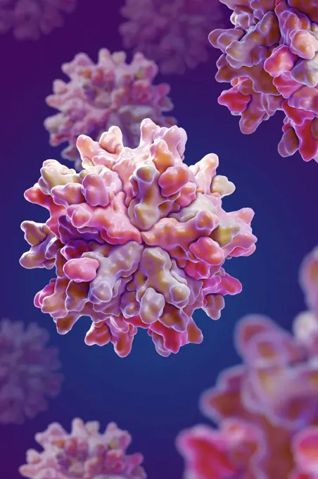
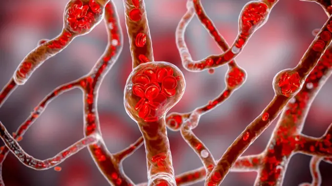
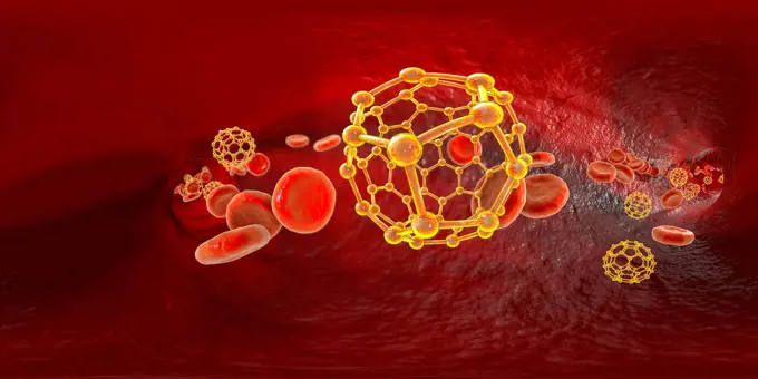
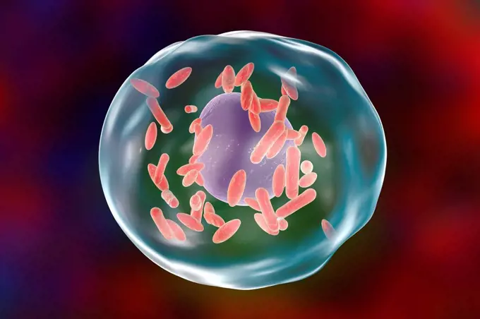
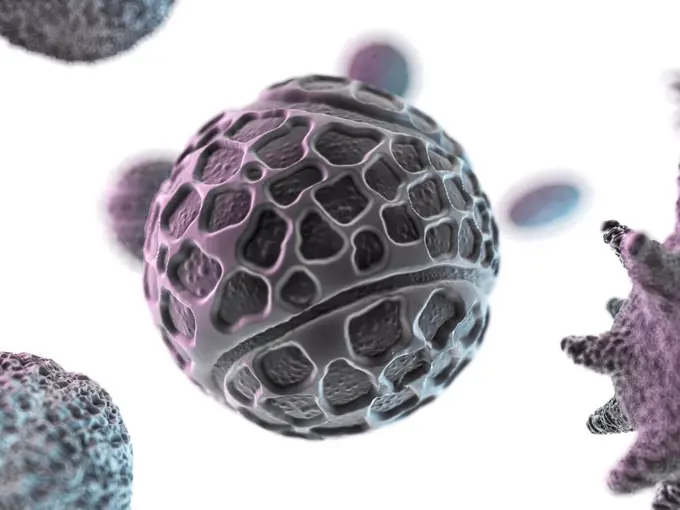
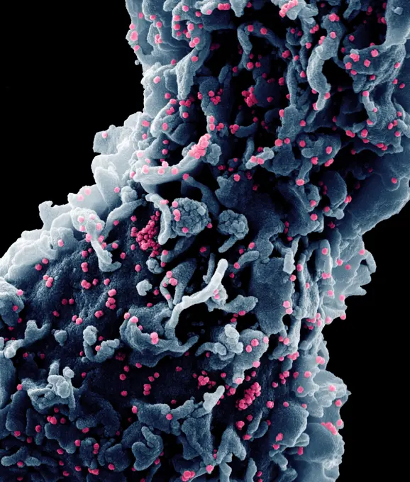
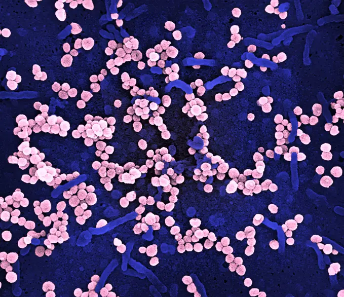
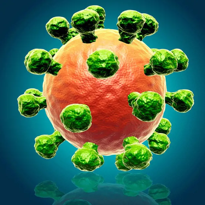
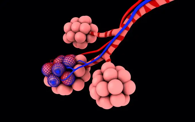
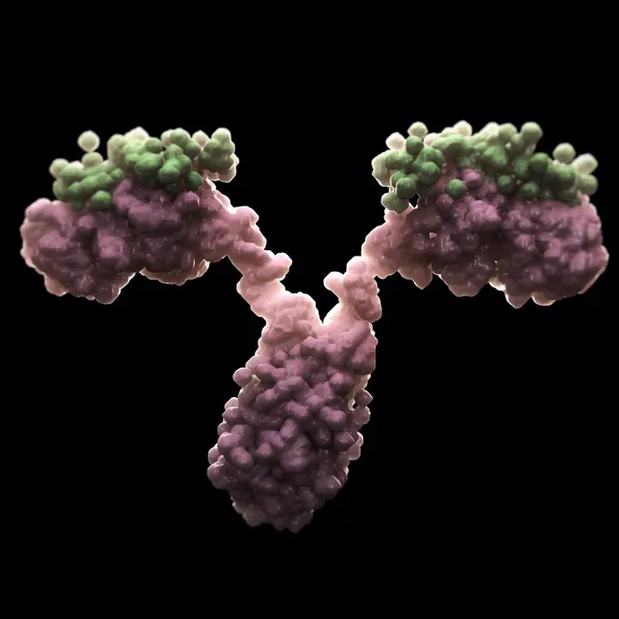
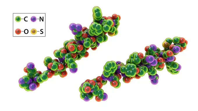
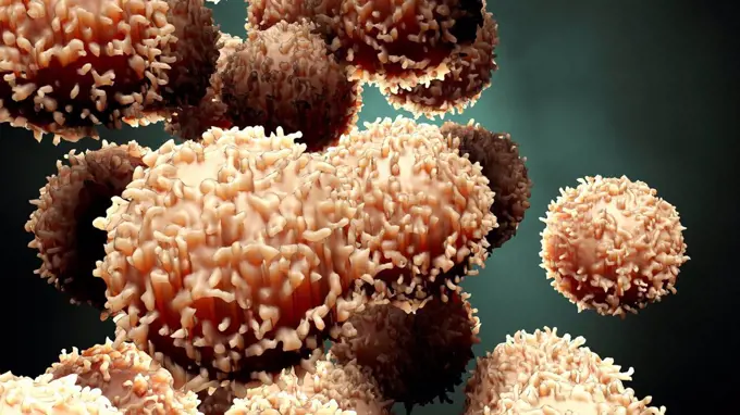
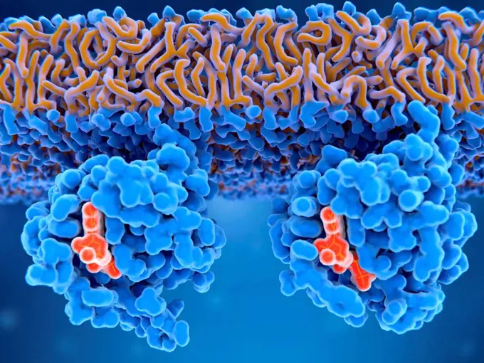
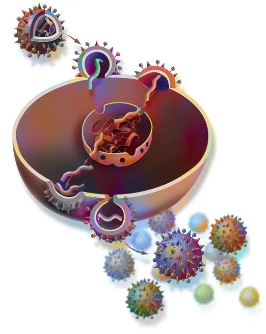
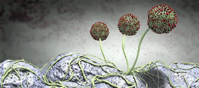
Dynamic illustrations of microscopic entities including cancer cells, bacteria, viruses, and molecules, showcasing vibrant colors and intricate details.

Bacterial pneumonia, illustration

Assets in this Story

4128-V58577055
4128R-15366362
Bacterial pneumonia, illustration
4128-15980256
Zika viruses, computer illustration. This is an RNA (ribonucleic acid) virus from the Flaviviridae family. It is transmitted to humans via the bite of...
4128-19358318
Molecule of patulin toxin, computer illustration. Patulin is a mycotoxin produced by mould fungi Aspergillus, Penicillium and Byssochlamys, found in r...
4128R-13372964
Insulin molecule. Computer model showing the structure of a molecule of the hormone insulin. Insulin plays an important role in blood sugar regulation...
1815-17861776
3D rendered illustration of micro plastic in blood stream
4128-28681152
Meningitis bacteria, illustration
4128R-14324169
Smallpox virus, illustration
4128-15979596
Lymphocytes white blood cells, computer illustration. Lymphocytes are involved in the immune system's defence mechanisms, lymphatic system, and antibo...
1525-27504862
Cancer outbreak and treatment for malignant cancer cells in a human body caused by carcinogens and genetics with a cancerous cell as an immunotherapy symbol and medical therapy as a 3D render.
4128R-15366388
Staphylococcal pneumonia, illustration
4128-15750294
Coronavirus particles, illustration
4128R-13023036
Dengue virus in blood, computer illustration.
4128-20040280
Insulin molecule, illustration
4128R-14060606
Western equine encephalitis virus, illustration
4128R-15366564
Gonorrhoeae bacteria inside neutrophils, illustration
4128-111495103
White blood cells, illustration.
4128R-15366379
Pneumococcal pneumonia, illustration
4128R-13022069
Adeno-associated viruses, computer illustration. Adeno-associated viruses (AAVs) are the smallest known viruses to infect humans. They do not cause di...
4128R-13710869
Mast cell releasing histamine during an allergic response, computer illustration. Mast cells are a type of leucocyte (white blood cell). They contain ...
4128R-15366391
Staphylococcal pneumonia, illustration
4128-20044429
Gamma-terpinene molecule, illustration
4378-20393262
Insulin 2HIU
4128R-13587440
Autophagy. Computer illustration of the degradation of cellular waste inside an autolysosome (large sphere). Autophagy (autophagocytosis) is the natur...
4128-18573671
Viral encephalitis, conceptual illustration
4128R-14846724
Transfusion transmitted virus particles, illustration
4239R-20483587
Microscipic view of pancreatic cancer cells.
4128R-15366483
Polyoma BK virus, illustration
4128-19056168
Fungi Paracoccidioides lutzii, illustration
4128-V58569140
1525-26275134
Metastasis for cancer that spreads to a different part of the body from where it 3D illustration. Metastasis for cancer that spreads to a different part of the body from where it
824-63223109
This scanning electron microscope image shows SARS-CoV-2 (yellow) also known as 2019-nCoV, the virus that causes COVID-19 isolated from a patient in the U.S. emerging from the surface of cells (blue/pink) cultured in the lab. Image captured and colorized at NIAID's Rocky Mountain Laboratories (RML) in Hamilton, Montana. Credit: NIAID
4128-20243907
Coronavirus particles, illustration
4128-V58577056
4239R-8325
Conceptual image of cell nucleus. The cell nucleus acts like the brain of the cell. It helps control eating, movement, and reproduction.
4128R-14181700
Nuclear division, illustration
4128R-15290441
Pneumocystis jirovecii fungi, illustration
4128-19054228
Flu virus, illustration
4128R-11311056
Antibodies attacking a virus, computer artwork.
4128-V58574351
4128-19489496
Monkeypox virus particles, illustration
4128R-12569
Virus, artwork
4128R-11473097
HIV (human immunodeficiency virus) infection, computer artwork.
4128-15819137
Bacteriophages infecting bacteria, illustration. A bacteriophage, or phage, is a virus that infects bacteria. It attaches to the surface of a bacteriu...
4128R-14324170
Smallpox virus, illustration
4128-48285244
T cells attacking cancer cells, illustration
4128R-15366495
West Nile virus particle, illustration
4128R-22205
Norovirus infection. Computer artwork of norovirus particles in the bloodstream.
4128R-22196
Hepatitis B infection. Computer artwork of Hepatitis B virus particles in the bloodstream.
4128-28767240
Prototheca wickerhamii algae, illustration
4128R-15366405
Blastocyst, illustration
4128R-13844625
Basophil white blood cell, illustration.
4239R-8241
Microscopic view of chlamydia. Chlamydia is a common sexually transmitted disease (STD) caused by a bacterium.
4128R-15405894
Human embryonic stem cells, illustration
1848-50846474
3D Image of three Flu Coronavirus Covid-19 concepts background. Concept with disease cell as a 3D renderer
4128R-15047304
Cancer cells, illustration
4128R-13587537
Human papilloma virus (HPV), computer illustration. HPV causes warts, which mostly occur on the hands and feet. Certain strains also infect the genita...
4128-20044464
Linalool molecule, illustration
4128-19054678
Madurella fungi, illustration
4197-63595678
Bacteria, cell and particle with abstract, render or illustration of blood virus. Immune system, micro biology and sick for science, microscope and medical research for sepsis or senolytics treatment
4128-20045722
High-density lipoproteins in the blood, illustration
4378-3376
Structure of Swine Vesicular Disease Virus (PDB 1OOP). SVDV is an Enterovirus of the family Picornaviridae that causes symptoms similar to those of foot-and-mouth disease.
4128R-14638064
Bacterial vaginosis, illustration
4128-48285239
Cancer cells dividing, illustration
4378-3216
Structure of Human Parvovirus B19 (PDB 1S58). B19 virus causes a childhood rash called fifth disease or erythema infectiosum which is commonly called slapped cheek syndrome.
1525-24586418
cloud of micro organizam made in 3d software
4128-111493557
Biofilm, conceptual illustration.
4128-28767193
Splendore-Hoeppli phenomenon, illustration
4128R-15366365
Bacterial pneumonia, illustration
4128-20044525
Terpinen-4-ol molecule, illustration
4378-20393403
Antibody 1IGT
4128-20044937
Molluscum contagiosum virus, illustration
4128-20041846
Monkeypox virus, illustration
4128-V58573988
4128R-11313025
Seneca Valley virus, computer artwork.
4128R-13763417
Human interferon alpha, molecular model. Interferons are proteins produced by white blood cells as part of the immune response to invading pathogens, ...
4128R-18525
Medical nanorobots, computer artwork.
4128-28575128
Mpox virus particles, illustration
4128-15750866
Babesia parasites inside red blood cell, illustration
4128-19054735
Microsporum canis fungus, illustration
4128R-11473228
Lymphocyte white blood cells, computer artwork.
4128-28575431
Ethylene glycol molecule, illustration
4128-20044380
Curcumin molecule, illustration
4128-19489525
Monkeypox virus particles, illustration
4128R-13682135
Ricin. Computer model of a molecule of the toxic protein ricin. It comprises two amino acid chains, termed A (upper left) and B (lower right). The A-c...
4128-19054670
Madurella fungi, illustration
824-65830895
Creative layout featuring 3D renderings of respiratory syncytial virus (RSV), a common contagious virus that infects the human respiratory tract. 2023 : NIH Celebrates FDA Approval of RSV Vaccine for People 60 Years of Age and Older.
4128R-14057585
Viruses and DNA, illustration
4128R-15366516
Cytomegalovirus infection, illustration
4128R-15366523
Eosinophil white blood cell, illustration
4128R-13763431
Interferon gamma, molecular model. Interferon gamma is produced by certain immune cells (T cells, dendritic cells and NK cells) as part of the immune ...
1815R-13530592
Monocyte immune system defense cells, 3D Rendering
4128-V58569565
4128R-13271891
Parechoviruses, computer illustration. RNA viruses from Picornaviridae family. There are two types of Parechoviruses: Parechovirus A (human Parechovir...
4128-18680943
Staphylococcus aureus bacteria, illustration
4197-63595674
Bacteria, blood and molecule with abstract, render or illustration of particle. Immune system, micro biology and sick for science, microscope and medical research for sepsis or senolytics treatment
4128-28575462
Muscimol molecule, illustration
4128-V58569498
4128-28767198
Splendore-Hoeppli phenomenon, illustration
4378-20393413
Antibody 1IGT
4128R-13619757
Blood cells, computer illustration. Red blood cells and white blood cells inside a blood vessel.
4128-18573409
Adeno-associated viruses, illustration
4128-20039454
Monkeypox virus particles, illustration
4128-28767194
Splendore-Hoeppli phenomenon, illustration
824-63225872
Colorized scanning electron micrograph of a cell (red) showing morphological signs of apoptosis, infected with SARS-COV-2 virus particles (yellow), isolated from a patient sample. Image captured at the NIAID Integrated Research Facility (IRF) in Fort Detrick, Maryland.
4128-19357687
Illustration of the internal and external structures of HIV (human immunodeficiency virus). Also seen are antibodies and T helper cells (large spheres...
4128-15818250
Human papilloma virus (HPV), computer illustration. HPV causes warts, which mostly occur on the hands and feet. Certain strains also infect the genita...
4128-48285246
CAR-T cell therapy, illustration
4128-28767226
Prototheca wickerhamii algae, illustration
824-63227257
Scanning electron micrograph of Lassa virus budding on a Vero cell.
4128-111612398
Macrophage foam cell, illustration. Foam cells are macrophage cells that contain lipid droplets and are components of atherosclerotic plaque.
4128R-13667007
Bacteria cells against white background.
4128R-13619820
Destruction of hepatitis B virus by silver nanoparticles, computer illustration. Conceptual image for hepatitis B treatment and prevention by using na...
4128R-13710868
Mast cell releasing histamine during an allergic response, computer illustration. Mast cells are a type of leucocyte (white blood cell). They contain ...
4378-1138
Stylized view of generic human or animal cells.
4128R-13844630
Lymphocyte white blood cell, illustration.
4128-19358668
Glucagon hormone molecule. Computer model showing the structure of the human hormone glucagon. Glucagon is involved in carbohydrate metabolism and is ...
4128-20044798
Polio virus binding to human cell, illustration
1428R-225
Close-up of cancer cells
4128R-13844709
Red blood cell structure, illustration. Haemoglobin molecule inside a red blood cell.
4128-17927948
Streptococcus pyogenes bacteria, illustration
4128-19357714
Illustration of a T helper cell infected with HIV (human immunodeficiency virus).
4128-V58569122
4378-3493
Structure of Infectious Bursal Disease Virus (IBDV) (PDB 2GSY). IBD is a highly contagious disease of young chickens causing severe mortality and is very damaging to the poultry industry worldwide.
6188-55651610
Flu, virus, pathogen, cold, cold, illness, diseases, pathogens, viruses ** flu, virus, bacterium, influenza, bacteriums 2oh-HHG
4128-20040245
Microaneurysms, illustration
4128-24795702
Fullerene nanoparticles in blood, illustration
4128R-14845748
Rotavirus particle, illustration
4128R-13161148
Low density lipoprotein in blood. Illustration of low density lipoprotein (LDL), or 'bad' cholesterol, in the bloodstream. This biochemical assembly c...
4128-28968835
Protein nanoparticles, illustration
4128-20041125
Growing mildew, illustration
4128R-13409390
Candida auris fungi, illustration
4128R-14638058
Bacterial vaginosis, illustration
4128-20045663
Molecule of tetanus neurotoxin, illustration
4128R-15366575
Neutrophil white blood cells, illustration
4128R-15515913
Illustration of a leucocyte and antibodies
4239-V53647337
824-65830857
Creative artwork featuring colorized 3D prints of HIV virus particles. The virus surface (red) is covered with proteins (dark blue) that enable the virus to enter and infect human cells, and additional proteins (teal spheres) that disguise the virus from the immune system. A colorized transmission micrograph of an H9 T cell (blue) appears in the background.
4128-20043182
RNA polymerase II transcribing DNA to mRNA, illustration
1899-65662453
Creative layout featuring 3D renderings of respiratory syncytial virus (RSV), a common contagious virus that infects the human respiratory tract.
4128R-15366387
Staphylococcal pneumonia, illustration
4128-30416625
Human cell, illustration
4128-16007407
Covid-19 coronavirus binding to receptors, illustration
4269-25095
AIDS virus. TEM (Transmission Electron Microscope)
4128-20038375
Engineered T-cell attacking a leukaemia cell, illustration
4128-111612461
Mycoplasma genitalium bacteria, illustration. M. genitalium is a small parasitic bacterium. It is a sexually transmitted organism that causes non-gono...
4128-19055955
Fertilisation, illustration
4239R-8059
Microscopic view of Streptococcus pneumoniae, also known as pneumococcus. Pneumococcus is a Gram-positive coccus shaped pathogenic bacteria which causes many types of pneumococcal infections in addition to pneumonia.
4128R-15366505
Human embryonic stem cells, illustration
4128-18798857
Flu virus, illustration
4128-28968557
Molecular model of amygdalin, illustration
4128R-13844623
Eosinophil white blood cell, illustration.
4128R-13282702
Q fever bacteria (Coxiella burnetii, green rods) inside a cell, computer illustration. Coxiella burnetiiis an obligate intracellular bacterium, meanin...
4128R-25593
Pollen grains, computer artwork.
824-63225906
Colorized scanning electron micrograph of a cell (blue) infected with a variant strain of SARS-CoV-2 virus particles (UK B.1.1.7 red), isolated from a patient sample. Image captured at the NIAID Integrated Research Facility (IRF) in Fort Detrick, Maryland.
824-63227046
Scanning electron micrograph of Lassa virus budding from a cell. Lassa virus (LASV) is an arenavirus that causes Lassa hemorrhagic fever, a type of viral hemorrhagic fever (VHF) in humans and other primates. Image requested from the NIAID Integrated Research Facility (IRF) in Fort Detrick, Maryland. Credit: NIAID
824-65830626
Colorized scanning electron micrograph of red blood cell infected with malaria parasites (teal). The small bumps on the infected cell show how the parasite remodels its host cell by forming protrusions called 'knobs' on the surface, enabling it to avoid destruction and cause inflammation. Uninfected cells (red) have smoother surfaces.
1525-22767779
Neisseria meningitidis or meningococcus is a bacterium that can cause meningitis and other forms of meningococcal disease. 3D illustration. Neisseria meningitidis bacteria
1525-23900160
Mutating virus concept and new coronavirus b.1.1.7 variant outbreak or covid-19 viral cell mutation and influenza background as dangerous flu strain medical health risk with disease cells as a 3D render.
4128R-14060556
Chitosan nanoparticles in blood, illustration
1525-27135604
Coronavirus. Background with viruses. Influenza viruses on colorful background. 3D illustration. Coronavirus outbreak and coronaviruses influenza background. 3D illustration.
4128R-15047058
Opioid and cannabinoid brain receptors, illustration
4128-V58567737
4128-30422380
Virus within blood stream, illustration
4128-18800774
Neurons in Tay-Sachs disease, illustration
824-63223179
MERS-CoV particles on camel epithelial cells. Credit: NIAID in collaboration with Colorado State University.
824-57657683
Colorized transmission electron micrograph of monkeypox virus particles (red) cultivated and purified from cell culture.
824-63223124
A dry fracture of a Vero cell exposing the contents of a vacuole where Coxiella burnetii are busy growing. Credit: NIAID.
4128R-13619822
Destruction of hepatitis B virus by silver nanoparticles, computer illustration. Conceptual image for hepatitis B treatment and prevention by using na...
4128R-9618
Virus particles, artwork
1848-49387913
Viruses Virus is an organic structure
4128R-14181652
Cancer cells, illustration
4128-19056176
Fungi Paracoccidioides lutzii, illustration
4128R-13193057
Zika viruses infecting neurons, computer illustration. This is an RNA (ribonucleic acid) virus from the Flaviviridae family. It is transmitted to huma...
4070-21285284
A blue light fluorescence photo of Jewel anemones (Corynactis viridis). The blue light source excites the fluorescent pigments in the anemones, which emit light of other colours. The muddy seabed and other marine life does not fluoresce, so is seen as black. Plymouth, Devon, England, Great Britain. Plymouth Sound, English Channel.
1525-56853457
The COVID-19 Inside Virus Structure 3D rendering. The COVID-19 Inside Virus Structure
4128-111495115
Cancer cells, illustration.
4128-30418001
Stachybotrys mould, illustration
4239-V53647340
4128R-15366591
Human embryonic stem cells, illustration
4128R-13575576
Flu virus. Illustration showing influenza virus with surface glycoprotein spikes hemagglutinin (HA, trimer) and neuraminidase (NA, tetramer). Hemagglu...
4128-20042849
Poliovirus, illustration
4128R-13282700
Q fever bacteria (Coxiella burnetii, green rods) inside a cell, computer illustration. Coxiella burnetiiis an obligate intracellular bacterium, meanin...
4128R-13763356
Tularaemia bacteria (Francisella tularensis), illustration. F. tularensis is Gram-negative, coccobacillus prokaryote. A zoonotic microorganism that ca...
4269-6698
Scanning electron micrograph (SEM) of a cancer cell. The spikes or pseudopodia that are characteristic of an invading cancer cell. Magnification x12000.
4239R-8287
Microscopic view of Methicillin-resistant Staphylococcus aureus (MRSA). MRSA is a bacterium responsible for several difficult-to-treat infections in humans. It is also called oxacillin-resistant Staphylococcus aureus.
4128R-14846182
Abstract molecule model, illustration
4128-30416519
Circulating blood cells, illustration
4128-15666273
Hiv virus, illustration
1525-26197948
epidemic and covid-19 outbreak concept - human silhouette in medical mask with 3d rendering of coronavirus cells on black background. human silhouette in mask with coronavirus cells
4128-111612381
Rhinoviruses, 360-degree panorama view, computer illustration. The rhinovirus infects the upper respiratory tract and is the cause of the common cold....
4128R-25395
Animal cells, computer artwork.
4128R-13844633
Macrophage white blood cell, illustration.
4128R-15535443
Illustration of Alzheimer's disease
4378-947
Cell receptors embedded on a cellular membrane.
5507-34404884
Virus in blood
4128-30416221
Human cell, illustration
4128R-13844641
Neutrophil white blood cell, illustration.
4128R-13725874
Computer illustration of a plasma cell (B-cell, left) secreting antibodies (white) against influenza viruses (right). Antibodies bind to specific anti...
1525-27094863
Viruses and dna close-up on a blue background. 3d render.
1899-61460422
Influenza virus. Viruses of the Orthomyxoviridae family. Avian plague virus. The flu virus is the cause of an infectious and contagious respiratory disease. It evolves into a major pandemic interspersed with small localized seasonal epidemics. Virus diameter: about 100 nm. Computer graphics on transmission electron microscope view
4128-19249539
Dopamine neurotransmitter, molecular model. Dopamine is a chemical released by the brain as a neurotransmitter and a hormone. It has been intensively ...
4128-18801072
Antibody, conceptual illustration
4128-20041022
Lymphocytes cells attacking a cancer cell, illustration
4128R-13575575
Flu virus. Illustration showing influenza virus with surface glycoprotein spikes hemagglutinin (HA, trimer) and neuraminidase (NA, tetramer). Hemagglu...
4128R-13575582
Flu virus. Illustration showing influenza virus with surface glycoprotein spikes hemagglutinin (HA, trimer) and neuraminidase (NA, tetramer). Hemagglu...
4128R-13844341
Fertilisation. Illustration of a sperm cell (yellow) fertilising an egg cell (ovum). Within the egg cell are mitochondria (green), which provide the c...
4128-17927471
Antibodies and covid-19 coronavirus, illustration
1525-24586421
cloud of micro organizam made in 3d software
4128R-13161165
Foetus and zika virus. Illustration of particles of the zika virus (purple) and a foetus (left). The zika virus is an RNA (ribonucleic acid) virus fro...
4128-20240075
Plasma cell, illustration
4128-16073788
Antibodies responding to covid-19 coronavirus, illustration
4128-20044988
Smallpox virus, illustration
4128R-14768527
Viral etiology of prostate cancer, conceptual illustration
4128R-11291696
Artwork of t-lymphocytes attacking cancer cells.
1815-18277974
Three dimensional render of heap of purple spheres
4128-18301080
Mucor mould, illustration
4128-20044533
Terpinen-4-ol molecule, illustration
4128-18800778
Neurons in Tay-Sachs disease, illustration
4128R-14057813
Cancer cell, illustration
4128R-15514887
T-cell receptors, illustration
4128R-15516415
Illustration of colon inflammation caused by gluten
4128R-23799
DNA molecule, computer artwork.
6188-55644454
Dangerous pathogens in the body Dangerous pathogens in the body
824-63223180
MERS-CoV particles on camel epithelial cells. Credit: NIAID in collaboration with Colorado State University.
4128R-13022160
Human papilloma virus (HPV) on a space background. Computer model showing the structure of the protein coat (capsid) surrounding a human papilloma vir...
6188-66486012
Mehrfarbige Antikörper oder Immunglobulin-Proteinstrukturen, die an Rezeptoren gebunden sind - 3d-Abbildung Multicolored antibodies or immunoglobulin protein structures attached to receptor- 3d illustration Copyright: xZoonar.com/ChristophxBurgstedtx 22600905
4128-V58565946
4128-20044542
Thymol molecule, illustration
4128-28575452
Hexaethylene glycol molecule, illustration
6188-56032703
virus,erreger,coronavirus,krankheitserreger,viren,corona,covid,covid 19,covid-19,covid19,sars cov2,sars-cov2 ** virus,bacterium,corona virus,bacteriums 2oh-gq6
4128R-14637478
Dendritic cell and T-lymphocytes, illustration
4128R-13410109
Rhinovirus, illustration
1525-25636387
Human antibody (immunoglobulin). Human antibody (immunoglobulin). 3D illustration
4239R-9279
Conceptual image of alveoli.
4378-1824
Antibody fighting infection.
4128-19358672
Glucagon hormone molecule. Computer model showing the structure of the human hormone glucagon. Glucagon is involved in carbohydrate metabolism and is ...
4128-30419046
Colon cancer, illustration
4128-V58569333
4128R-13844339
Fertilisation. Illustration of a sperm cell fertilising an egg cell (ovum). Within the egg cell are mitochondria (green), which provide the cell with ...
1899-53511490
Abdominal x-ray by barium enema of colorectal cancer of the sigmoid colon.
824-63224723
Abdominal x-ray by barium enema of colorectal cancer of the sigmoid colon.
4128-28968401
Antibody bound to an amyloid beta peptide, illustration
4128-20044484
Linalool molecule, illustration
6145-29297347
NK T-cell lymphoma is a highly aggressive cancer of a specific type of immune cell called lymphoid cells, and is associated with the Epstein Barr virus (glandular fever). In later stages of the disease, the lymphoma can spread to the lymph nodes, as in this case.
4128R-15514886
Tau protein in Alzheimer's disease, illustration
1525-26235478
Antibodies destroy an infected cell by a virus, immun defense kill the infected cell 3D illustration. Antibodies destroy an infected cell by a virus, immun defense kill the infected cell
4128-18498559
Tumour growth, illustration
4128-48285234
Botulinum toxin in neurone, illustration
4128-20041848
Monkeypox virus, illustration
4128-16073806
Covid-19 infection, illustration
4128-V58573421
4128R-13844782
Computer artwork of a bone structure and nanomaterial. A slice of cortical bone structure and nanomaterial is depicting bone regeneration.
4239R-20483342
Conceptual image of alveoli.
1848-49194847
Cell division illustration
1848-61038302
Coronavirus disease (COVID-19) outbreak and coronaviruses influenza background
4128-111494727
White blood cells, illustration.
4128R-14847017
Inactive and active Ras proteins, illustration
824-63165026
Replication of influenza virus from a host cell.
1848-50872620
Asian flu ncov coronavirus over Earth background and its blurry hologram. Concept of cure search and spreading disease. 3d rendering toned image. Elements of this image furnished by NASA
4128-111612387
Rhinoviruses, 360-degree panorama view, computer illustration. The rhinovirus infects the upper respiratory tract and is the cause of the common cold....
6188-65540225
Mikroskopische Bild der wachsenden Schimmel oder Schimmelpilz und Sporen - 3d Illustration Microscopic image of growing molds or mold fungus and spores - 3d illustration Copyright: xZoonar.com/ChristophxBurgstedtx 22577794
1525-27208321
Corona Virus pandemic deadly virus health risk and coronavirus public disease and flu outbreak or influenza background as dangerous viral strain medical concept with cells in a 3D illustration style..
4128-19054259
Haemagglutinin from H3N2 1968 influenza virus, illustration
4128-111612384
Hepatitis B viruses, 360-degree panorama view, computer illustration. This is a species of the genus Orthohepadnavirus, which is part of the Hepadnavi...
1525-56516484
Corona virus vaccination, Healthcare cure concept
4128-111495133
Cancer cell spreading, illustration.
4128-30419038
Adrenal gland tumour, illustration
4128-16171799
Bacteria from a human beard. Coloured scanning electron micrograph (SEM) of bacteria cultured from a human beard. A European study has found that the ...
824-65830279
Herpes virus (HSV. HSV is the cause of cold sores, genital herpes, chickenpox, infectious mononucleosis, roseola, sometimes shingles. They are involved in certain cancers including lymphoma by Burkitt. Electron microscopy image.
824-63223155
Colorized scanning electron micrograph of Ebola virus particles (blue) both budding and attached to the surface of an infected VERO E6 cell (yellow). Image captured and color-enhanced at the NIAID Integrated Research Facility in Fort Detrick, Maryland. Credit: NIAID.
4128R-14181695
Viral encephalitis, illustration
1525-56853724
Autoimmune attack on thyroid cells 3D rendering. Autoimmune attack on thyroid cells
4128R-14588020
Histamine binding to receptor, illustration
4128R-13591277
Brochosomes, illustration.
4239R-9278
Conceptual image of alveoli.
4128R-15290622
Japanese encephalitis, illustration
824-63191248
Colorized image of HIV-infected H9 T-cell.
824-63223162
Scanning electromicrograph of an HIV-infected H9 T cell. Credit: NIAID.
824-63227220
This human T cell (blue) is attacked by HIV (yellow), the virus that causes AIDS. The virus targets T cells, which play a vital role in the body's immune response against invaders like bacteria and viruses.
4128R-28133
Tumour, computer artwork. Tumours are caused by the uncontrolled growth of previously normal cells. The resulting growth (blue) can invade and damage ...
4128-18929695
Hand reaching for a coronavirus particle, illustration
1525-27702246
Drugs and negative effects on fertility as prescription and recreational drug objects as marijuana alcohol and pills causing infertility with a sperm and human egg as a 3D illustration.
4128R-15515935
Illustration of fat inside of an artery
1525-27187720
Hand with a medical thermometer on 3d generated COVID-19 background. Concept of corona virus.. Concept of corona virus.
4128-28575145
Mpox virus particles, illustration
4128R-13587467
Aspergillus fungus, computer illustration. This is a toxic fungus that causes diseases in humans. These include fungal ear, lung and skin infections (...
4128-30419043
Stomach tumour, illustration
1899-65662328
Scanning electron micrograph of a human H9 T cell (blue/green) infected with HIV virus particles (yellow).
6188-66486349
Querschnitt durch ein HIV-Virus, das das Immunschwächesyndrom verursacht, mit Beschriftung der Bestandteile - 3d-Illustration Cross section of a HIV virus causing immunodeficiency syndrome with inscriptions of the components - 3d illustration Copyright: xZoonar.com/ChristophxBurgstedtx 22602955
4128R-13575583
Flu virus. Illustration showing influenza virus with surface glycoprotein spikes hemagglutinin (HA, trimer) and neuraminidase (NA, tetramer). Hemagglu...
4128R-11473198
Coloured scanning electron micrograph (SEM) of Schizosaccharomyces pombe yeast. S. pombe is a single-celled fungus that is studied widely as a model o...
4128-20043196
Cross-section of a synechococcus, illustration
4128-30419036
Mammary gland tumour, illustration
1525-24614957
health, safety and pandemic concept - young african american man wearing reusable protective mask over coronavirus virions on black background. african american man wearing black reusable mask
4128-19358565
Cancer in situ and invasive cancer, illustration.