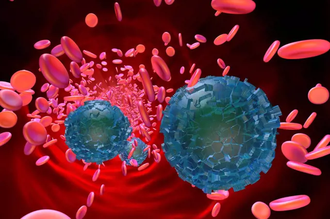
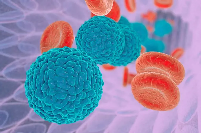
Colorized scanning electron micrograph of red blood cell infected with malaria parasites (teal). The small bumps on the infected cell show how the parasite remodels its host cell by forming protrusions called 'knobs' on the surface, enabling it to avoid destruction and cause inflammation. Uninfected cells (red) have smoother surfaces.
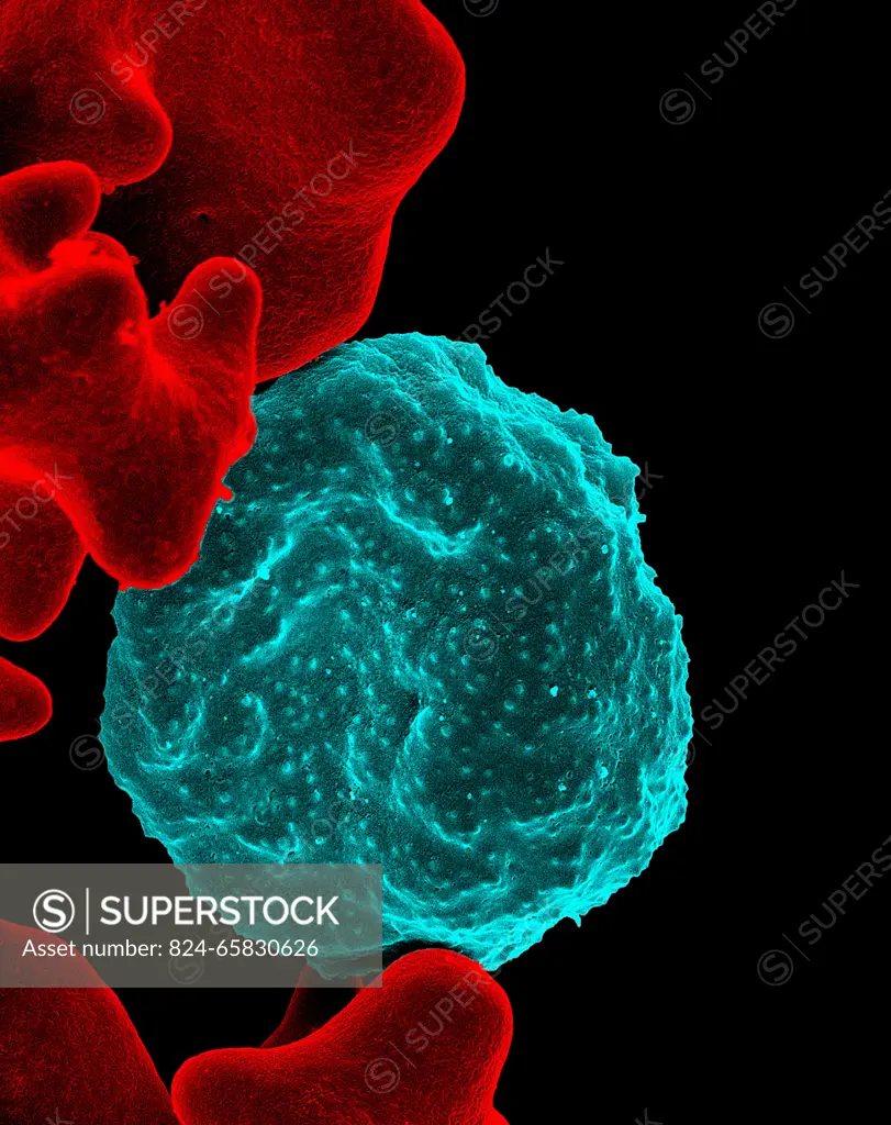
SuperStock offers millions of photos, videos, and stock assets to creatives around the world. This image of Colorized scanning electron micrograph of red blood cell infected with malaria parasites (teal). The small bumps on the infected cell show how the parasite remodels its host cell by forming protrusions called 'knobs' on the surface, enabling it to avoid destruction and cause inflammation. Uninfected cells (red) have smoother surfaces. by NIH-NIAID/IMAGE POINT FR/BSIP is available for licensing today.
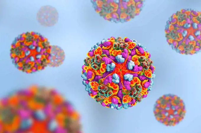
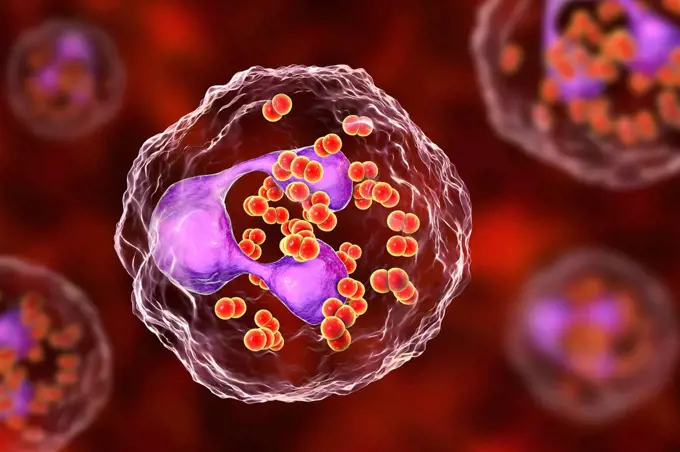
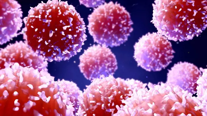
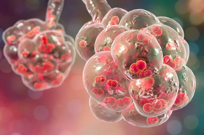
Visually Similar More from Microscopic Biological Structures story


Looking for a license?
Click here, and we'll help you find it! Questions? Just ask!
Click here, and we'll help you find it! Questions? Just ask!
DETAILS
Image Number: 824-65830626Rights ManagedCredit Line:NIH-NIAID/IMAGE POINT FR/BSIP/SuperStockCollection:BSIP Story:Microscopic Biological StructuresContributor:NIH-NIAID / IMAGE POINT FR / BSIP Model Release:NoProperty Release:NoResolution:3020×3810