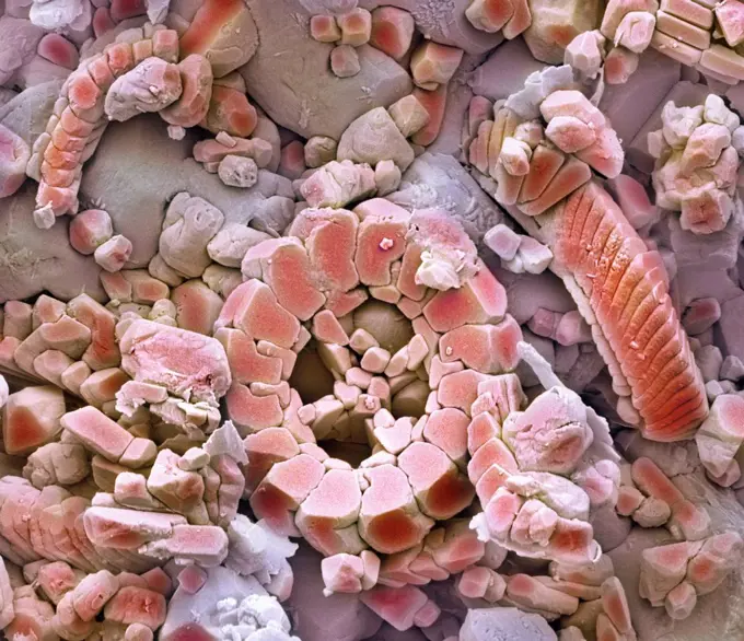
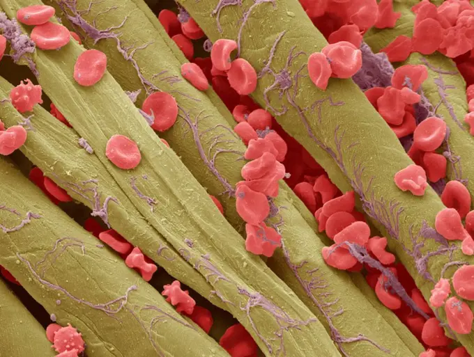
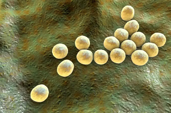
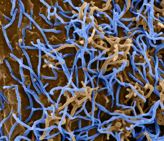
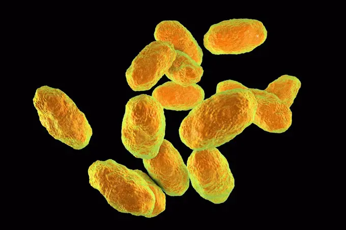
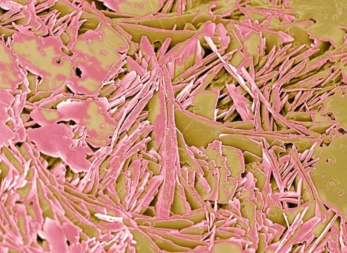
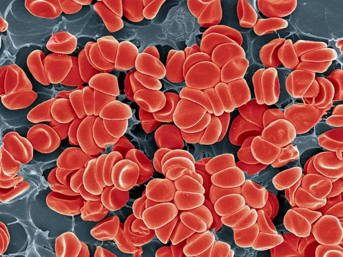
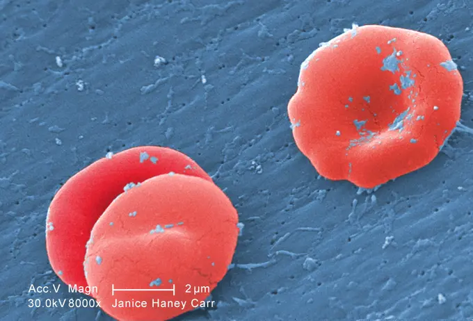
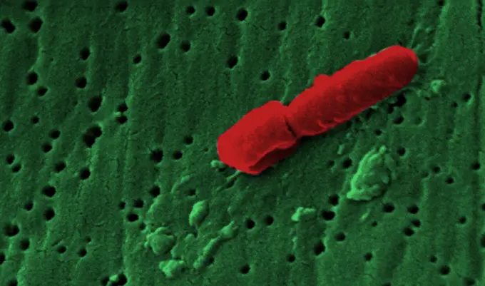
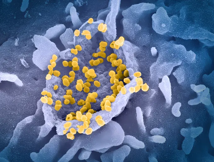
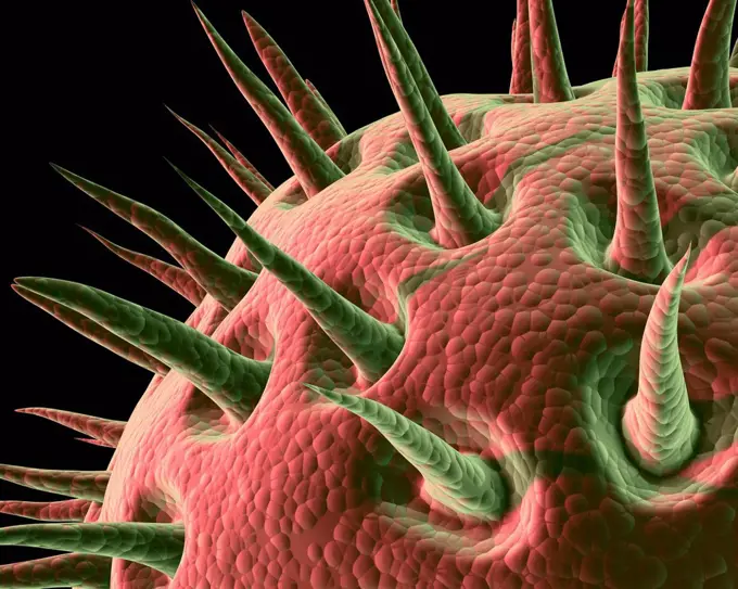
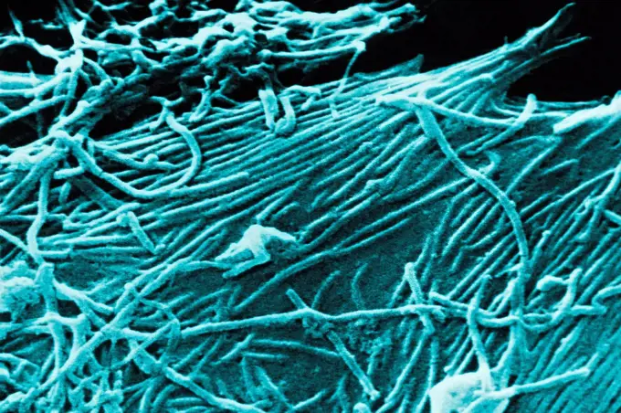
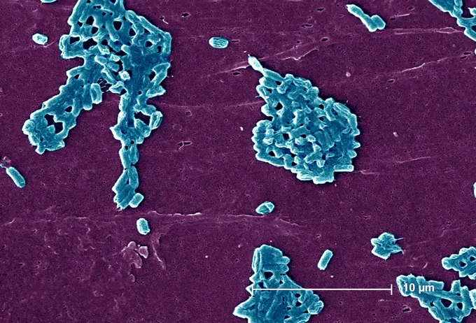
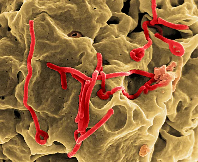
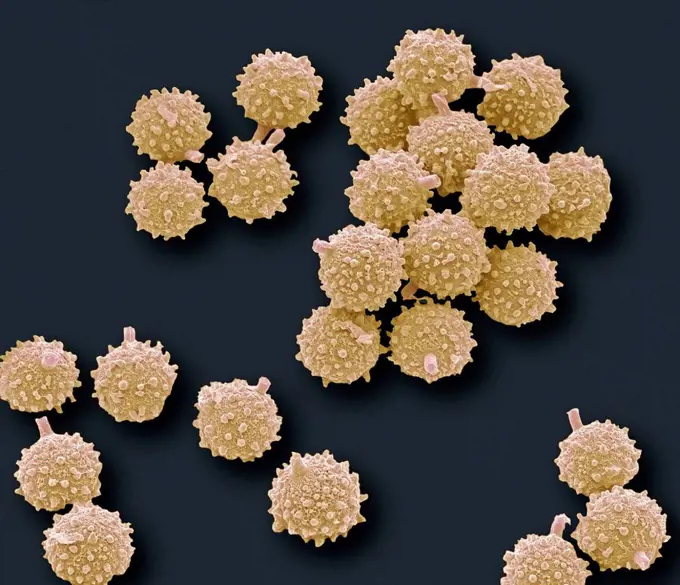
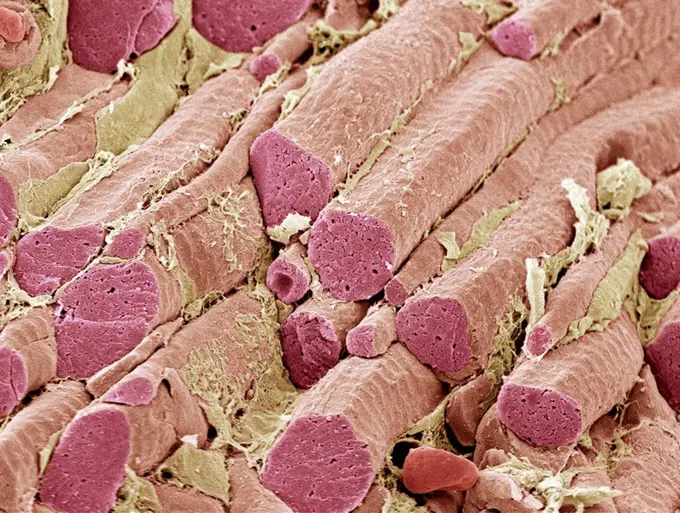
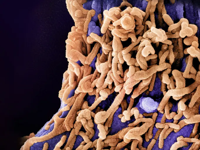
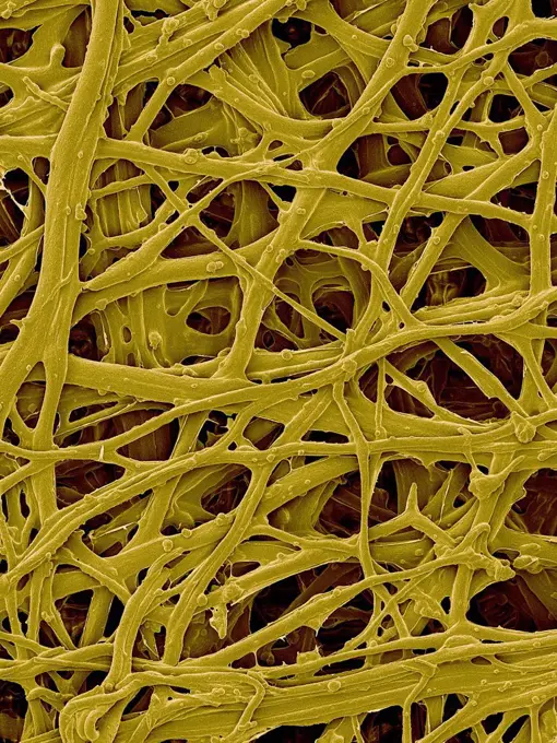
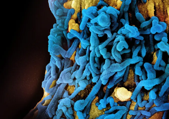
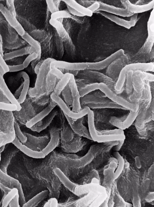
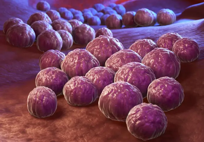
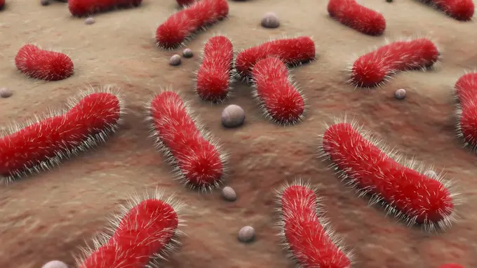
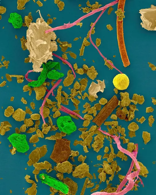
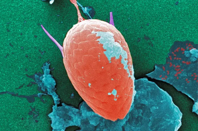
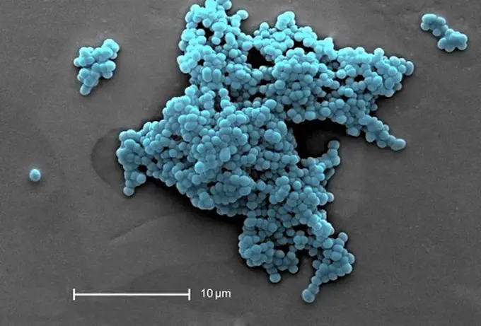
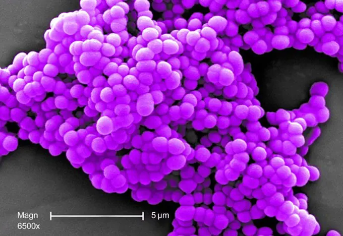
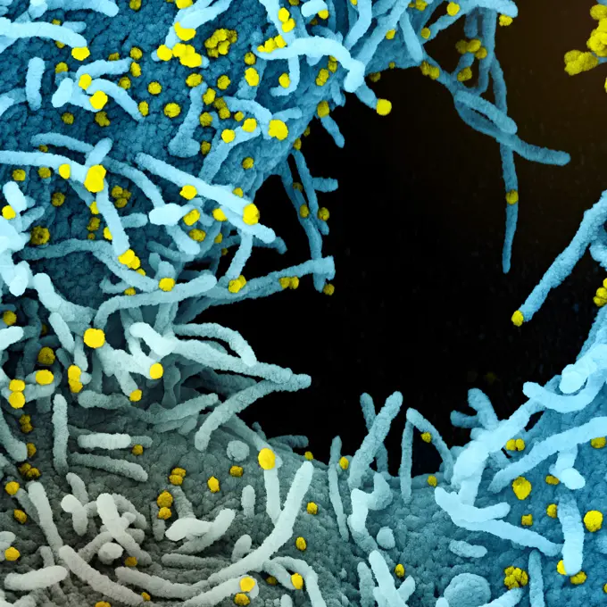
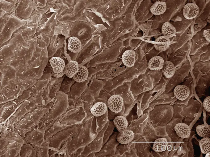
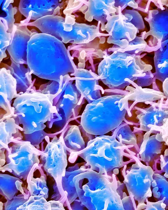
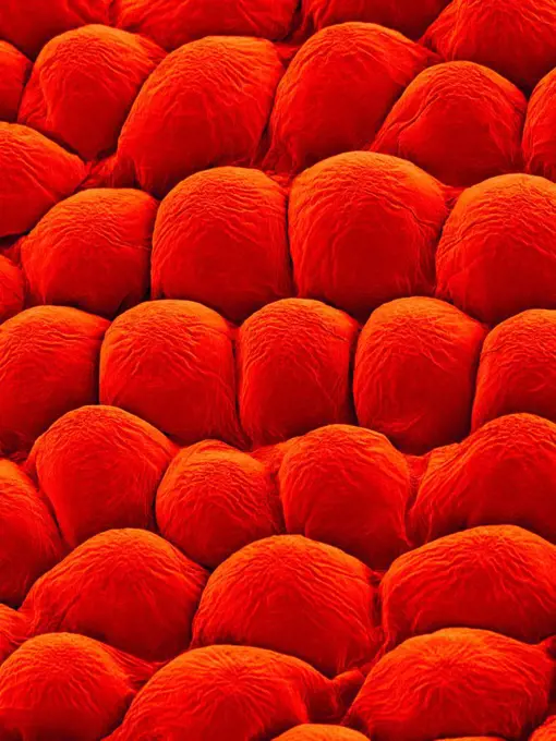
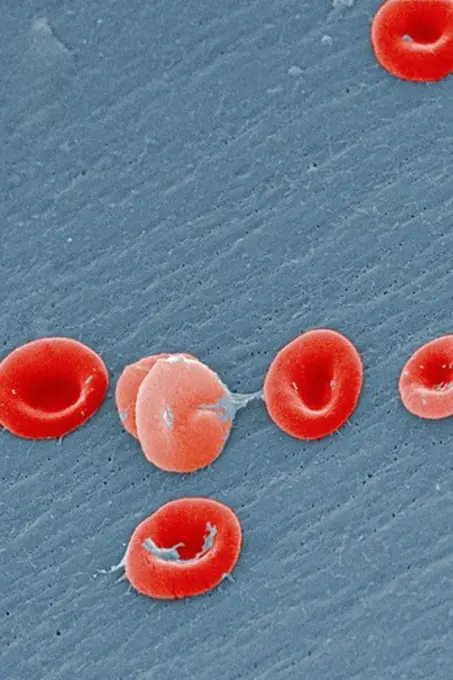
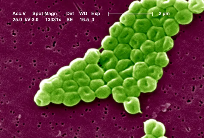
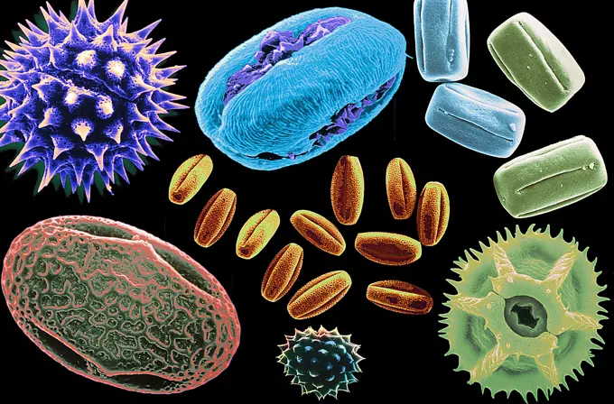
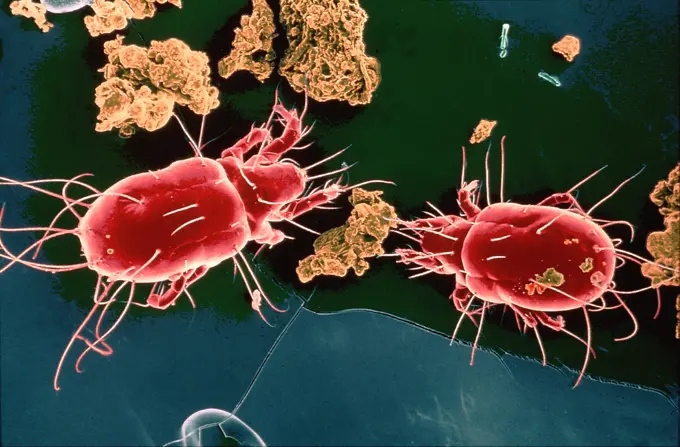
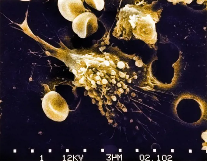
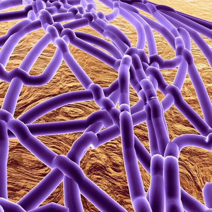
Fine ArtVintageFootageSubscriptionBoardsFree ResearchPricing
  • Fine Art
  • Vintage
  • Footage
  • Subscription
  • Boards
  • Free Research
  • Pricing

Microbial Structures under Microscope

Colorized scanning electron micrographs revealing intricate details of cells and bacteria, showcasing infections and immune responses.

Foot bacteria, SEM
Foot bacteria, SEM
179 assets in this story
4128R-13469
Fossil debris in chalk. Coloured scanning electron micrograph SEM of a fossilised fragment of a coccolithotrope skeleton.
4128R-7514
Used surgical swab, SEM
4128-28575390
MRSA bacteria, illustration
824-63223147
Colorized scanning electron micrograph of Ebola virus particles (blue) found both as extracellular particles and budding particles from chronically infected African green monkey kidney cells (brown). Image captured and color-enhanced at the NIAID Integrated Research Facility in Ft. Detrick, Maryland. Credit: NIAID.
1899-61460446
Colorized scanning electron micrograph of monkeypox virus (green) on the surface of infected VERO E6 cells (red)
4128R-13763907
Computer illustration of Haemophilus influenzae, coccobacillus bacteria. These bacteria cause a number of disease including meningitis in children, pn...
4128R-7594
Gallstone, SEM
4128R-8066
Blood clot, SEM
4128-V58557852
824-63194917
Under a high magnification of 8000X, this scanning electron micrograph (SEM) revealed some of the ultrastructural morphology displayed by red blood cells (RBCs) in a blood specimen of a 6 year old male patient that has sickle cell with hereditary persistence of fetal hemoglobin ( S-HPFH). In these individuals, the presence of the persistent fetal hemoglogin reduces the severity of the consequences of the sickle cell disease, thereby, reducing the degree of cellular deformity, ie sickling, seen in the sickled cells.
4128R-11476149
Protein molecules, computer artwork.
4297-1784
Scanning Electron Micrograph (SEM) of methicillin-resistant Staphylococcus aureus bacteria, commonly known as MRSA
4128-V58557907
4128R-15366406
Blastocyst, illustration
824-63123778
This SEM depicts a number of Gram-negative, non-motile Acinetobacter baumannii bacteria, Mag: 12365x. Members of the genus Acinetobacter are nonmotile rods, 1.0-1.5m in diameter, and 1.5-2.5m in length, becoming spherical during their stationary growth phase, they're widely distributed in nature, and are normal flora on the skin. Some members of the genus are important because they are an emerging cause of hospital acquired pulmonary, ie pneumoniae, hemopathic, and wound infections.
824-63128592
This scanning electron micrograph (SEM) revealed some of the ultrastructural morphologic features of a Klebsiella pneumoniae bacterium. K. pneumoniae is a non-motile, Gram-negative rod, and a facultative anaerobe, which means that it is able to adapt to an existence in an oxygenated or deoxygenated environment. It is an encapsulated bacterium that is commonly found in the human digestive tract, and is often the cause of hospital acquired, or nosocomial infections involving the urinary and pulmonary systems. See image n1305605 on the site.
4128R-15466815
Bacteria found on mobile phone, SEM
824-63227076
This scanning electron microscope image shows SARS-CoV-2 (round gold particles) emerging from the surface of a cell cultured in the laboratory. SARS-CoV-2, also known as 2019-nCoV, is the virus that causes COVID-19. Image captured and colorized at Rocky Mountain Laboratories in Hamilton, Montana. Credit: NIAID
4128R-20585
Virus, artwork
4384-182
This scanning electron micrograph (SEM) depicts a number of Ebola virions. Ebola hemorrhagic fever (Ebola HF) is a severe, often-fatal disease in humans and nonhuman primates (monkeys, gorillas, and chimpanzees) that has appeared sporadically since its initial recognition in 1976. The disease is caused by infection with the Ebola virus.  The virus is one of two members of a family of RNA viruses called the Filoviridae. There are four identified subtypes of Ebola virus. Three of the four have cau
824-63194432
Scanning electron micrograph of a murine macrophage infected with Francisella tularensis strain LVS. Macrophages were dry-fractured by touching the cell surface with cellophane tape after critical point drying to reveal intracellular bacteria. Bacteria (colorized in green) are located either in the cytosol or within a membrane-bound vacuole.
824-63123757
This scanning electron micrograph (SEM) depicts a number of rod-shaped Escherichia coli bacterium, some of which have formed colonial groupings, while others have remained isolated as single cells; Magnification 3607x. Escherichia coli is a Gram-negative
4128R-11476219
Protein molecules, computer artwork.
4128-V58562086
824-63194380
Scanning electron micrograph of Ebola virus budding from the surface of a Vero cell (African green monkey kidney epithelial cell line). Produced by the National Institute of Allergy and Infectious Diseases (NIAID), under a very-high magnification, this digitally-colorized scanning electron micrograph (SEM) depicts a number of filamentous Ebola virus particles (red) that had budded from the surface of a VERO cell (brown) of the African green monkey kidney epithelial cell line. Ebola hemorrhagic fever (Ebola HF) is one of numerous Viral Hemorrhagic Fevers. It is a severe, often fatal disease in humans and nonhuman primates (such as monkeys, gorillas, and chimpanzees). Ebola HF is caused by infection with a virus of the family Filoviridae, genus Ebolavirus. When infection occurs, symptoms usually begin abruptly. The first Ebolavirus species was discovered in 1976 in what is now the Democratic Republic of the Congo near the Ebola River. Since then, outbreaks have appeared sporadically.
4128R-12899
Puffball fungus spores, coloured scanning electron micrograph SEM. These are the reproductive cells of the fungus. Magnification: x3000 when printed a...
4128-18799453
Biofilm of Mycobacterium tuberculosis bacteria, illustration
4128R-4019
Skeletal muscle fibres, SEM
824-63123779
This SEM depicts a highly magnified cluster of Gram-negative, non-motile Acinetobacter baumannii bacteria, Mag: 13331x. Members of the genus Acinetobacter are nonmotile rods, 1-1.5m in diameter, and 1.5-2.5m in length, becoming spherical in shape while in their stationary phase of growth. Acinetobacter spp. are widely distributed in nature, and are normal flora on the skin. Some members of the genus are important because they are an emerging cause of hospital acquired pulmonary, ie pneumoniae, hemopathic, and wound infections.
4128-V58562079
4128R-1948
Passion flower pollen, SEM
4128R-5280
Bladder stone, SEM
4128R-13619932
Used wax dental floss with dental plaque (yellow), bacteria (orange) and cheek cells (pink) on the dental floss fibres (blue), coloured scanning elect...
4128-V58557898
4128-111494476
Malaria infection, illustration.
824-65830615
Colorized scanning electron micrograph of Marburg virus particles (orange) both budding and attached to the surface of an infected VERO E6 cell (purple).
4128R-14057538
Gonorrhoeae bacteria, illustration
4128-V58557917
4128R-13620190
Coloured scanning electron micrograph (SEM) Chicken eggshell inner membrane (Gallus gallus domesticus). Shown here is the eggshell inner membrane that...
1899-65662310
Colorized scanning electron micrograph of Marburg virus particles (blue) both budding and attached to the surface of an infected VERO E6 cell (gold).
4128R-7108
Lactobacillus bulgaricus bacteria
1525-56278078
Chlamydia trachomatis, an obligate intracellular human pathogen, is one of four bacterial species in the genus Chlamydia. 3D illustration. Chlamydia Bacterium
824-63225928
Colorized scanning electron micrograph of chronically infected and partially lysed cells (blue) infected with a variant strain of SARS-CoV-2 virus particles (green), isolated from a patient sample. Image captured at the NIAID Integrated Research Facility (IRF) in Fort Detrick, Maryland.
4239-18641705
Conceptual image of the Bacillus bacteria.
4128R-13620322
Coloured scanning electron micrograph (SEM) of House dust. Components such as fibre, hair, pollen, insect faecal matter and scale (animal dander) caus...
4384-169
Under a moderate magnification of 1000X, this digitally-colorized scanning electron micrograph (SEM) of an untreated water specimen extracted from a wild stream mainly used to control flooding during inclement weather, revealed the presence of unidentified organisms, which included bacteria, protozoa, and algae. In this particular view, a single copepod-like microorganism was seen occupying the field of view, which seemed to be encased in an outer shell of armour-like plates, or scales. Looking
4297-1431
Scanning electron micrograph revealed a number of clusters of Gram-positive beta-hemolytic Group C Streptococcus spp. bacteria
4297-1428
Beta strep Group C, scanning electron micrograph (SEM) at a magnification of 6,500X, showing a cluster of Gram-positive, beta-hemolytic Group C Streptococcus sp
4128-V58557844
4128R-14057535
Gonorrhoeae bacteria, illustration
824-63227333
Colorized scanning electron micrograph of a cell heavily infected with SARS-CoV-2 virus particles (yellow), isolated from a patient specimen. The black area in the image is the extracellular space between cells. Image requested from the NIAID Integrated Research Facility (IRF) in Fort Detrick, Maryland.
4128R-13619950
Cotton bud with ear wax embedded in fibres, coloured scanning electron micrograph (SEM). A cotton bud is a small stick with a cotton swab at one or bo...
1439-57940077
Microscopic view of cells and structures with scale indicator.
4128R-15404811
Activated platelets, SEM
4128R-11476147
Protein molecules, computer artwork.
4128R-13622329
Papillae on the upper surface of a rose flower petal (Rosa sp), coloured scanning electron micrograph (SEM). Papillae are projections from epidermal c...
1899-53513333
Colorized scanning electron micrograph of Ebola virus particles (yellow) both budding and attached to the surface of infected VERO E6 cells (green). Image captured and color-enhanced at the NIAID Integrated Research Facility in Fort Detrick, Maryland.
4384-148
This scanning electron micrograph (SEM) revealed some of the ultrastructural morphology displayed by red blood cells (RBCs) in a blood specimen of a 6 year old male patient with sickle cell anemia, which was accompanied by hereditary persistence of fetal hemoglobin (HPFH). In these individuals, the presence of the persistent fetal hemoglogin reduces the severity of the consequences of the sickle cell disease, thereby, reducing the degree of cellular deformity, i.e., sickling, seen in the sickled
824-63123780
This SEM depicts a highly magnified cluster of Gram-negative, non-motile Acinetobacter baumannii bacteria, Mag: 13331x. Members of the genus Acinetobacter are nonmotile rods, 1-1.5m in diameter, and 1.5-2.5m in length, becoming spherical in shape while in their stationary phase of growth. Acinetobacter spp. are widely distributed in nature, and are normal flora on the skin. Some members of the genus are important because they are an emerging cause of hospital acquired pulmonary, ie pneumoniae, hemopathic, and wound infections.
4128R-13022180
Panch phoran. Coloured scanning electron micrograph (SEM) of panch phoran or indian five spice. It is a spice blend commonly used in Eastern India and...
4128R-7970
Cancer cells, SEM
4128R-13620421
Coloured scanning electron micrograph (SEM) of Dust mite (Dermatophagoides pteronyssinus). Millions of dust mites inhabit the home, feeding on dead hu...
4128R-13941152
Cell nucleus, illustration.
4128R-6396
Avian influenza virus, computer artwork
1899-53513328
Colorized scanning electron micrograph of Ebola virus particles (red) in extracellular space between infected African green monkey kidney cells. Image captured and color-enhanced at the NIAID Integrated Research Facility in Ft. Detrick, Maryland.
4269-1218
Scanning electron microscope view of different pollens, responsible for respiratory allergies.
4269-24585
Dust mite. SEM (Scanning Electron Microscope) of dust mite (magification x 200). They can cause allergic reactions in some people.
4297-1742
SEM image of the Death of a Cancer Cell (step 1 of a 6 step sequence)
4128R-11474533
Ebola virus, computer artwork.
4128-111494389
Vaginal bacteria. Coloured scanning electron micrograph (SEM) of bacteria on the vaginal wall. A healthy vaginal flora protects the body against uroge...
824-63191236
Scanning electron micrograph of just-divided HeLa cells. Zeiss Merlin HR-SEM.
4128R-13620432
Coloured scanning electron micrograph (SEM) of Dust mite (Dermatophagoides pteronyssinus) with dust mite faecal pellets (light brown), pollen (yellow)...
824-63194406
Scanning electron micrograph of HIV-1 budding from cultured lymphocyte. Multiple round bumps on cell surface represent sites of assembly and budding of virions.
4128R-4408
Kidney glomeruli, SEM
4128R-15514879
CAR T cell cancer immunotherapy, illustration
4128R-13928413
Parasitic Amoeba protozoan engulfing bacteria, computer illustration. The amoeba will engulf and digest the prey in a process called phagocytosis.
4128-16224384
Snake red blood cells, SEM
4297-1430
Scanning electron micrograph revealed a small clustered group of Gram-positive beta-hemolytic Group C Streptococcus spp. bacteria
4378-20013628
Tsetse Fly
PREVIOUS
of 2
NEXT
2401 S. Ervay, Suite 206
Dallas, Texas 75215
United States
Browse
CategoriesGalleriesFree Research
For Creators
License Your ContentSuperStock For StudentsFrame of Mind BlogContributor PortalNew Image SpotlightLicense AI Imagery
Resources
About UsLicensingTerms & ConditionsAPI AccessPrivacyPricing
Contact
+1 866 236 0087help@superstock.com Contact form
©2026 SuperStock. All Rights Reserved. -A
This site is protected by reCAPTCHA and Google Privacy Policy and Terms of Service apply.