Fine ArtVintageFootageSubscriptionBoardsFree ResearchPricing
  • Fine Art
  • Vintage
  • Footage
  • Subscription
  • Boards
  • Free Research
  • Pricing

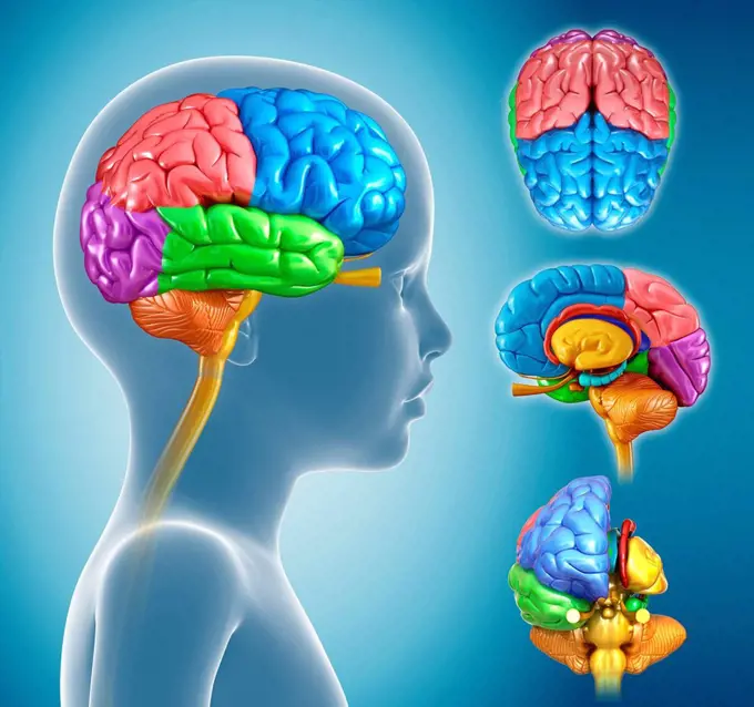
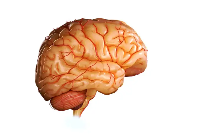
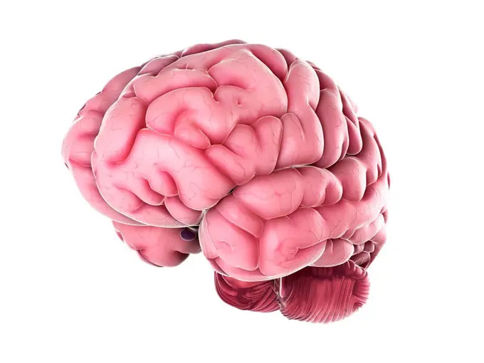
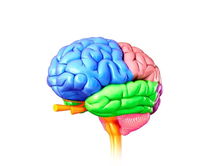
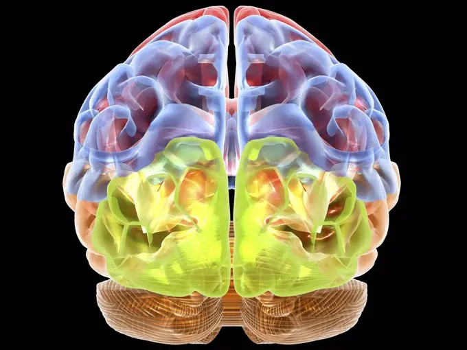
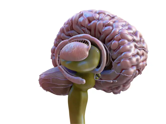
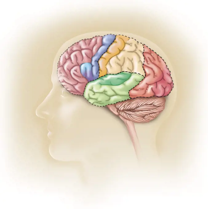
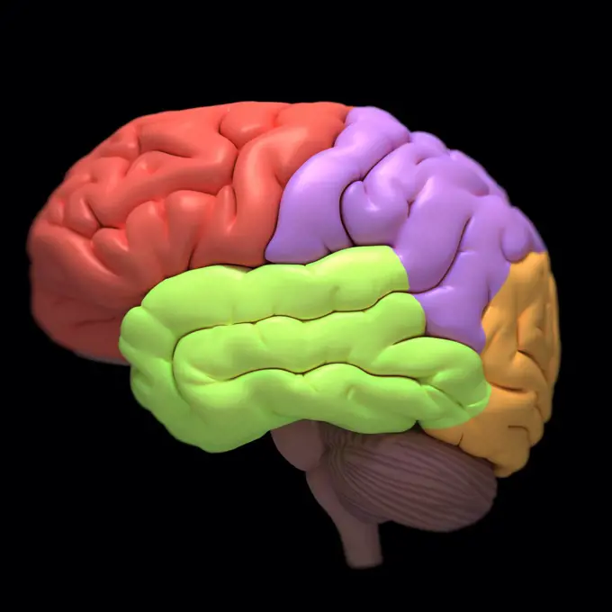
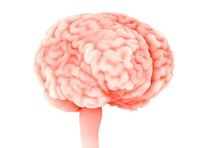
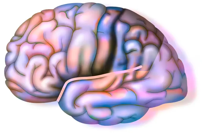
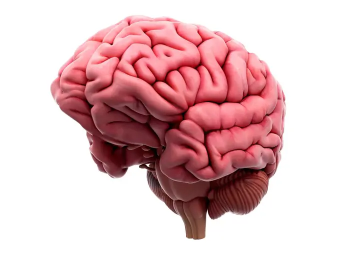
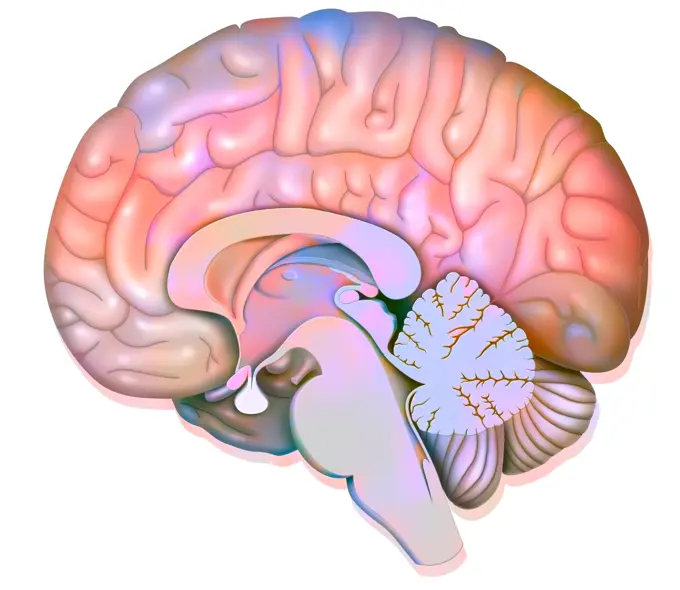
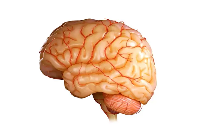
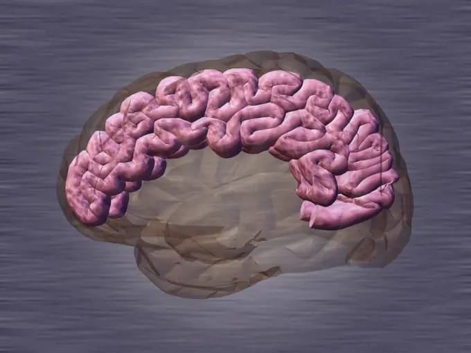
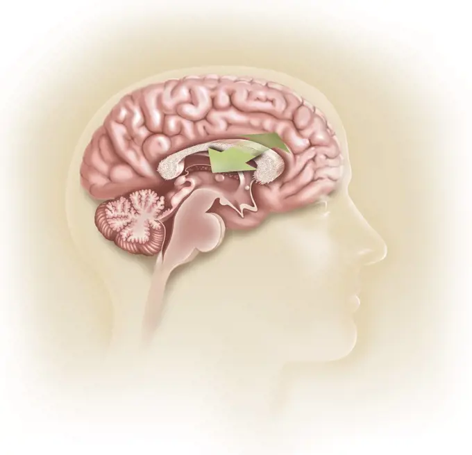
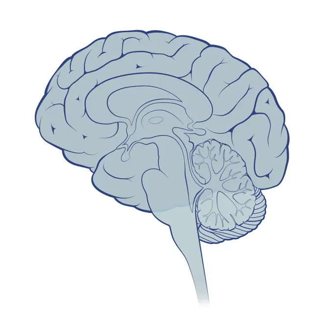
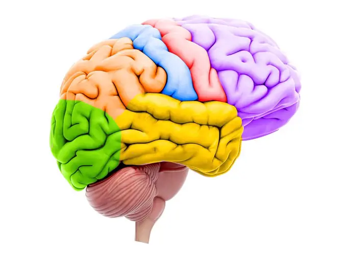
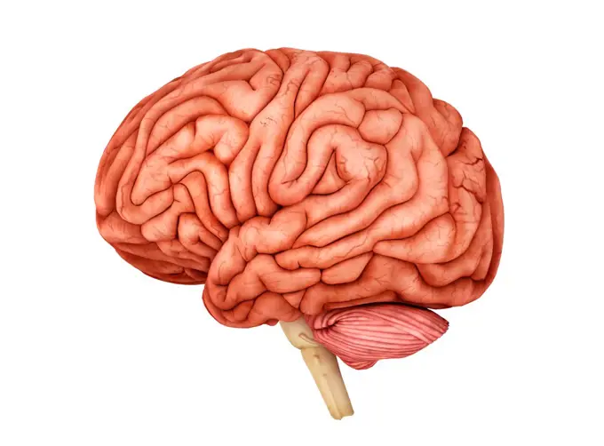
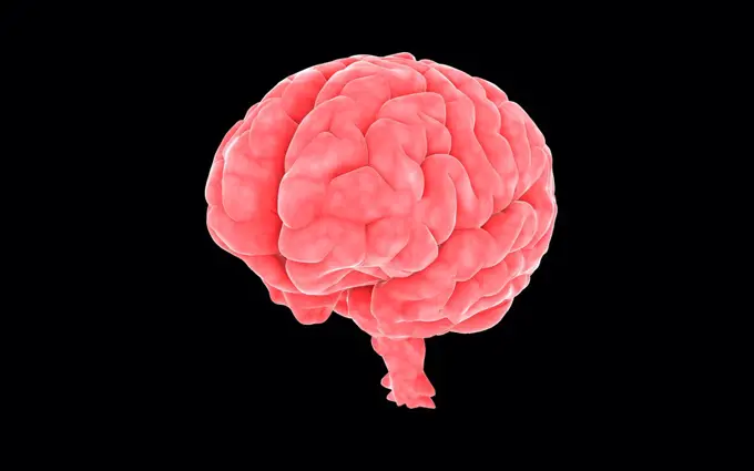
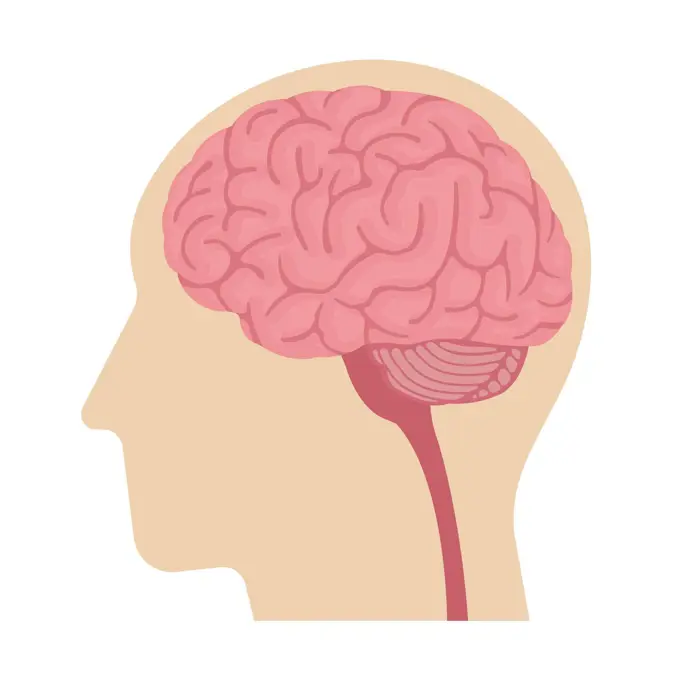
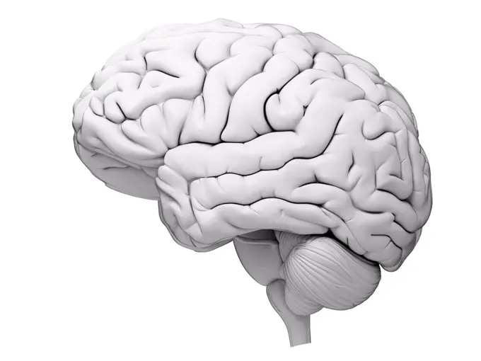
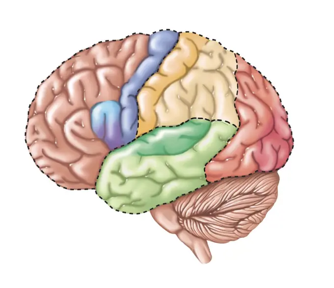
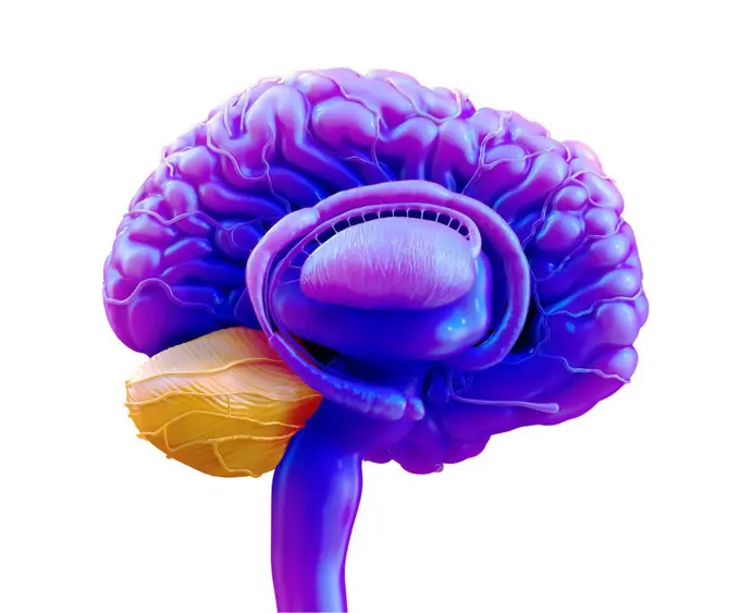
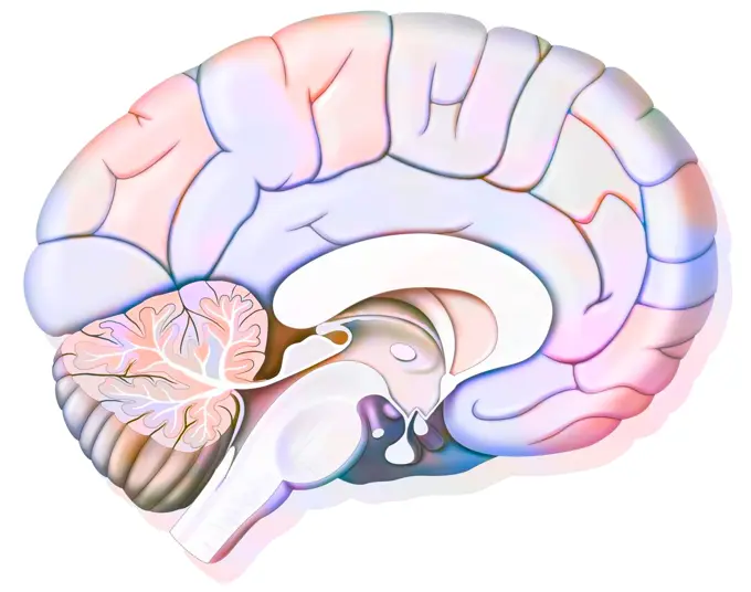
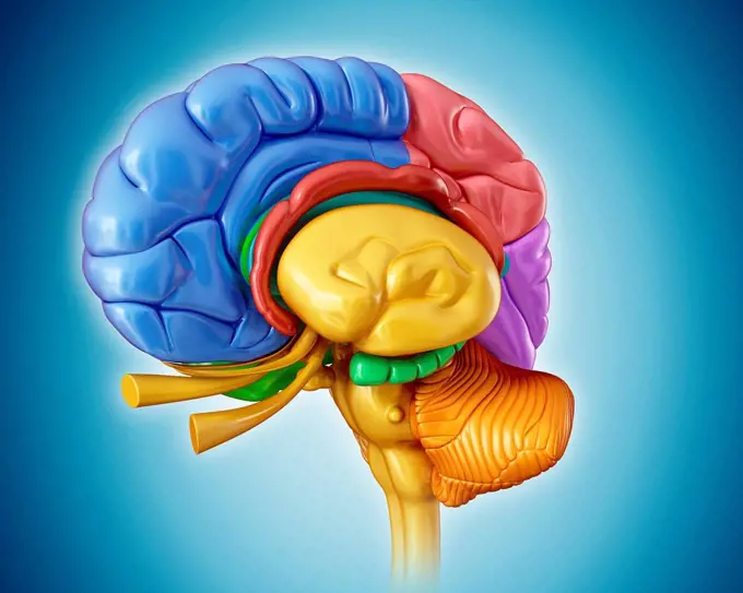
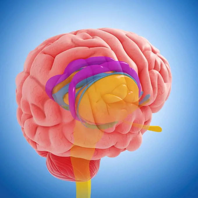
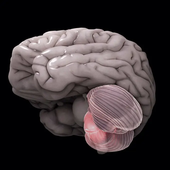
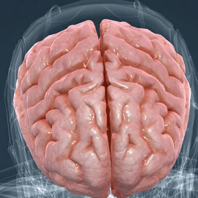
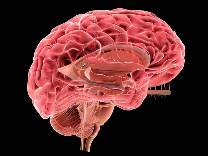
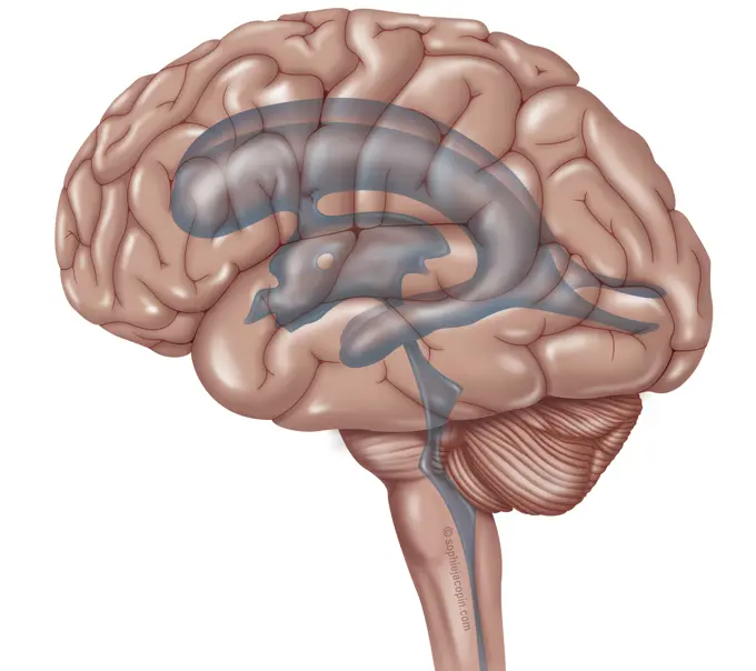
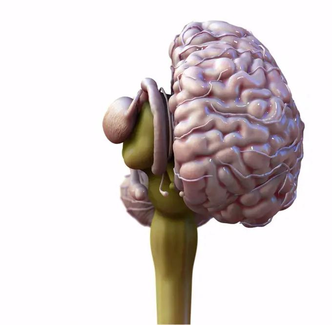
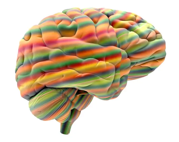
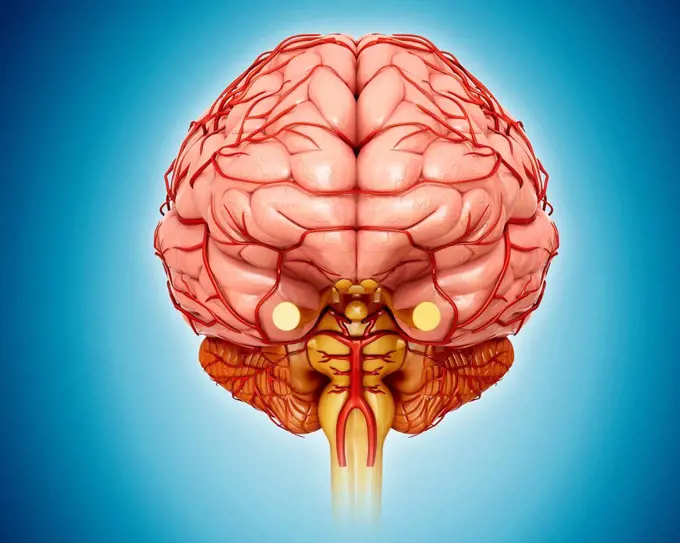
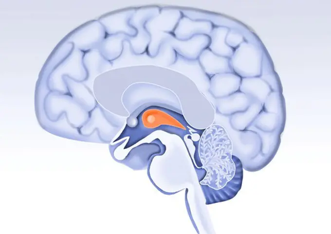
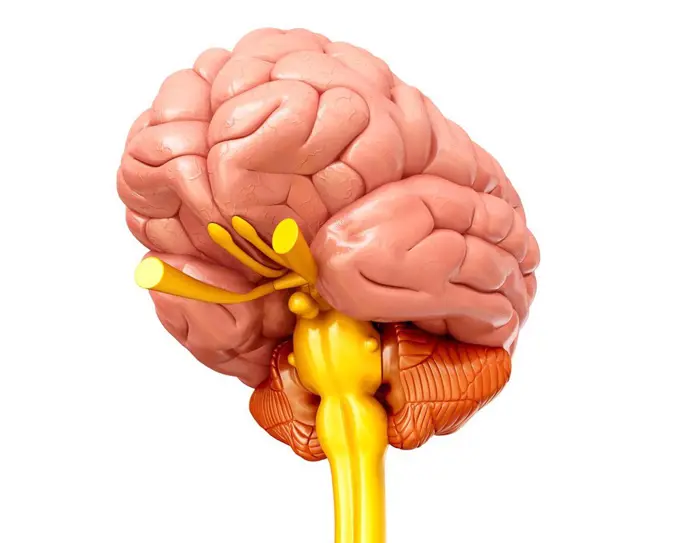
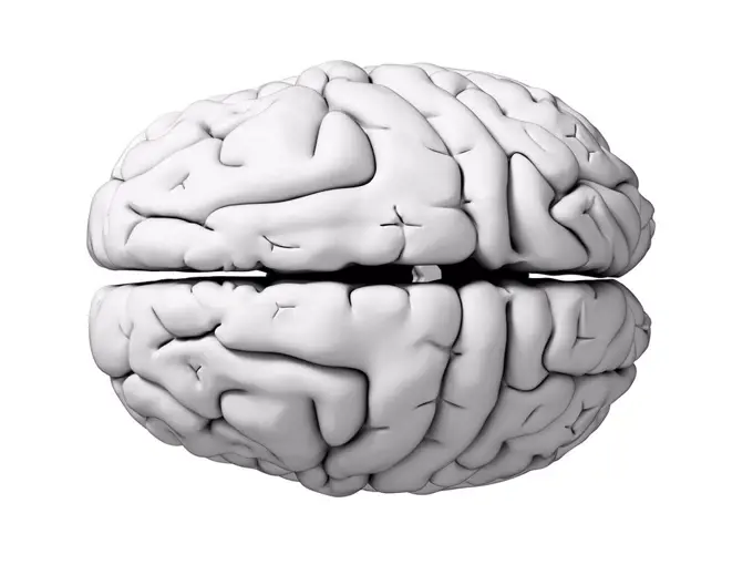
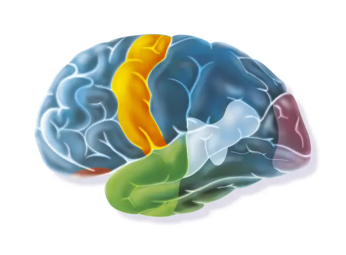
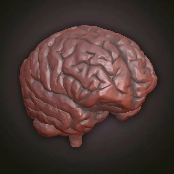
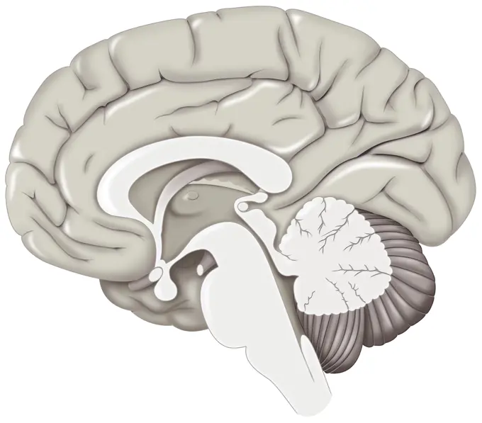
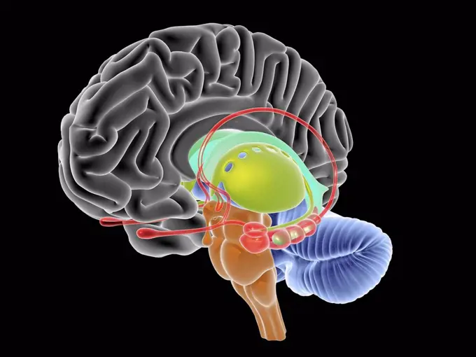
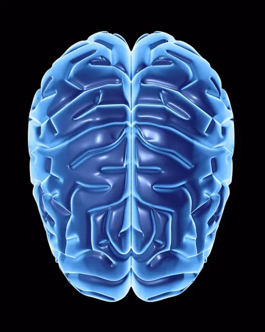
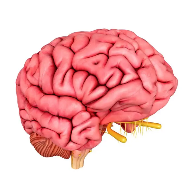
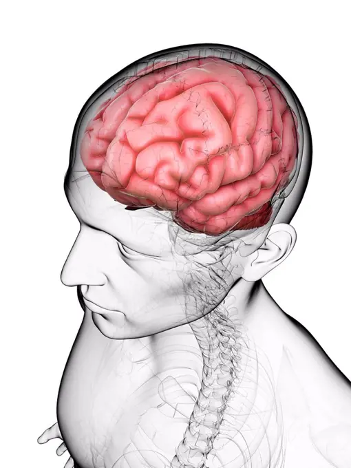
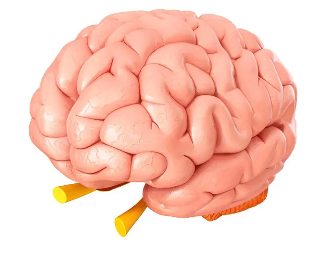
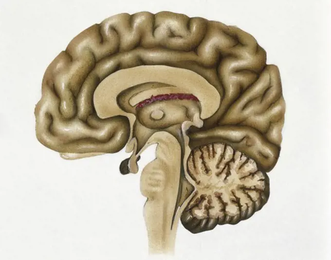
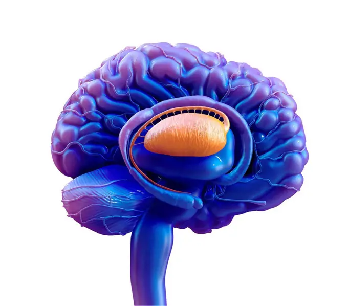
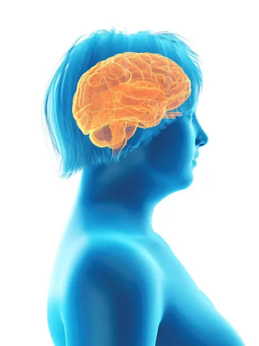
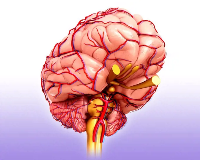
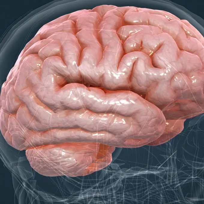
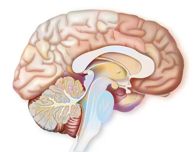
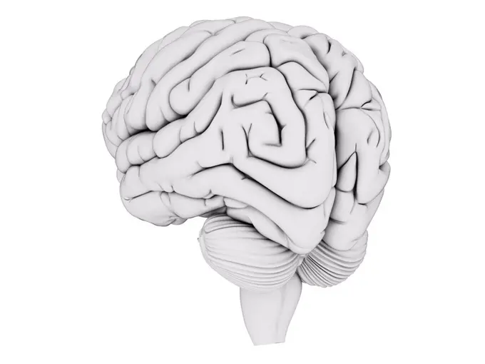
Human Brain Illustrations

Computer-generated and artistic representations of the human brain, displaying its anatomy and various regions in vibrant colors.

Human Brain
Human Brain
374 assets in this story
4378-5677
Human Brain
4128R-25966
Human brain, computer artwork.
4128-19055746
Illustration of a human brain
4128R-13725062
Illustration of human brain regions.
4128R-13375564
Human brain
4128R-20578
Human brain anatomy, artwork
4128R-14323974
Brain anatomy, illustration
4239-20481104
Side view of the human head showing the functional lobes of the human brain.
4128R-13723654
Illustration of human brain regions.
1525-20518328
human brain
4128-30421629
Human brain, illustration
824-63164613
Brain, with the two cerebral hemispheres, the cerebellum and the brainstem.
4378-1687
A model of the human brain, isolating different lobes with color.
4128R-13586396
Illustration of human brain anatomy.
4128R-14323996
Brain, illustration
824-63164667
Normal brain with Sylvian fissure and parietal lobe.
4378-20014162
Human Brain
4128R-13371393
Human brain, illustration.
824-63164027
Mid sagittal section of the human brain showing the hypothalamus.
4378-5353
Human Brain
4128R-13587287
Human brain, illustration.
4128R-14181189
Human brain regions, illustration
4128-V58567372
4128R-11296194
Human brain anatomy, computer artwork.
1428-174
Close-up of a human brain
824-72488644
The occipital lobe of the brain. The occipital lobe of the brain corresponds to the visual cortex: primary visual area at the end, surrounded by the associative visual areas.
4239-20481102
Sagittal view of human brain showing the corpus callosum.
824-63224053
Sagittal section of human brain on white background.
4128-19055732
Illustration of a human brain
824-63164379
Brain with occipital, parietal, temporal lobes, cerebellum.
4128R-15582432
Illustration of the brain sections
4239R-8486
Anatomy of human brain, side view.
1525-57069522
Illustration of an abstract digital human brain
4128-15980535
Human brain, computer illustration.
4239R-7968
Conceptual image of human brain.
5507-43624213
Vector Illustration of human head anatomy structure ( no text )
4128R-14323991
Brain, illustration
1525-27207301
Close up Realistic Brain Concept on black background. 3D Render.
4128-19055792
Illustration of a human brain
4128R-13375573
Human brain
4239-20481103
Side view of the human brain showing the functional lobes.
4128R-14323979
Brain cerebellum, illustration
824-63163908
Mid sagittal section of the human brain showing the hypothalamus.
4128-30421638
Human brain, illustration
4128R-13387039
Illustration of human brain anatomy.
4128R-13371381
Human brain, illustration.
4128R-24251
Brain anatomy, computer artwork.
4128-19055811
Illustration of a human brain
4378-1674
A stylized model of a human brain.
1525-56183616
The brain controls thoughts, memory and speech, arm and leg movements and the function of many organs within the body. 3D illustration. Detailed anatomy of human brain and nervous system closeup
4128R-13446707
Illustration of human brain structures and regions.
4128-15980536
Human brain, computer illustration.
4128R-29537
Human brain, computer artwork.
824-63224369
Ventricular system of the brain in a brain in external profile view. The ventricles are a collection of cavities. They are located inside the brain. And are continuous with the channel of the ependyma of the spinal cord. They participate in the secretion and circulation of cerebrospinal fluid (or cerebrospinal fluid). The cerebral ventricles are four in number. They are represented in transparency in bluish gray in this profile view of the brain. The two lateral ventricles are connected to the third ventricle in the center of the brain through the interventricular foramen. Then the structure descends downwards through the midbrain aqueduct. The fourth ventricle is located slightly above and in front of the cerebellum. Then finally, the structure descends into the brainstem. It forms the canal of the spinal cord.
4128R-13371395
Human brain, illustration.
1899-53513439
Sagittal section of the encephalon, brain, cortex, diencephalon, pituitary, cerebellum, brainstem. The brain is made up of two hemispheres. One is on the right (right hemisphere) and the other on the left (left hemisphere). This sagittal or median cut is made in the center of the brain. It emphasizes the internal view of the right hemisphere. The brain is made up at the periphery of the cerebral cortex. In the center is found diencephalon and pituitary gland. Down in front is the brainstem followed back by the cerebellum. (Photo by: JACOPIN/BSIP/UIG)
824-63224361
Sagittal section of the encephalon, brain, cortex, diencephalon, pituitary, cerebellum, brainstem. The brain is made up of two hemispheres. One is on the right (right hemisphere) and the other on the left (left hemisphere). This sagittal or median cut is made in the center of the brain. It emphasizes the internal view of the right hemisphere. The brain is made up at the periphery of the cerebral cortex. In the center is found diencephalon and pituitary gland. Down in front is the brainstem followed back by the cerebellum.
4128-30421646
Human brain, illustration
4128R-14323973
Brain anatomy, illustration
4128R-12576729
Brain. Abstract computer artwork of a human brain. The front of the brain is at right. The brain is the nerve centre that controls bodily function and...
4128R-13410122
Human brain, illustration
4252-4462
Brain thalamus
6188-62318390
Central Organ of Human Nervous System Brain Anatomy 3D Illustration Concept of Central Organ of Human Nervous System Brain Anatomy Copyright: xZoonar.com/magicminex 21748153
4128R-13711049
Illustration of human brain anatomy.
1525-20518370
Creative concept of the human brain
4128R-11542178
Human brain, computer illustration.
824-63180663
Illustration of the areas of the brain that are active while taking a written test. The reading area (light blue area) is activated and the prefrontal cortex (small red area) activates the working memory, reasoning and language. The temporal lobe (green area) activates short-term memory. The visual cortex (pink area) is activated to a greater or lesser extent depending on the test and the motor cortex (orange area) activates the hand.
4378-2337
Clay model showing the anatomical structure of a brain.
824-63205883
Illustration of the brains frontal lobe, seen in a profile view from the left hemisphere side.
824-63164700
Areas of the brain of the right hemisphere: cortex (visual, parietal, sensory, motor, premotor) and auditory area.
824-63188692
Mid-saggital plane illustration of the brain showing the central structures that link the right and left hemispheres.
4128R-11479465
Computer artwork of a section through a healthy brain. The highly folded area in grey is the cerebrum, which is responsible for conscious thought, emo...
4128R-7071
Human brain, artwork
4128R-15514930
Illustration of the human brain
4128R-13574376
Illustration of brain veins and anatomy.
824-63180660
Illustration of the areas of the brain that are active while memorising a lesson. The visual cortex (pink area) and the reading area (light blue area) are active when decrypting information. They then pass the baton to the temporal love (dark green area) and the dorsolateral prefrontal cortex (light green area) that allows short-term memorisation. If the lesson is being learnt by repeating out loud, then Broca's cortex is active (purple area). The limbic system (yellow area) allows long-term memorisation, by associating information to memories and emotions.
4128R-13723524
Illustration of human brain anatomy.
4128R-13409572
Brain's blood supply, artwork
4128R-27660
Male brain, computer artwork.
4128R-13711030
Illustration of human brain anatomy.
1788-39818
Medicine: encephalon
4128R-11322884
Human brain, computer artwork.
4128-V58567438
4128R-14323963
The basal ganglia , illustration
4128R-5244
Blood vessels of the brain, artwork.
4128R-13710977
Illustration of human brain regions.
4128R-15515678
Illustration of an obese woman's brain
4128R-13711052
Illustration of the anatomy of the human brain and its arteries.
824-63208866
Illustration of the areas of the brain affected in the case of transient global amnesia (TGA), a pathology affecting short-term memory. Two types of memory are concerned in short-term memory, controlled and coordinated by the pre-frontal cortex (green), the central administrator: -phonological memory, represented by white arrows, affecting the Brocas area (purple, production of spoken words) and Wernickes area (pink, auditory and oral language, comprehension of these words) -visuo-spatial memory represented by the occiptal cortex (blue, treatment of visual and spatial information).
4128R-14637804
Human brain, illustration
1525-56183620
The brain controls thoughts, memory and speech, arm and leg movements and the function of many organs within the body. 3D illustration. Detailed anatomy of human brain and nervous system closeup
1899-53509228
Areas of the brain involved when we are afraid.
824-63164302
Areas of the brain involved when we are afraid.
4128-19055796
Illustration of a human brain
824-72488619
Illustration of Alzheimer's disease (damage to the cortex and hippocampus).
824-757
Human Brain Showing Highlighted Area For The Sense of Taste
4128R-13387033
Illustration of human brain anatomy.
4128R-13371391
Human brain, illustration.
4128R-11542175
Human brain, computer illustration.
4128R-13409796
Brain anatomy, illustration
PREVIOUS
of 4
NEXT
2401 S. Ervay, Suite 206
Dallas, Texas 75215
United States
Browse
CategoriesGalleriesFree Research
For Creators
License Your ContentSuperStock For StudentsFrame of Mind BlogContributor PortalNew Image SpotlightLicense AI Imagery
Resources
About UsLicensingTerms & ConditionsAPI AccessPrivacyPricing
Contact
+1 866 236 0087help@superstock.com Contact form
©2026 SuperStock. All Rights Reserved. -A
This site is protected by reCAPTCHA and Google Privacy Policy and Terms of Service apply.