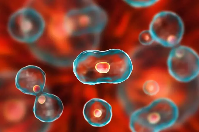
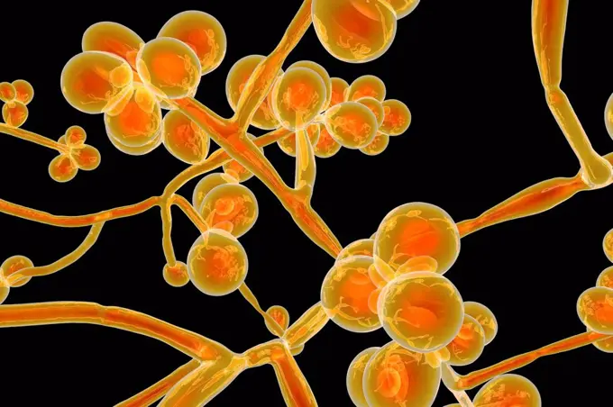
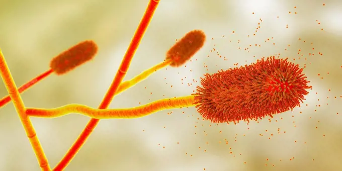
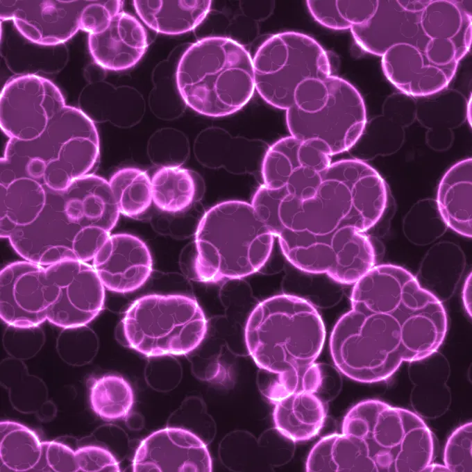
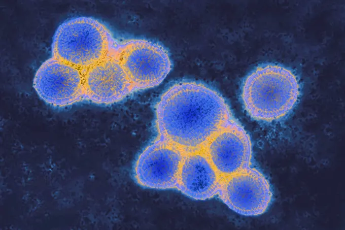
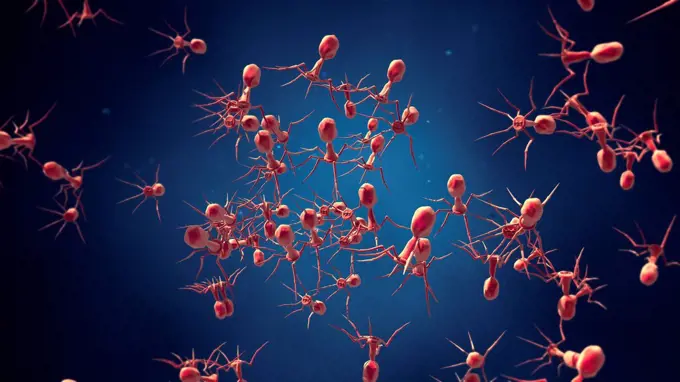
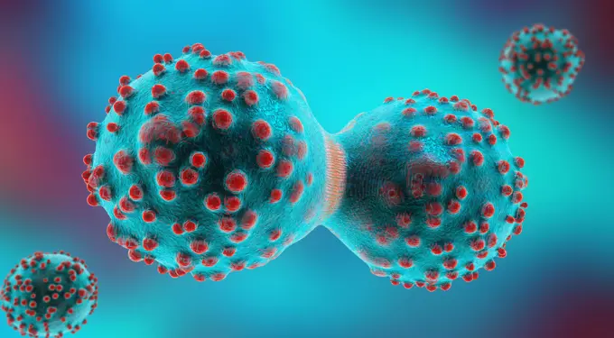
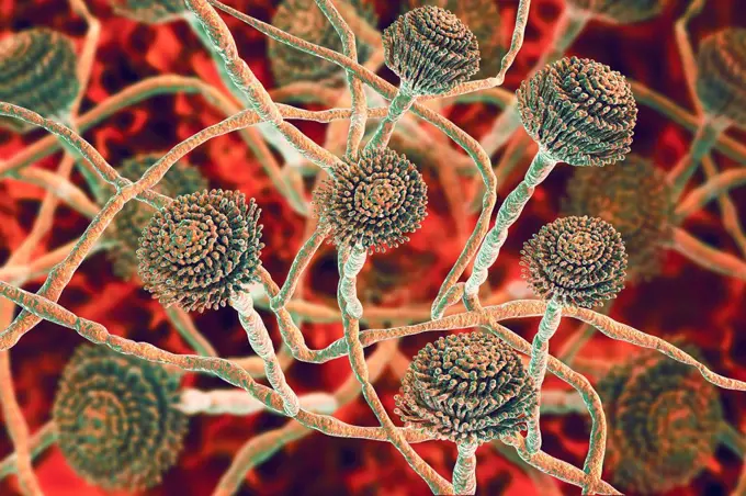
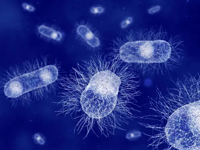
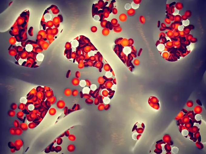
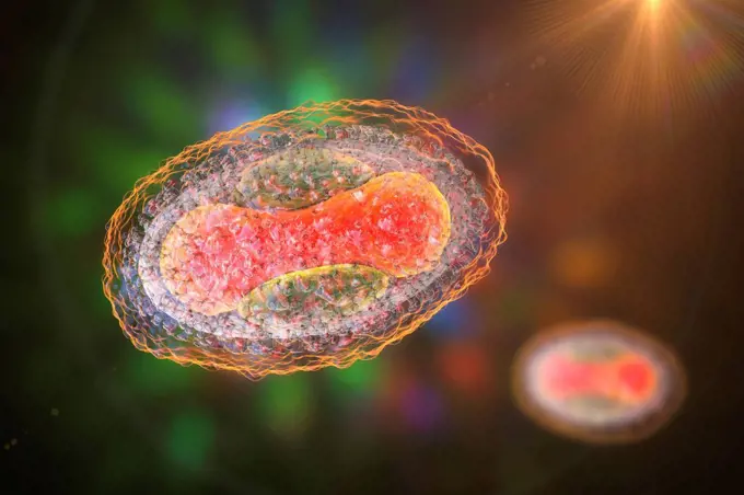
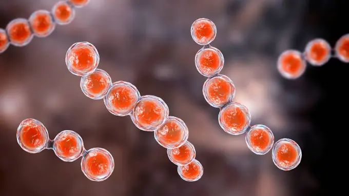
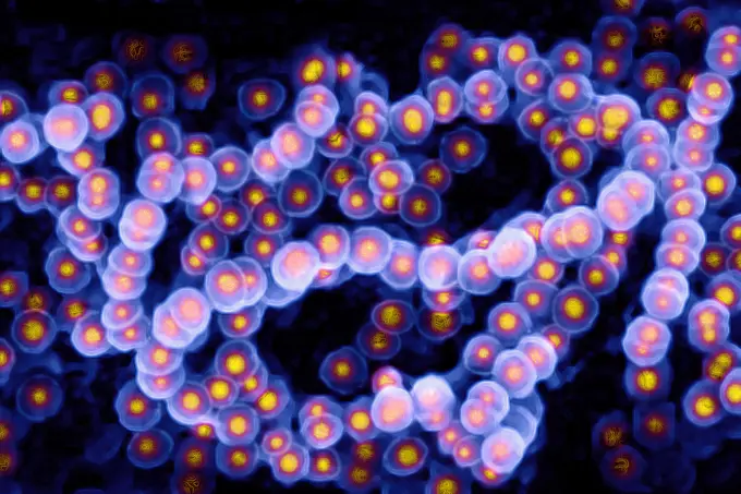
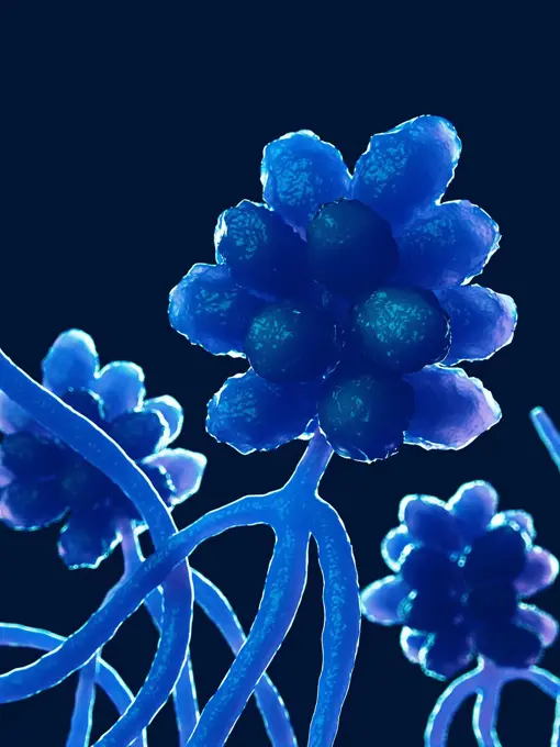
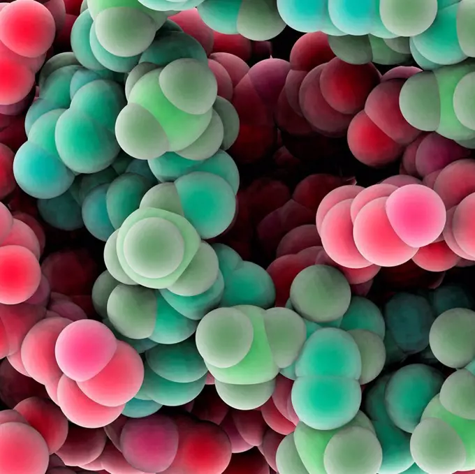
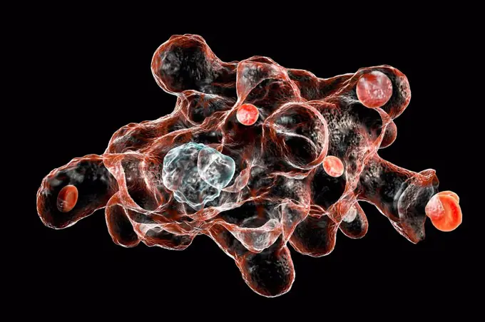
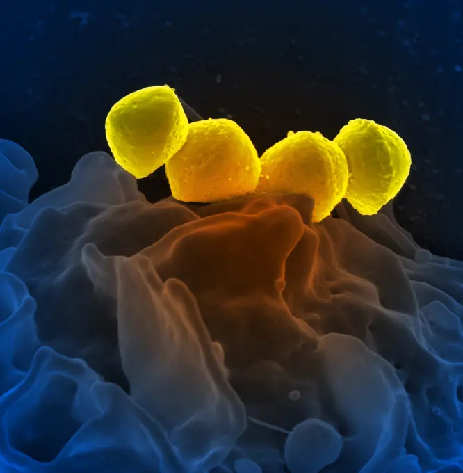
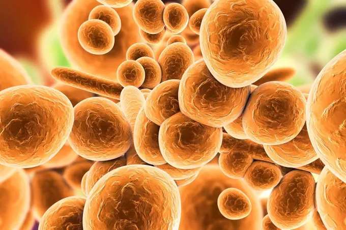
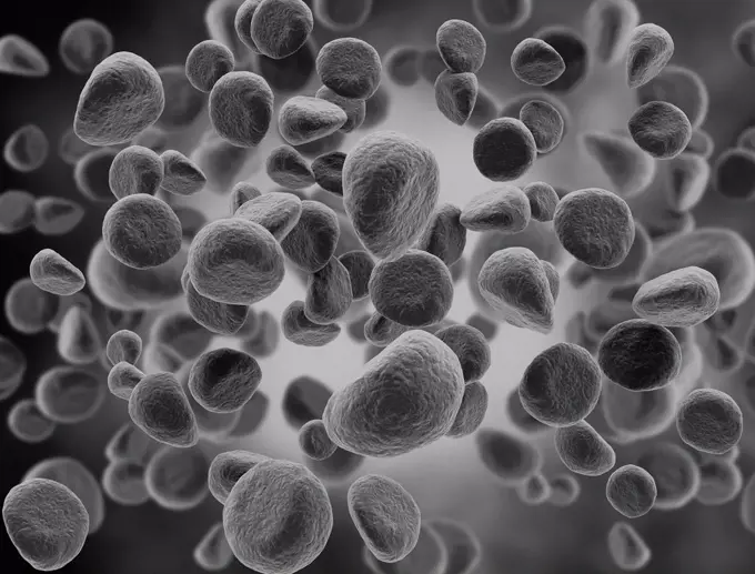
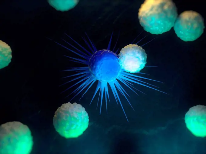
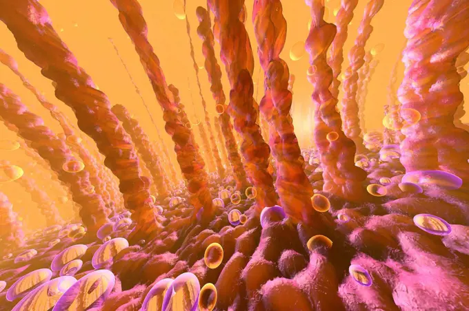
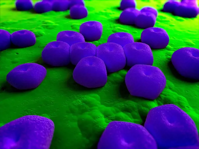
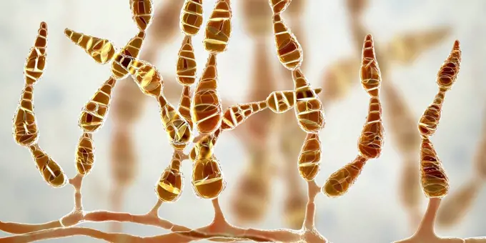
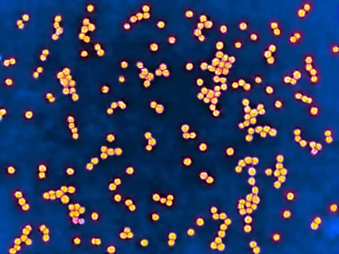
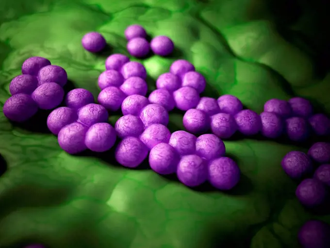
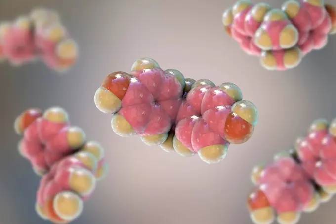
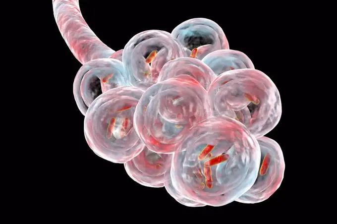
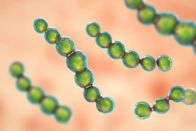
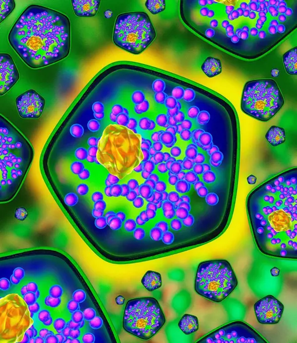
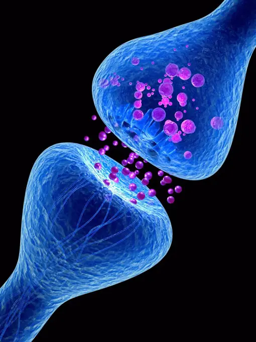
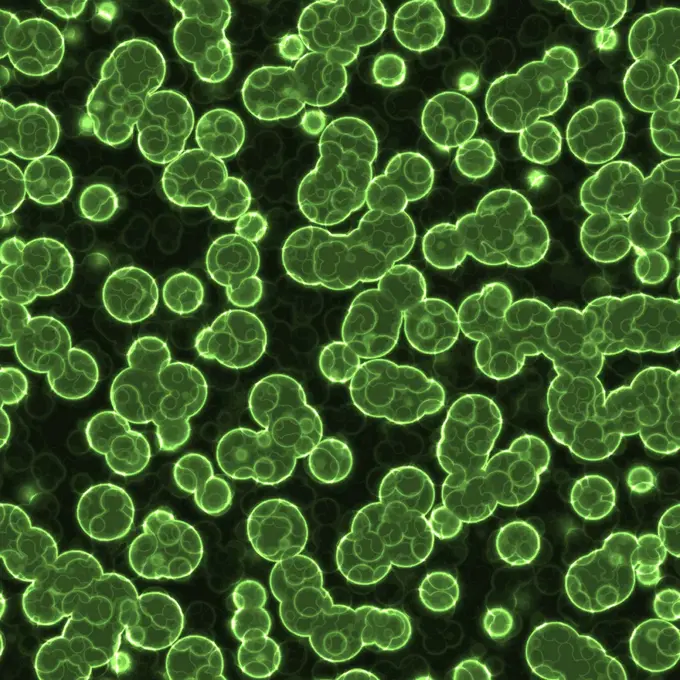
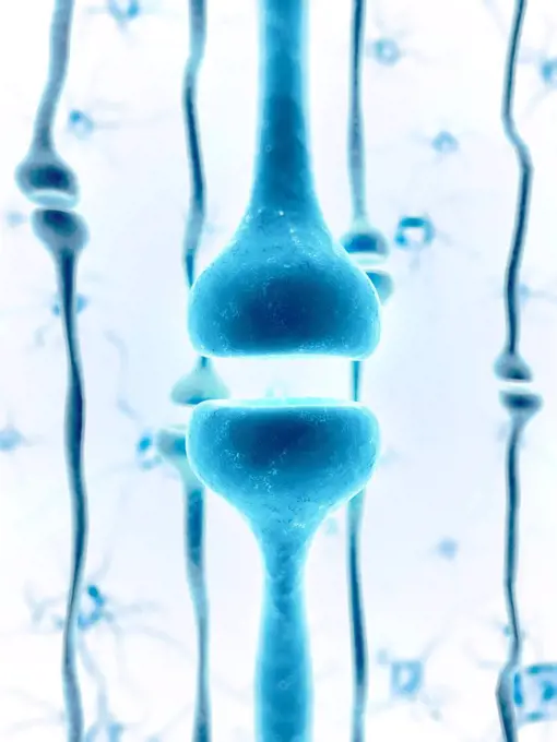
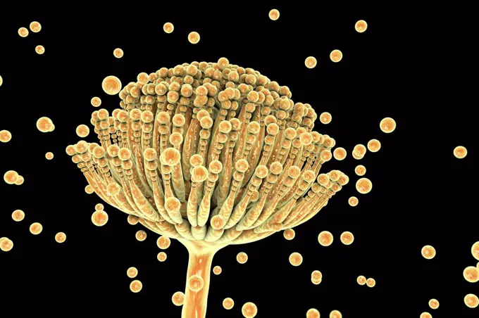
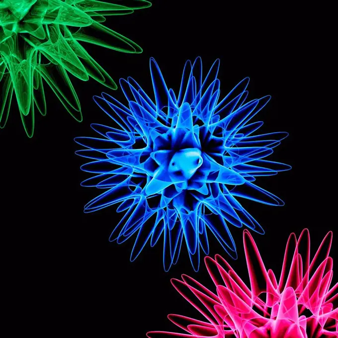
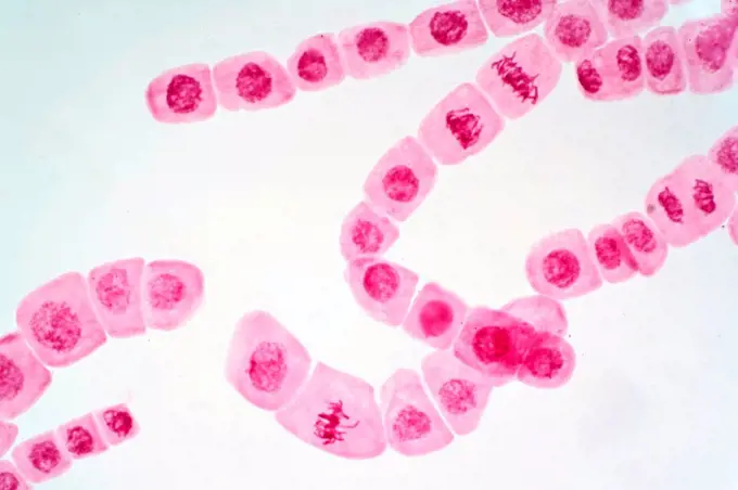
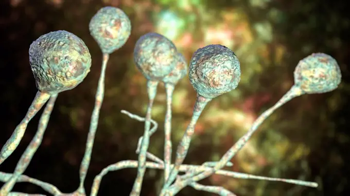
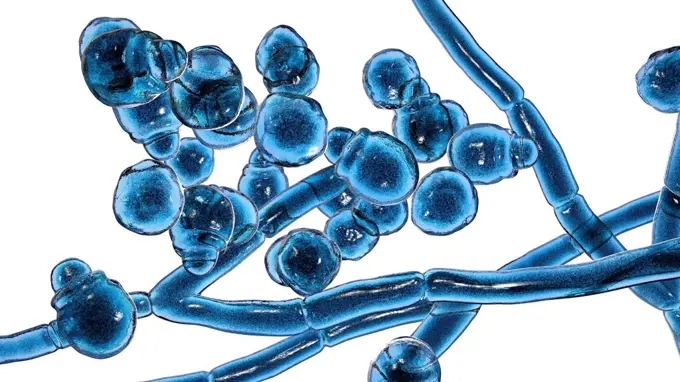
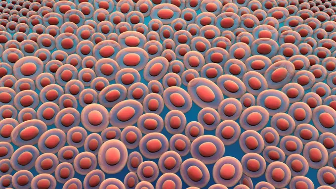
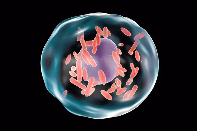
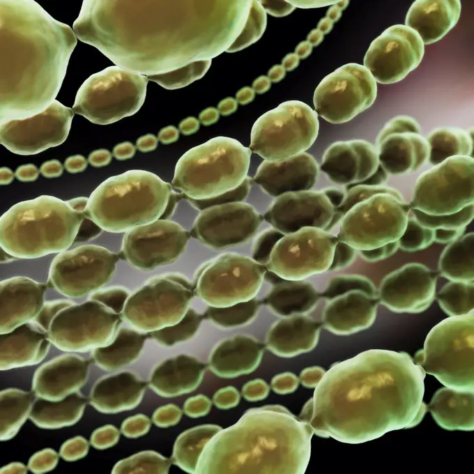
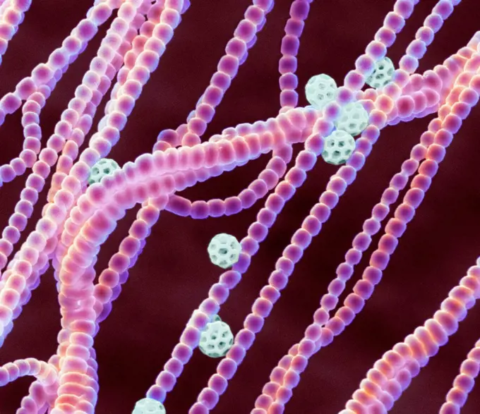
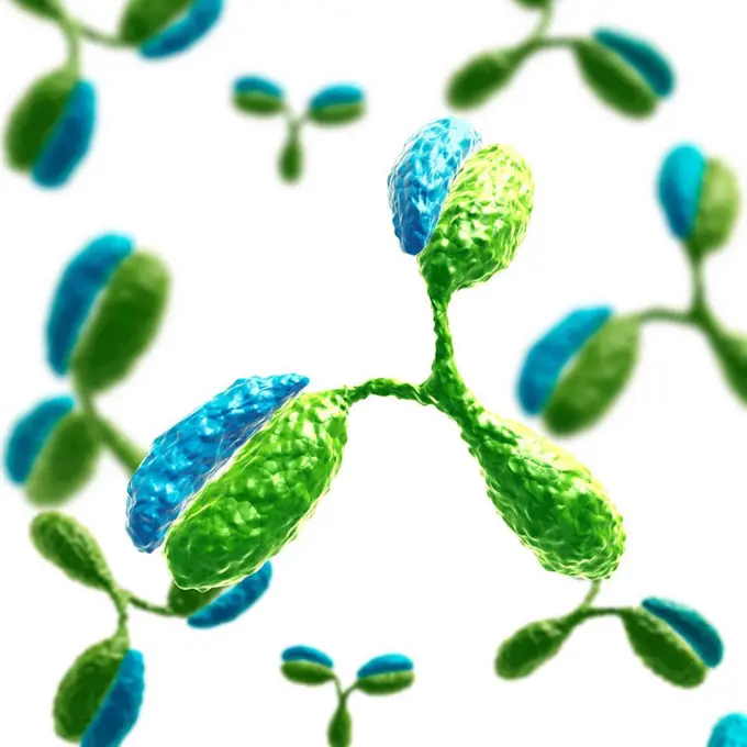
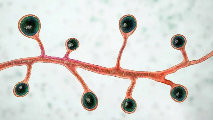
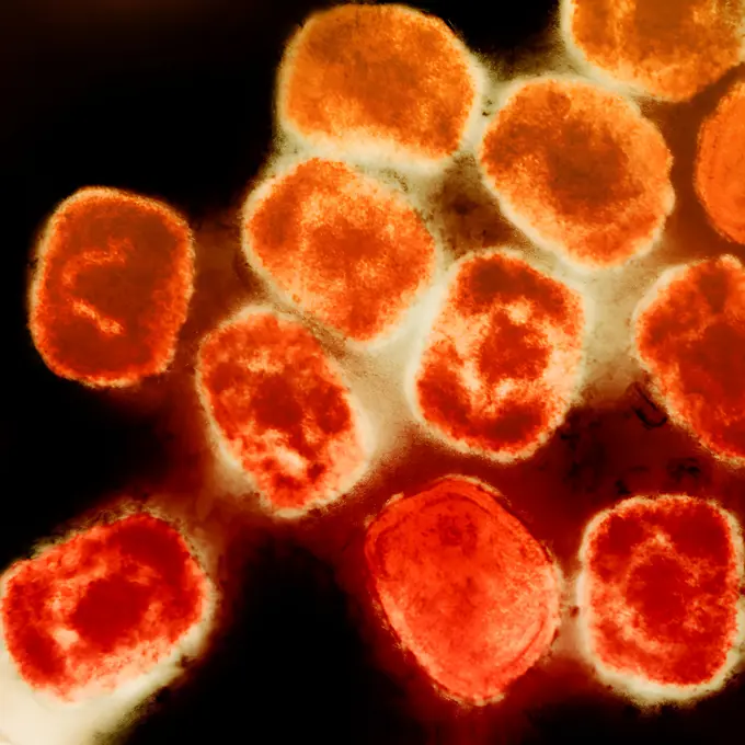
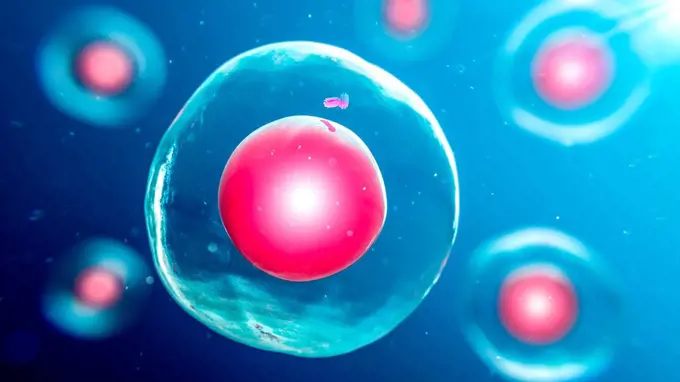
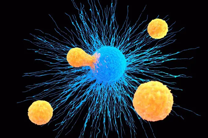
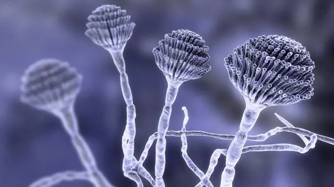
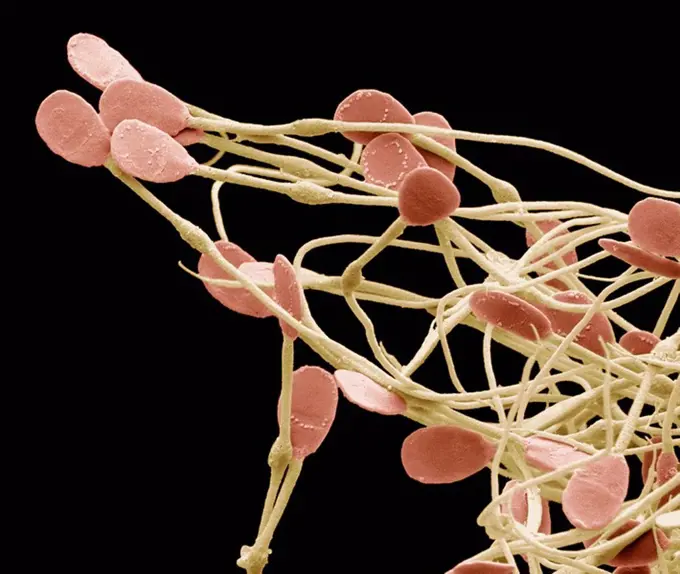
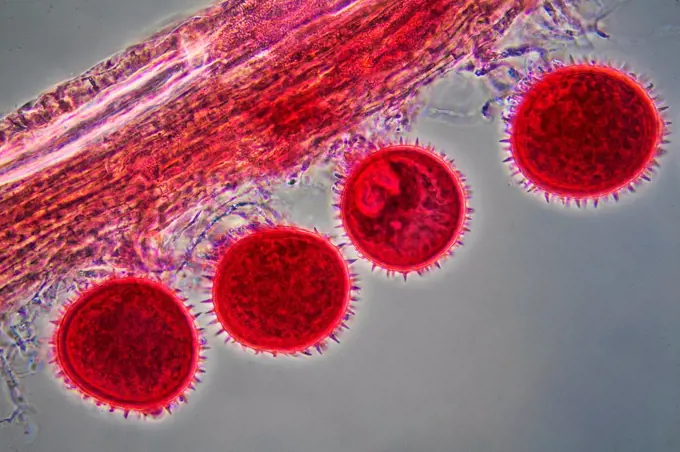
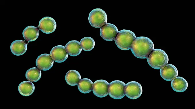
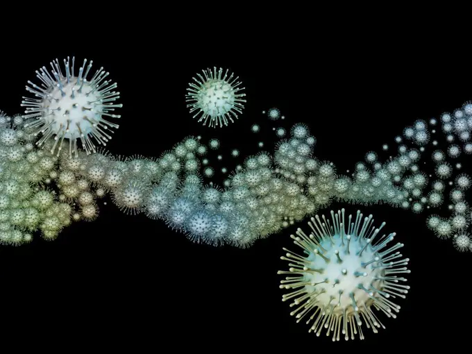
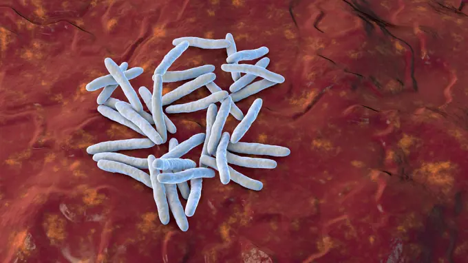
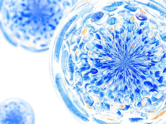
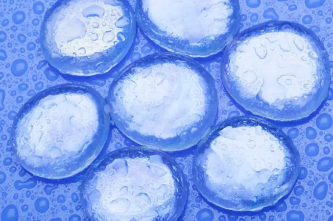
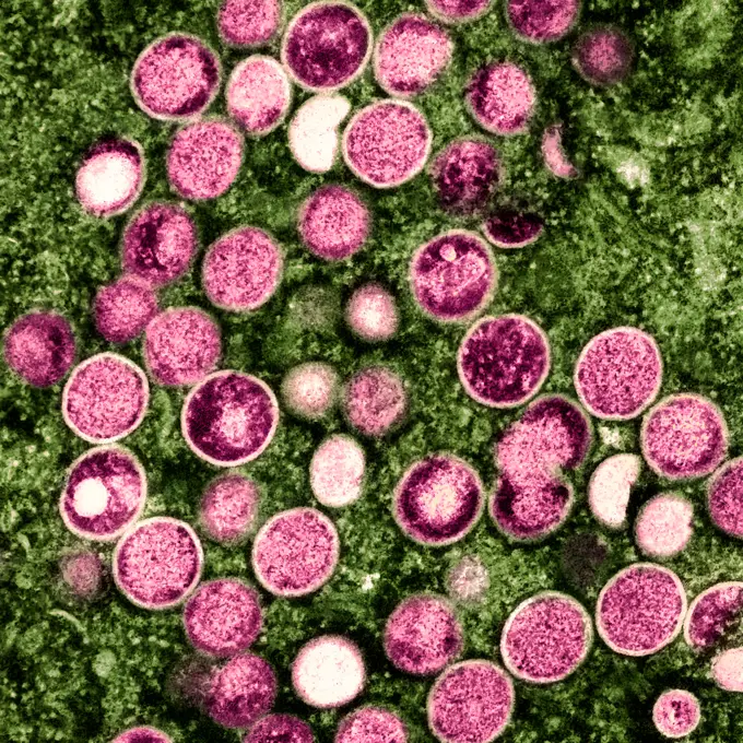
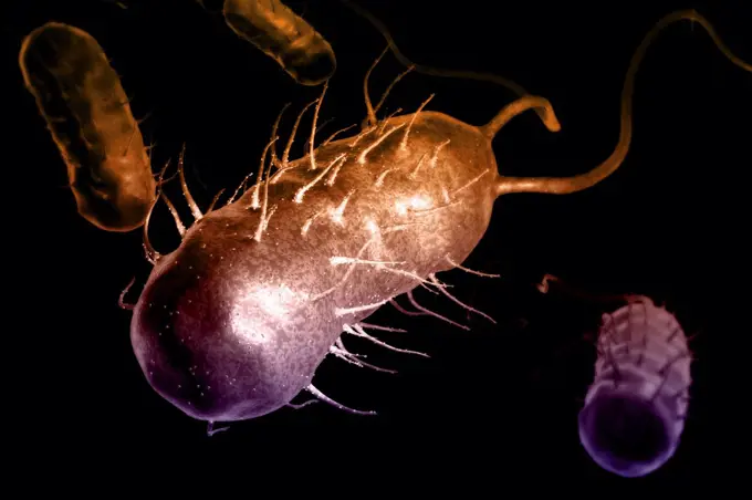
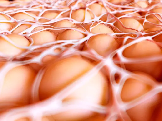
Fungal and Bacterial Illustrations

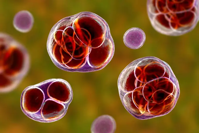
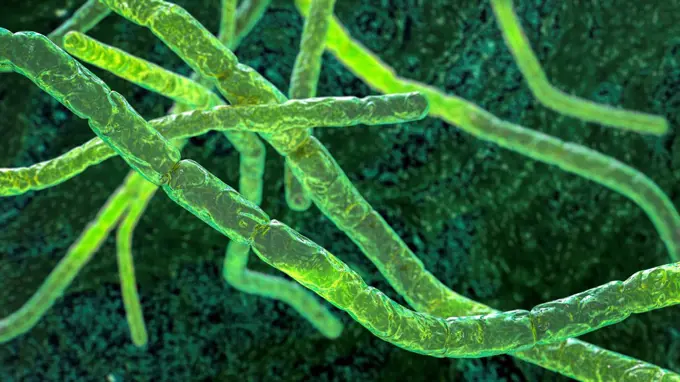
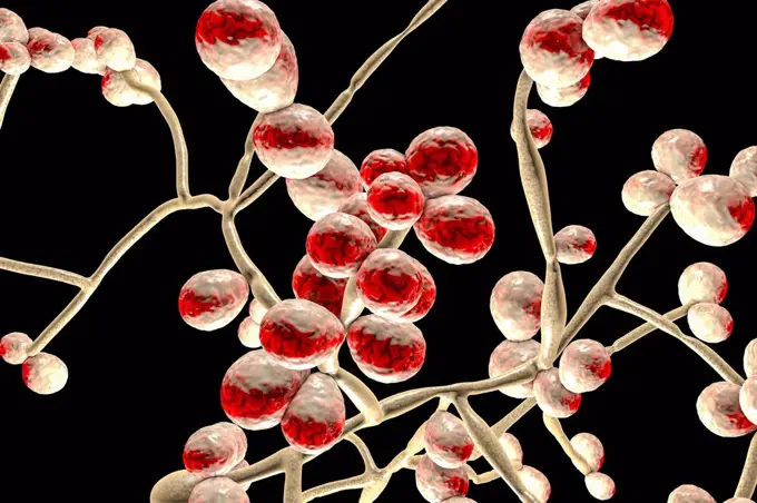
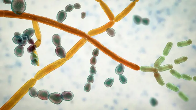
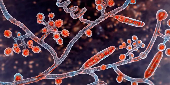
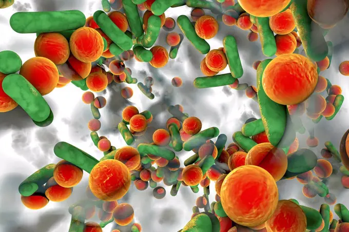
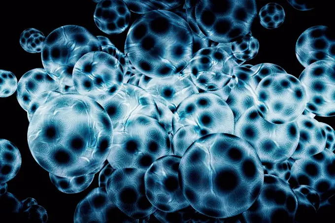
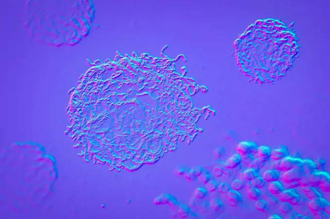
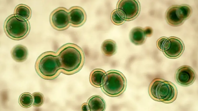
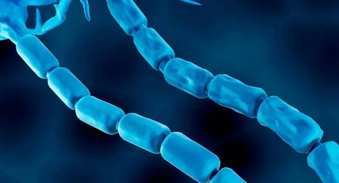
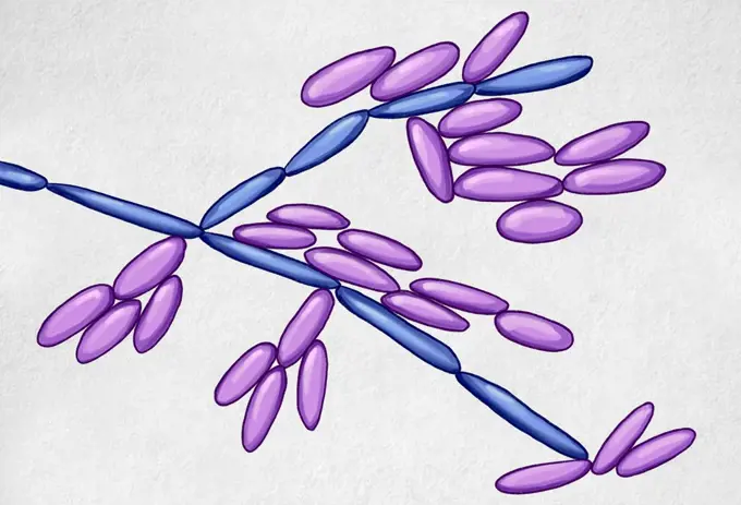
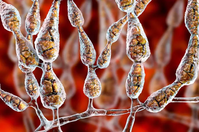
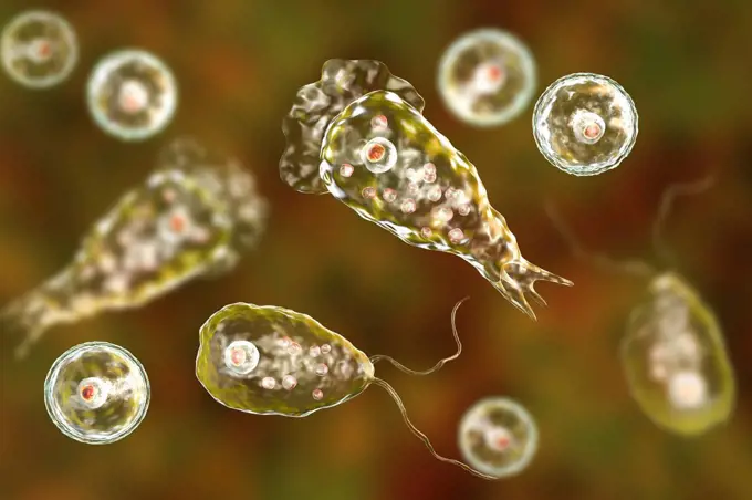
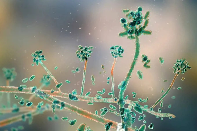
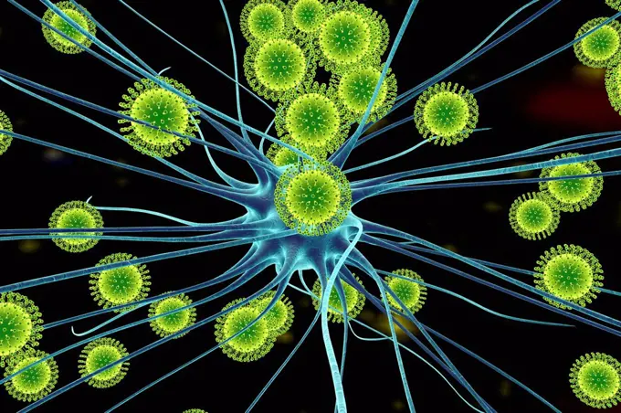
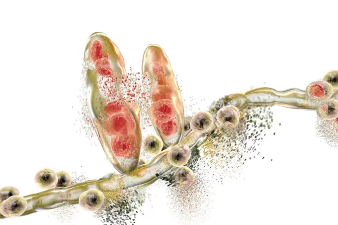
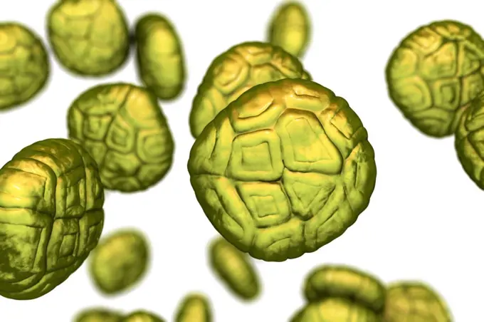
Computer-generated images of yeast, fungi, and bacteria in vibrant colors illustrating their structures. The mood is educational and intriguing.

Candida auris fungi, illustration

Assets in this Story

4128-16506791
Fungus Sporothrix schenckii, causative agent of the infection sporotrichosis, computer illustration. Fungal threads of the vegetative mycelium are see...
4128R-15405925
Yeast Saccharomyces cerevisiae, illustration
4128R-13682066
Spherical bacteria and rod-shaped bacteria inside biofilm with quorum sensing molecules (small red circles), illustration. A biofilm is a colony of ba...
4128R-15405892
Candida auris fungi, illustration
4128-19056144
Fungi Paracoccidioides braziliensis, illustration
4128-28767231
Prototheca wickerhamii algae, illustration
4128R-15350
Bacteria, conceptual computer artwork.
1848-54783080
Streptococcus bacteria, a dangerous pathogen, illustration
1848-61039601
Fake microorganisms. microbiology. Bacteria, cells, viruses, germs, microorganisms
4128-111493633
Cellular communication, conceptual illustration.
4128R-13940893
Mycoplasma bacteria, computer illustration. Mycoplasmas are the smallest cellular organisms known (diameter 0.3-0.9 microns). They resemble bacteria i...
4128-20041206
Thymol molecule, illustration
4128R-14181703
Nuclear division, illustration
4128R-15405891
Candida auris fungi, illustration
4128-19358267
Aspergillus clavatus mould fungus, computer illustration. A soil microscopic fungus, causes allergic aspergillosis, lung infections, endocarditis, ext...
4128R-14846661
Peptococcus bacteria, illustration
6188-65590988
micro bio An illustration of a black and purple micro bio Copyright: xZoonar.com/magannx 1120103
4128-V58569645
4128-28767237
Prototheca wickerhamii algae, illustration
824-57657644
Coronavirus (CoV). viruses of the Coronaviridae family and the Orthocoronavirinae subfamily. It is a pathogen of respiratory syndromes. Viewed from a transmission electron microscopy (TEM) image. Viral diameter 80nm to 100nm.
1525-56324715
abstract light blue background with oil circles . bubbles of water close up . oil bubbles in the water macro.. abstract light blue background with oil circles . bubbles of water close up .
4128-18799605
Nocardia bacteria, illustration
4128R-15405881
Candida albicans yeast and hyphae stages, illustration
4128-19490109
Trichosporon fungus, illustration
4128-30420521
Lacazia loboi fungi, illustration
4128-24796398
Emmonsia fungi, illustration
4128-20044381
Curcumin molecule, illustration
4128-19489836
Blastomyces fungus, illustration
1899-61460418
Coronavirus (CoV). viruses of the Coronaviridae family and the Orthocoronavirinae subfamily. It is a pathogen of respiratory syndromes. Viewed from a transmission electron microscopy (TEM) image. Viral diameter 80nm to 100nm
4128-111494713
Bacteriophages, illustration.
6188-66486085
3d-Illustration einer Krebszelle im Prozess der Mitose 3d illustration of a cancer cell in the process of mitosis Copyright: xZoonar.com/ChristophxBurgstedtx 22601106
4128-19358659
Glucagon hormone molecule. Computer model showing the structure of the human hormone glucagon. Glucagon is involved in carbohydrate metabolism and is ...
4128R-15366403
Aspergillus fumigatus fungus, illustration
824-63227043
Scanning electron micrograph of Lassa virus budding from a cell. Lassa virus (LASV) is an arenavirus that causes Lassa hemorrhagic fever, a type of viral hemorrhagic fever (VHF) in humans and other primates. Image requested from the NIAID Integrated Research Facility (IRF) in Fort Detrick, Maryland. Credit: NIAID
4128R-15405863
Balamuthia amoeba, illustration
1848-51348804
Shape of bacterial cell: cocci, bacilli, spirilla bacteria background
4128-V58562026
4128-V58562069
4128-18498474
Leaf epidermis, light micrograph
4128R-11291613
Artwork of the E. coli bacteria.
4128R-13345
Molecular model, computer artwork.
4128-19358163
Bone marrow, illustration. Bone marrow contains stem cells which, through the process of haematopoiesis, generate red and white blood cells and platel...
4128-20044925
Molluscum contagiosum virus, illustration
4128-30420520
Lacazia loboi fungi, illustration
4128-19054696
Scopulariopsis brevicaulis fungus, illustration
1525-22331109
Close-up of bubbles in water
4128-19489870
Blastomyces fungus, illustration
1899-61460535
Naegleriasis (or amoebic meningoencephalitis or AMP) is a fatal infection of the brain by the single-celled, free-living eukaryote Naegleria fowleri
824-63217835
Picornavirus (picornavirid), responsible for: Colds, gastroenteritis, poliomyelitis and meningitis. Image made from a transmission electron microscopy view.
4128-V58573459
4128-19054724
Microsporum canis fungus, illustration
4128-20040230
Blood vessels, illustration
1525-24143571
Fake microorganisms. microbiology. Bacteria, cells, viruses, germs, microorganisms.
4413-88867
Cross section of flower bud Cotton
4128-30417369
Stomach cancer, illustration
4128R-26270
MRSA bacteria. Computer artwork of methicillin-resistant Staphylococcus aureus (MRSA) bacteria.
4128-28767330
Saksenaea microscopic fungi, illustration
4128-111579946
Group of viruses, computer illustration.
4128-30416511
Circulating blood cells, illustration
1899-65662492
Colorized scanning electron micrograph of a cell (teal) infected with the Omicron strain of SARS-CoV-2 virus particles (yellow), isolated from a patient sample.
4128R-13682499
Vibrio cholerae and flagella, illustration.
4128-20041301
Athlete's foot fungus, illustration
4128R-13682059
Spherical bacteria and rod-shaped bacteria inside a biofilm, illustration. A biofilm is a colony of bacteria that forms a coating on a surface. Common...
4128-15663085
Quantum foam, conceptual illustration
4128R-13682125
Close-up view of molecules, computer illustration.
4128R-10080
Tuberculosis bacteria, artwork
4128-20244185
Epidermophyton floccosum dermatophyte fungus , illustration
4128-19489874
Blastomyces fungus, illustration
4128-20044969
Smallpox virus, illustration
824-63190270
Staphylococcus epidermidis, an opportunistic bacterium. It is responsible for acute cystitis in its staphylococcus saprophyticus form. Image taken with an optical microscope X 1000.
824-63191240
Scanning electron micrograph of Streptococcus pyogenesis bacteria (yellow) bound to a human neutrophil (blue).
4128-19489919
Blastomyces fungus, illustration
824-63208130
Streptococci (Streptococcus pyogenes). Image made from a microscope view.
4128-30417999
Stachybotrys mould, illustration
4128R-12699902
Coloured scanning electron micrograph (SEM) of fungal cells. At centre is a sporangiaum, which houses the fungus' reproductive cells (spores). Magnifi...
4128R-11476177
Protein molecules, computer artwork.
6214-V64217903
4128R-13928459
Parasitic amoeba (Entamoeba histolytica) engulfing red blood cells, computer illustration. This single-celled organism causes amoebic dysentery and ul...
1848-53914872
Viruses, illustration
4141-16145
green alga volvox aureus senescent mother colony releasing daughter colonies
4297-1755
Colorized scanning electron micrograph of a single Gram-negative Escherichia coli bacterium
6188-56042222
cells a large background image of cells or bacteria under the microscope Copyright: xZoonar.com/PhilxMorleyx 1191676
4269-21307946
Drug-resistant Salmonella bacteria. 3D computer illustration of drug-resistant non-typhoidal Salmonella sp. bacteria, showing their flagella (long filaments) and fimbriae (hair-like). Non-typhoidal Salmonella usually causes diarrhoea, fever, and abdominal cramps. Some infections spread to the blood and can have life-threatening complications. These bacteria are showing resistance to multiple classes of drugs including ceftriaxone and ciprofloxacin.
4128-18498476
Leaf epidermis, light micrograph
4128R-15536506
Illustration of human receptors
4128-28968444
Saccharomyces yeast, illustration
1899-53513335
Scanning electron micrograph of Group A Streptococcus (Streptococcus pyogenes) bacteria on primary human neutrophil.
824-63227407
Streptococcus pyogenesis bound to a human neutrophil.
4128R-13409393
Candida auris fungi, illustration
4128R-13666942
Cancer cells, illustration.
4128R-14862
Immune response to cancer. Computer artwork of white blood cells green attacking a cancerous cell blue.
4128-20039389
Monkeypox virus particles, illustration
4128-19054302
Cancer cells, illustration
4128-19489896
Blastomyces fungus, illustration
4128-19357730
Illustration of damaged myelin sheath in multiple sclerosis (MS).
4128-19358300
Candida kefyr (formerly Candida pseudotropicalis) yeasts, computer illustration. A yeast fungus with its teleomorph currently recognized as Kluyveromy...
4128-V58572063
4128R-22436
Multicellular organism, conceptual computer artwork.
4128-V58574251
6188-65540567
Kette von Aminosäure- oder Biomolekülen, genannt Protein - 3d-Illustration Chain of amino acid or bio molecules called protein - 3d illustration Copyright: xZoonar.com/ChristophxBurgstedtx 22579614
824-63178979
Bacillus anthracis. This bacterium causes anthrax with malignant lesions that turn into eschars. If the contamination is not treated the illness spreads through the lymphatic system and the septicemia is rapidly deadly. Optical microscope x 2000.
4128-28515295
Monkeypox virus particles, illustration
4128-20040240
Microaneurysms, illustration
4128-111612480
Filamentous allergenic fungus Alternaria alternata, computer illustration. Alternaria is a dematiaceous (phaeoid) fungus commonly isolated from plants...
4128R-14845699
Naegleria brain-eating amoeba forms, illustration
4128-19358241
Acremonium (formerly Cephalosporium) mould fungus , computer illustration. Acremonium is a hyaline hyphomycete (glassy, lightly pigmented conidial fun...
4128R-13193044
Zika viruses infecting neurons, computer illustration. This is an RNA (ribonucleic acid) virus from the Flaviviridae family. It is transmitted to huma...
4378-2655
Clusters of 'superbug' (MRSA) bacteria.
4128-28515071
Bacteria cultured on Petri dish
1525-24125058
chocolate round donuts with multicolored sugar sprinkles levitate in a cloud of brown cocoa on a black background. Powder flies up
4128R-13763521
Synapse. Computer illustration of a synapse, the junction between two neurons. Synapses transmit electrical signals from one nerve cell to the next. W...
824-63227206
The purple pods you see in this scanning electron micrograph are the H5N2 avian influenza virus, an averted threat to the poultry and egg industry and, in very rare cases, a risk to human health. However, these particular pods are unlikely to infect anything as they are trapped in a gray mesh of carbon nanotubes. Made by hanging carbon atoms in a cylindrical pattern, these nanotubes are about 10,000 times smaller than the width of a human hair.
1815R-14647093
3D Illustration of lung or liver cells with oxygen bubbles floating within
4128R-15652
Acinetobacter sp. bacteria, computer artwork.
4128-111612478
Filamentous allergenic fungus Alternaria alternata, computer illustration. Alternaria is a dematiaceous (phaeoid) fungus commonly isolated from plants...
824-63179063
Staphylococcus (Staphylococcus aureus), the bacterium that causes food poisoning, localised suppurating infections, and in some extreme cases, septicemia. Optical microscopy x 2000.
1848-50209050
Christmas Illuminations at Galeries Lafayette, Boulevard Hausmanns in Paris, France, Europe
4378-3712
A single mitochondrion. These organelles act as cell power plants as they generate adenosine triphosphate (ATP), a source of chemical energy.
4128R-11544227
Bacteria, computer illustration.
1899-53511535
Plasmodium falciparum is a malaria-causing parasite or malaria, transmitted by the bite of female anopheles which releases merozoites into the blood which enter red blood cells. Optical microscope view.
4413-153999
Air bubbles in the ice France
4128R-13385222
Mrsa bacteria, illustration.
4128-28767087
Bisphenol A molecule, illustration
4239-18641826
3D illustration of a fluconazole-resistant species from the genus of fungal organisms, Candida. In this view, you will note that there were clusters, as well as single blastoconidia, which are the asexually produced reproductive cells that are produced through the process of budding, that were attached to the septate hyphal filaments. This artistic recreation is based upon scanning electron microscopic (SEM) imagery.
4128R-15366366
Bacterial pneumonia, illustration
4128-30420517
Lacazia loboi fungi, illustration
4128R-13023001
Illustration of rod-shaped bacteria. Rod-shaped bacteria include Escherichia coli, Salmonella, Shigella, Klebsiella, Citrobacter, Enterobacter, Yersin...
824-63191193
3D illustration made from SEM imagery. This illustration depicts a three-dimensional (3D) computer-generated image of a fluconazole-resistant species from the genus of fungal organisms, Candida. In this particular view, you'll note that there are clusters, as well single blastoconidia, which are the asexually-produced reproductive cells that are produced through the process of budding, which are attached to the septate hyphal filaments. The artistic recreation was based upon scanning electron micrographic imagery. Some Candida strains are increasingly resistant to first-line and second-line antifungal treatment agents. Recent data demonstrate a marked shift among infections towards Candida species with increased resistance to antifungal drugs including azoles and echinocandins. CDC conducts multicenter surveillance for antifungal resistance in the United States, candidal infections, their economic impact, and possible areas where prevention and control strategies can be focused.
4413-174714
Stamens and stigma of horsebean by light microscopy
1899-61461211
Meningococcus or meningococcal disease or Neisseria meningitidis, is a bacteria involved in meningitis. These bacteria are the cause of mild pharyngitis and only one person in approximately 400 becomes the victim of a serious meningococcal infection, most often in the form of acute purulent meningitis. Image produced from optical microscopy
4128-V58566845
4128-28970687
Cancer cells dividing, illustration
4128R-10271
Plant cells, artwork
4128R-13763505
Synapse. Computer illustration of a synapse, the junction between two neurons. Synapses transmit electrical signals from one nerve cell to the next. W...
5507-41010492
cells
1525-20461319
sperm and egg cell on monitor in laboratory
4269-7094
Colorized transmission electron micrograph of avian influenza A H5N1 viruses (seen in drak) grown in Madin-Darby canine kidney (MDCK) cells (seen in pale).
4378-20013727
Legionella Pneumophila Bacteria
4128-V58573455
4128-V58562013
4128R-11477607
Human synapse, compute artwork.
6188-63385894
Kiwischeiben Farbvariante Kiwischeiben als Farbvariante Copyright: xZoonar.com/angetax 5575340
1525-24066977
Jellyfish floating in the ocean. Macro view. Jellyfish floating in ocean
4378-20365103
Anopheles Mosquito
4128R-13587432
Aspergillus fungus, computer illustration. This is a toxic fungus that causes ear, lung and skin infections (lobomycosis and pulmonary aspergillosis, ...
4128R-12577703
Pollen, computer illustration.
4128-18680655
Mitosis, light micrograph
4128-18301070
Mucor mould, illustration
4128-15818412
Malassezia skin fungus. Computer illustration of spore clusters from the fungus Malassezia furfur. Malassezia fungi are naturally found on the skin su...
4128-111495126
Tumour, illustration.
1525-23993819
Cellular Division and Growth of some cell like lifeform.. Cellular Division and Growth
4128R-13282698
Q fever bacteria (Coxiella burnetii, green rods) inside a cell, computer illustration. Coxiella burnetiiis an obligate intracellular bacterium, meanin...
4378-1896
Circular-shaped bacteria (coccus).
4128R-13474
Evening primrose pollen. Coloured scanning electron micrograph (SEM) of evening primrose (Oenothera sp.) pollen, showing the characteristic threads se...
4128-V58562080
4128R-13447006
Illustration of antibody 1IGT molecules.
4128-19489832
Blastomyces fungus, illustration
824-57657684
Colorized transmission electron micrograph of monkeypox virus particles (orange) cultivated and purified from cell culture.
4128R-14846674
Peptostreptococcus bacteria, illustration
4128-20042823
Parent cell, illustration
4239-18641849
3D illustration of a group of gram-negative, Chlamydia psittaci bacteria. This artistic recreation is based upon scanning electron microscopic (SEM) imagery.
4128-20042765
Thiomargarita magnifica compared to Vibrio sp., illustration
4128-20038753
Egg cell surrounded by sperm cells, illustration
4378-1834
Genetic determining human chromosomes.
4128-19358184
Embryonic stem cells, illustration.
4128R-13928414
Parasitic Amoeba protozoan engulfing bacteria, computer illustration. The amoeba will engulf and digest the prey in a process called phagocytosis.
1525-24199562
bright bubbles water with drops
5507-49090870
a close up view of an orange and blue neuron
4128R-13619908
Destruction of athlete's foot fungus Trichophyton mentagrophytes by nanoparticles, computer illustration. Conceptual image for Athlete's foot treatmen...
4201-66380
Garden Four-o'clock (Mirabilis jalapa) SEM close-up view of pollen at 210x magnification
4128R-13763900
Computer illustration of Haemophilus influenzae, coccobacillus bacteria. These bacteria cause a number of disease including meningitis in children, pn...
4128-30416212
Human cell, illustration
4128-19358171
Helicobacter pylori bacteria, illustration. Helicobacter pylori is a gastrointestinal bacterium that can affect the stomach lining and cause ulcers or...
6188-54819430
structure, pattern and crystals of newly formed ice structure, pattern and crystals of newly formed ice called the nylas black ice, skin Copyright: xZoonar.com/MaximilianxBuzunx 13446079
4128R-14060531
Toxoplasma gondii parasites, illustration
1848-61037820
Electric flash effect. Thunderbolt light and storm sparks, realistic Energy lighting voltage power charge and electricity strike
4128-28970689
Cancer cells dividing, illustration
4128-19056134
Fungi Paracoccidioides braziliensis, illustration
4128R-14324172
Smallpox virus, illustration
1848-53914848
Bacteria strung together, illustration
1788-20761632
Pollen grains of winter jasmine (Jasminum nudiflorum) viewed under a microscope.
4128-16073936
T-cell attaching to cancer cell, illustration
4128-19358297
Aspergillus fungus, computer illustration. This is a toxic fungus that causes ear, lung and skin infections (lobomycosis and pulmonary aspergillosis, ...
4128R-10581
Immature dog sperm, SEM
4128R-14717
Chromosomes, computer artwork.
4413-134995
Mallow Stigma flat with pollen germinating
4128R-10375
Bronchial tree, artwork
4128R-13620011
Giardia lamblia, a protozoan (cyst stage) found in water contaminated by animal faeces, coloured scanning electron micrograph (SEM). Causes the protoz...
4269-24897
Fertilization. Ovule et spermatozoon;Sperms Computer-generated image
4128R-13048927
Computer illustration of Trichophyton mentagrophytes, the cause of athlete's foot (tinea pedis) and scalp ringworm (tinea capitus). Both of these cont...
4128-30420515
Lacazia loboi fungi, illustration
4128-V58562475
1899-61460369
Colorized transmission electron micrograph of monkeypox particles (pink) found within an infected cell (green), cultured in the laboratory
1525-27143777
Coronavirus Micro World. Viral Epidemic series. 3D Illustration of Coronavirus particles and micro space elements relevant for virus, epidemic, infection, disease and health
4128-28575401
Tuberculosis bacteria, illustration
4128-111553082
Cell, conceptual illustration
4239-69154252
Conceptual biomedical illustration of the fungal infection mucormycosis.
4128-18182220
Helicobacter pylori bacterium, illustration
4128-111493730
Streptococci bacteria, illustration. Streptococcus species are responsible for pharyngitis, meningitis, bacterial pneumonia, endocarditis and necrotiz...
4128-24796391
Emmonsia fungi, illustration
4128R-14057481
Hepatitis A viruses in intestine, illustration
4239-V53647277
1525R-76465
Water droplets
824-57657562
Colorized transmission electron micrograph of monkeypox particles (pink) found within an infected cell (green), cultured in the laboratory.
4378-967
Scanning electromicrograph styled rod shaped bacteria.
4128R-13587298
Fatcells, illustration.
4128-V58557871
4128-18300720
Mucor mould, illustration
4128-30420528
Lacazia loboi fungi, illustration
4128R-13048917
Computer illustration of staphylococci bacteria (Staphylococcus aureus).
1525-24477785
Electric flash effect. Thunderbolt light and storm sparks, realistic Energy lighting voltage power charge and electricity strike
4128R-15290703
Ebola virus, illustration
4128R-13941172
Acetobacter bacteria, illustration.
4128R-13818070
Leishmania sp. protozoa, computer illustration. This parasite causes the tropical disease leishmaniasis. This can take several forms, causing open sor...
4128R-13763534
Synapse. Computer illustration of a synapse, the junction between two neurons. Synapses transmit electrical signals from one nerve cell to the next. W...
4128-19489292
Monkeypox virus emerging from cell, illustration
4128-18799877
Tissue infected with pathogenic microorganisms
6188-67192055
Close up of small, multi -year tomatoes that hang in bubbling water. The background is dark. There are many air bubbles to be seen. Close up of small, multi -year tomatoes that hang in bubbling water. The background is dark. There are many air bubbles to be seen.
6188-65540163
Mikroskopische Bild der wachsenden Schimmel oder Schimmelpilz und Sporen - 3d Illustration Microscopic image of growing molds or mold fungus and spores - 3d illustration Copyright: xZoonar.com/ChristophxBurgstedtx 22576025
1525-22389673
Goggles
4128R-20192
Nerve cells, computer artwork.
4128R-21200
Giardia protozoan, artwork
4128-111493979
Computer illustration of tardigrades surrounded by algae. Tardigrades can feed on algae.
4128R-13620012
Giardia lamblia, a protozoan (cyst stage) found in water contaminated by animal faeces, coloured scanning electron micrograph (SEM). Causes the protoz...
4128R-15405515
Pollen grains of Mimosa flower, illustration
4128R-11310281
Helicobacter pylori bacterium, computer artwork.
4128R-13619900
Destruction of athlete's foot fungus Trichophyton mentagrophytes, computer illustration. Conceptual image for Athlete's foot treatment and prevention....
1848-50833028
Air bubbles in the ice, Germany, Europe
1525-24240806
Abstract molecular structure from shiny glass spheres. Vector realistic set of 3d molecules, chemical or biotechnology models with transparent crystal balls isolated on blue background. Abstract molecules from shiny glass spheres
4269-12753
Food supplement capsules.
4128R-15405514
Pollen grains of Mimosa flower, illustration
4128R-22571
Chromosome, computer artwork.
4128-20244038
Microbes, conceptual illustration
4128R-14846753
Veillonella bacteria, illustration
4128R-13282754
Trichina spiralis roundworm (formerly called Trichinella spiralis), computer illustration. These parasites infect mammals, including humans, pigs, dog...
4128R-11310225
Lyme disease bacteria (Borrelia burgdorferi), computer artwork.
1525-26586493
Blood spatters realistic bloodstains. Blood splatters isolated on black. Blood splashes drops and trail.
1848-54783076
Chromosomes during research, genetic testing, illustration
4128-V58560637
1848-54798932
Illustration, walnuts
1525-20588779
Chromosome as a human biology x structure containing dna genetic information as a medical symbol for gene therapy or microbiology genetics research.
4128-30418017
Scabies mite on human skin, illustration
4128-17927500
Cross-reactive HIV antibody fragment, illustration
4128-20244039
Alien microbe from space, conceptual illustration
4239R-8004
Microscopic view of HIV virus, cross section.
4128-15666335
Gallbladder, illustration
4128R-13282752
Trichina spiralis roundworm (formerly called Trichinella spiralis), computer illustration. These parasites infect mammals, including humans, pigs, dog...
1525-22197423
cell division
4128-111493995
Computer illustration of giardiasis, an infection in the small intestine caused by the parasite giardia lamblia. The condition can spread via contact ...
4128R-14846635
Nerve cells, computer illustration and micrograph
4128-19489824
Blastomyces fungus, illustration
4128R-13575506
Destruction of Candida fungi, computer illustration. Image can be used to illustrate antifungal properties of different types of nanoparticles.
1848-49273084
The synaptic cleft is the space between two neurons. It connects the presynaptic neuron with the postsynaptic neuron and ensures that the transmission of excitation can take place between the two
4128-15665343
Liver tumour, illustration
4413-109860
Microscopic view of vinegar worm
4239-69154274
Conceptual biomedical illustration of Streptobacillus moniliformis bacteria, on black background.
1525-25604755
Human Arm. Hand Model, 3D Geometric Design. Can Be Used for Science, Technology, Medicine, Hi-Tech, Sci-Fi. Human Arm. Hand Model, 3D Geometric Design. Can Be Used for Science, Technology, Medicine, Hi-Tech, Sci-Fi - Illustration Vector
824-65830675
Colorized electron micrograph of Treponema pallidum, the bacteria that cause syphilis. Several spiral-shaped bacteria have been highlighted in gold.
6188-55642962
Kieselalgen Kieselalgen Diatomeen im Wasser Copyright: xZoonar.com/Dr.xLangex 13381632
4128-19056108
Corynebacterium diphtheriae, illustration
4128R-12923321
Gall stones, illustration
4128-V58567136
4378-4507
Bronchioles
1525R-7278
4128R-22261
Computer virus, conceptual computer artwork.
4378-4461
Birch Tree Pollen
4128R-13575561
Illustration of Helicobacter pylori bacterium. It is a curved, or spiral, Gram-negative bacilli with flagella at one end (seen here) that make it high...
1525-27373088
Virus realistic vector banner template. Virology research. Infection pathogen 3d mock up design. Bacteria cell. Microbiology advertisement horizontal printable flyer, brochure with text space. Virus realistic vector banner template
4128R-13326021
White blood cell engulfing bacteria, illustration.