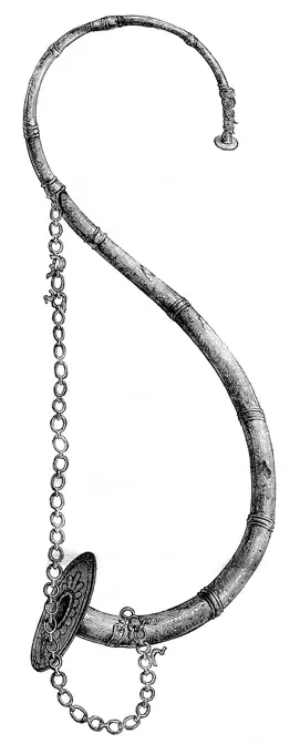
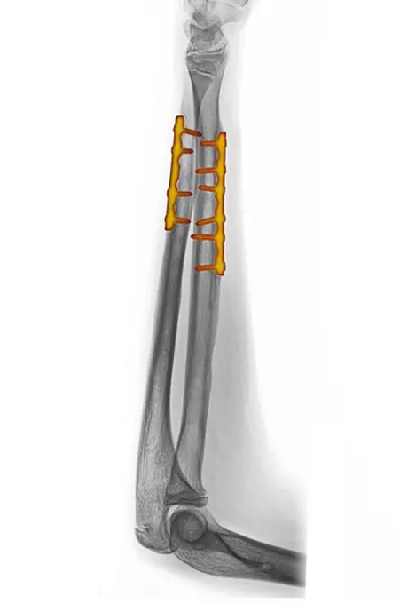
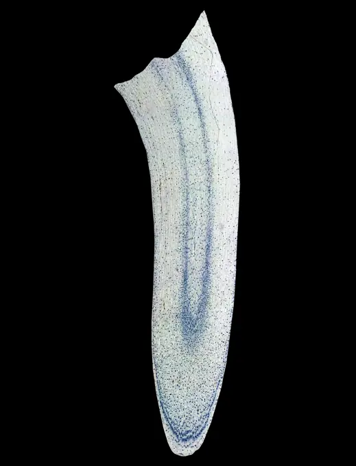
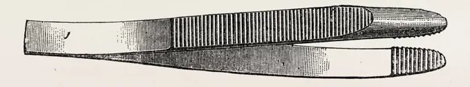
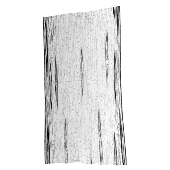
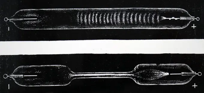
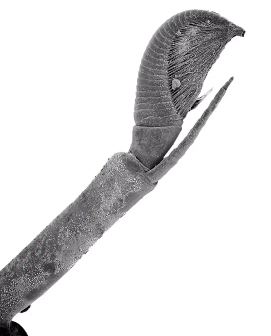
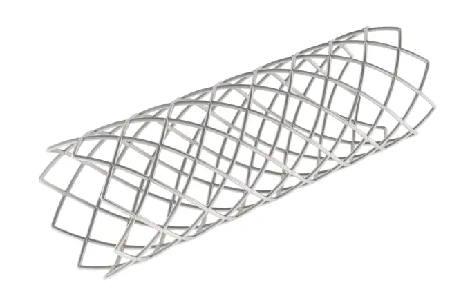
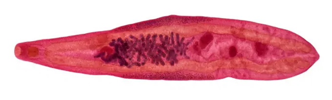
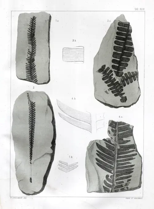
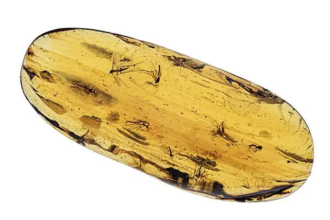
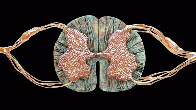
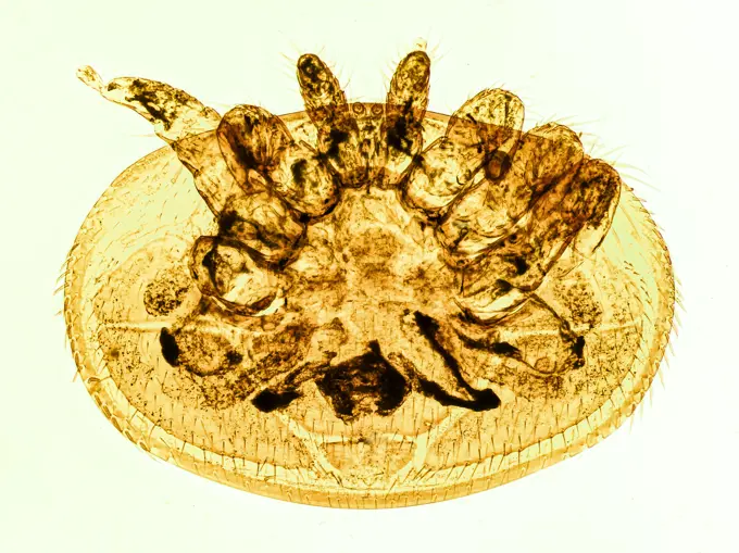
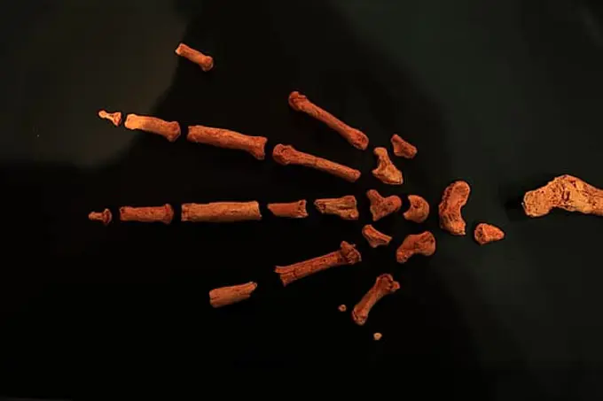
Botanical and Medical Illustrations

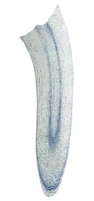
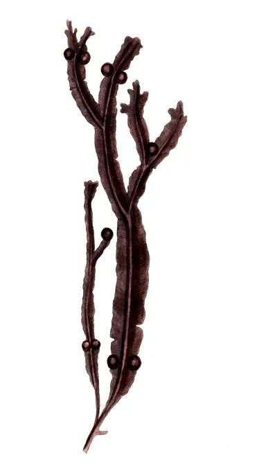
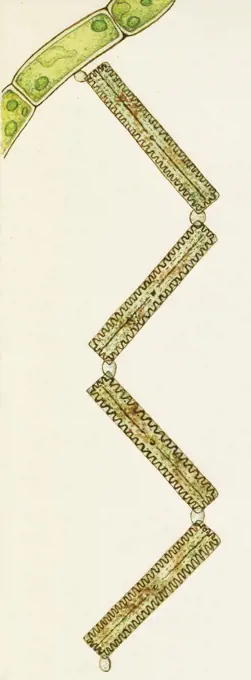
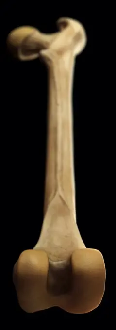
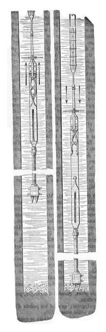
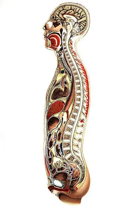
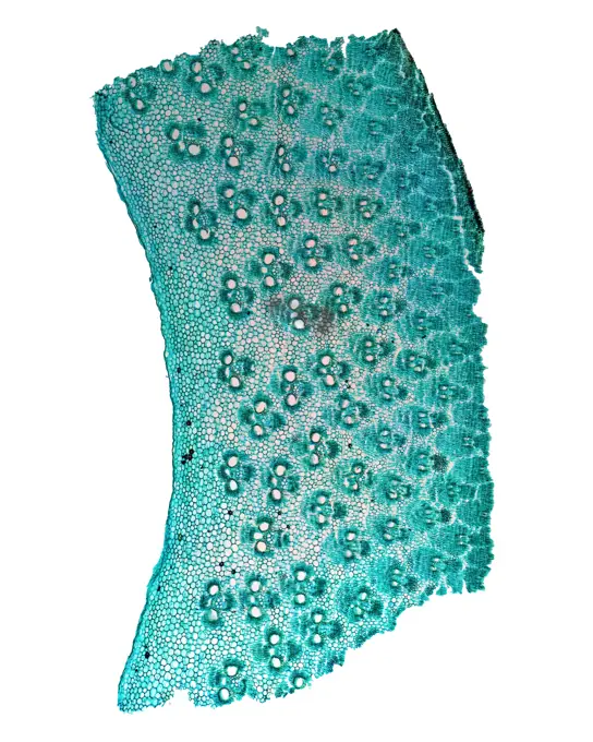
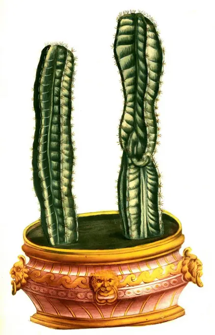
Detailed illustrations of plants and medical equipment from historical sources, highlighting their scientific importance and aesthetic beauty.

Colorized x-ray of the forearm of a 14 year old boy showing the surgical repair of a fractured ulna and radius
The ship worm (Teredo navalis), vintage engraved illustrationRoot hair star, Rhizocrinus loffotensis, Historic, digitally restored reproduction from a 19th century originalsyme's aneurysm needle, medical equipment, surgical instrument, history of medicineAngons Franks, vintage engraved illustration. Magasin Pittoresque 1861Papyrus plant (Cyperus papyrus) Ancient Egypt History. Old 19th century engraved illustration from El Mundo Ilustrado 1879Wheat boats, Fishmonger fresh water, Boatman passer, vintage engraved illustration. Magasin Pittoresque 1846Museum of Copenhagen, The Lur, Scandinavian trumpet, vintage engraving. Museum of Copenhagen, The Lur, Scandinavian trumpet, vintage engraved illustration. Magasin Pittoresque 1876. Copyright: xZoonar.com/PatrickxGuenettex 10679681Colorized x-ray of the forearm of a 14 year old boy showing the surgical repair of a fractured ulna and radiusAngons Franks, vintage engraved illustration. Magasin Pittoresque 1861.Breast bone, illustrationColorized x-ray of the forearm of a 14 year old boy showing the surgical repair of a fractured ulna and radiusTitan (T28) Viewed by Cassini's Radar-- April 10, 2007Sketch of plate tectonics, formation of new crust at oceanic ridges, plates movement and subduction areas.Corn root tip micrograph Light photomicrograph of Corn root tip cross section seen through microscope Copyright: xZoonar.com/ClaudioxDiviziax 7886860stout dissecting forceps, medical equipment, surgical instrument, history of medicineHuman leg bones, computer illustration.Isolated muscle, vintage engraved illustration. from Zoology Elements from Paul Gervais.Illustration of a neuron. The cell body contains a nucleus and its peripherie is made up od dendrites. It is linked to the axon cone by the axon sheathed in Schwann cells. The nerve fiber ends in terminal synaptic buttons.An engraving depicting the Von Welsbach Gas Mantle. Dated 19th centuryCorn root tip micrograph Light photomicrograph of Corn root tip cross section seen through microscope Copyright: xZoonar.com/ClaudioxDiviziax 6580644Pointed spear of flint, vintage engraved illustration. From the Universe and Humanity, 1910.Snuffers, vintage engraved illustration. Magasin Pittoresque 1853Fucus vesiculosus, bladdering, digital reproduction of a historical template, between 1790 and 1810, botanica pharmaceutica by Andreas Friedrich HappeZig-zag colony, Diatoma vulgare, Diatom algae, Bacillariophyceae. Drawing.Model of the right femur bone.Black and white Corn stem micrograph Light photomicrograph of Corn stem longitudinal section seen through microscope in black and white Copyright: xZoonar.com/ClaudioxDiviziax 7777972Blue Whale (Balaenoptera musculus)Historic illustration, tablet 12, title Talamophora, name Miliola, 12a/ Vertebralina insignis, Milioloda familiy, Ernst Haeckel, Kunstformen der Natur, "Artforms of Nature"Istorical illustration, tablet 4, title Diatomea, name Triceratium, Plagiogramma barbadense, plants, Ernst Haeckel, Kunstformen der Natur, "Art Forms in Nature"Engraving depicting a Geissler tube, an early gas discharge tube used to demonstrate the principles of electrical glow discharge, similar to modern neon lighting. Invented by Heinrich Geißler (1814-1879) a German glassblower, physicist and inventor. Dated 19th CenturyCommon dandelion (Taraxacum officinale), X-rayVoltaic pile invented by the italian physicist Alessandro Volta (1745-1827). Colored engraving.Sperm Whale (Physeter macrocephalus)Electric spark plug invented by Jules Célestin Jamin (1818-1886) French physicist, professor at the Faculty of Sciences in Paris. Inventions of the nineteenth century. Old 19th century engraved illustration from El Mundo Ilustrado 1879Right-handed Hermit Crab (Paguras sp) SEM close-up view of antenna at 21x magnificationThis portion of a Magellan radar image strip shows a small region on Venus 20 km (12.4 mi.) wide and 75 km (50 mi.) long on the east flank of a major volcanic upland called Beta Regio. The image is centerd at 23 degrees north latitude and 286.7 degrees east longitude. The ridge and valley networkin the middle part of the image is formed by intersection faults which have broken the Venusian crust into a clomplex, deformed type of surface called tessera, the Latin word for tile. The parallel mountains and valleys resemble the Basin and Range Province in the western United States. The irregular dark patch near the top of the image is a smooth surface, probably formed by lava flows in a region about 10 km (6 mi.) across. Similar dark surfaces within the valleys indicate lava flows that are younger than the tessera giving an indication of the geologic time relationships of the events that formed the present surface. The image has a resolution of 120 meters (400 feet).Cervical spine, computer artwork.Ibis mummy kept in the Museum of Natural History, vintage engraved illustration. Magasin Pittoresque 1852.Stent, Koronarstent. 3D-Rendering Stent, coronary stent. 3D rendering isolated on white background Copyright: xZoonar.com/natatravelx 22874436Liver fluke. Light micrograph (LM) of the liver fluke Clonorchis sinensis, which infects millions of people in the Far East. The fluke's digestive sys...A790890 U12G TELEMETRY TRANSMITTER, RECEIVER SATTLER (Project Engineer) DEC 12 79EG&G/NTS PHOTO LAB Publication Date: 12/12/1979  EDGERTON, GERMESHAUSEN & GRIER; EG&G; G-TUNNEL; NEVADA; NEVADA TEST SITE; NTS; NUCLEAR ENERGY TECHNOLOGY; PIPES; RECEIVERS; TELEMETRY; TELEMETRY TRANSMITTER/RECEIVER; TEST SITES; TRANSMITTERS; TUNNELS; TUNNELS BY USAGE; UGT; UNDERGROUND TESTING; WIRE & CABLES; TELEMETRY TRANSMITTER RECEIVER  historical images. 1972 - 2012. Department of Energy. National Nuclear Security Administration. Photographs Related to Nuclear Weapons Testing at the Nevada Test Site.Flora, fossilis, formationis, oolithicae, Padova, Tip. del Seminario, 1856/1868-1873/1885, Jurassic, paleobotany, The illustration showcases a collection of fossilized fern prints along with a few stylized representations of their features. Positioned in a grid layout, the upper two sections highlight detailed impressions of fern fronds etched into rock surfaces. The first features elongated, pointed leaves arranged in a symmetrical pattern, while the second showcases a more robust, broader-leaved fern, both indicating the rich diversity of prehistoric vegetation. Below, the two smaller depictions offer simplified outlines of the ferns, enhancing the understanding of their shapes and structures. This representation combines scientific documentation with artistic interpretation, emphasizing the significance of these ancient plants in Earth's geological history.3D rendering of a t rex dinosaur abstract model isolated on white background 3D rendering of a t rex dinosaur abstract model isolated on white background silhouette. Copyright: xZoonar.com/HugoxKurkx 16699803Cervical spine, computer artwork.Ibis mummy kept in the Museum of Natural History, vintage engraved illustration. Magasin Pittoresque 1852Maneuver the bit with the slider Kind, vintage engraving. Maneuver the bit with the slider Kind, vintage engraved illustration. Industrial encyclopedia E.-O. Lami - 1875. Copyright: xZoonar.com/PatrickxGuenettex 10648707Spinal BonesMuscles of Caterpillar Goat-MothIllustration of Skeleton of Diplodocus (neck and head)Poppy seed heads, X-rayManeuver the bit with the slider Kind, vintage engraved illustration. Industrial encyclopedia E.-O. Lami - 1875.DNA molecule pictureWorm at the end of the Third Age, vintage engraving. Worm at the end of the Third Age, vintage engraved illustration. Magasin Pittoresque 1845. Copyright: xZoonar.com/PatrickxGuenettex 10673102Stratified deposits at mouth of river. Colour illustration.Human body blood vessel anatomy Human body blood vessel anatomy Copyright: xZoonar.com/StanislavxRishnyakx 15124627DNA molecule pictureGhost Mantis (Phyllocrania paradoxa) male, Analamazaotra MadagascarEngraved diagram showing the method of work in the Hoosac Tunnel, the first rock tunnel in America.Mitochondrial cristae within a mitochondrion. Drawing.Illustration depicting a compound microscope by Adams of London. Dated circa 1788.Worm at the end of the Third Age, vintage engraved illustration. Magasin Pittoresque 1845Many of the internal organs of the human body according to the state of knowledge 1895, digitally restored reproduction of an original 19th-century modelIllustration of yellow bone marrow located in the medullary cavity of the diaphysis of a femur. Yellow marrow is a large reserve of chemical energy.Salsola sativa, common saltwort, digital reproduction of a historical model, between 1790 and 1810, (Botanica Pharmaceutica by Andreas Friedrich Happe).Human Louse (Pediculus humanus) eggs on hair, GermanyGamme chromatique bleu, Des couleurs et de leurs applications aux arts industriels à l'aide des cercles chromatiques, Chevreul, M. E. (Michel Eugène), 1786-1889, Digeon, René, Steel engraving, 1864, Printed in colorBreathing apparatus of Saccobranche, vintage engraved illustration. Zoology Elements from Paul Gervais.Engraving depicting a regenerative x-ray tube, which by two lightbulbs, allowed a more efficient vacuum to be created. Dated 19th CenturyChromosomes during research, genetic testing, illustrationBamboo stem micrograph Light photomicrograph of Bamboo Stem cross section seen through microscope Copyright: xZoonar.com/ClaudioxDiviziax 8010416Anatomy of an earthworm.Brachiopod of the Upper Cretaceous, vintage engraved illustration. From the Universe and Humanity, 1910.Zoology, Insects, Coleoptera. Larva of the Tiger beetle (Cicindelinae), illustrationColoured scanning electron micrograph (SEM) of Tack nail. Tacking nails are often used for carpet attachment to wood flooring. A nail is a pin-shaped,...Historic illustration, tablet 24, title Desmidiea, marine algae, name Staurastrum, 10/ Closterium costatum, Ernst Haeckel, Kunstformen der Natur, "Artforms of Nature"Mosquitoes in Amber 40 Million Years Old, Dominican Republic, Central AmericaVertebral compaction. Color enhanced sagittal computed tomography (CT) scan of the spine showing vertebral compaction due to osteoporosis. The erosion of the bone structure of the vertebrae causes its deformation, and the reduction of the intervertebral space (yellow).Close up of the bones of a 85-foot-long blue whale skeleton.AE. PantikapaionNine Months with Signs of the Zodiac: May with the Sign of Gemini, Jan Kamphuysen, Giambattista Maderni, 1790 - 1791Illustration showing a cross section on one of Alfred Nobel's submersible mines. Cross-section of the spinal cord, illustrationGerm black icon. Microbe symbol. Biology signStereo photograph by Romanus Schmehlik, 1912, showing Trichine specimen at scale 200 on silver gelatin paper for microscopic study.Varroa mite (Varroa destructor)Ectoparasitic mite of the honey bee on a white backgroundAddis Ababa, in the Ethnological Museum, illustration of Luci, Lucy refers to the partial skeleton of an individual of the species Australopithecus afarensis, Ethiopia, discovered in the Afar Triangle in 1974 and interpreted as female, AfricaRe-construction of a Wing structure, based on a drawing in CODEX ATLANTICUS- FOLIO 858 r. (1478 -1519), by Italian polymath Leonardo da Vinci (1452-1519). Chromosomes in mitosis or cell division, illustrationRhipsalis baccifera (Cereus quadrangularis), quinque angula, a genus of plant (cactaceae) in the family, Historical, digitally restored reproduction f...Gamme chromatique jaune-vert, Des couleurs et de leurs applications aux arts industriels à l'aide des cercles chromatiques, Chevreul, M. E. (Michel Eugène), 1786-1889, Digeon, René, Steel engraving, 1864, Printed in colorSpinal cord and nerve roots.Gamme chromatique vert, Des couleurs et de leurs applications aux arts industriels à l'aide des cercles chromatiques, Chevreul, M. E. (Michel Eugène), 1786-1889, Digeon, René, Steel engraving, 1864, Printed in colorStratigraphic column, illustrationScanning electron microscopic image of castor bean plant pollenHuman arm nerves, computer illustration.Re-construction of a Ball Bearing, based on a drawing by Da Vinci, in the Madrid Codex I. folio 20 v. His invention was the result of research linked to the yield optimisation of machines in motion, and the limitation of wear caused by friction. Leonardo designed this rolling contraption with perpetual motion, with evenly spaced bearings, which allowed an entire stage to rotate during a theatrical performance of Orpheus for Charles d'Amboise in Milan. Leonardo da Vinci (1452-1519), Italian artist and polymathClose-up of a dropper in a test tubeBelemnite restored, vintage engraved illustration. Earth before man a 1886.