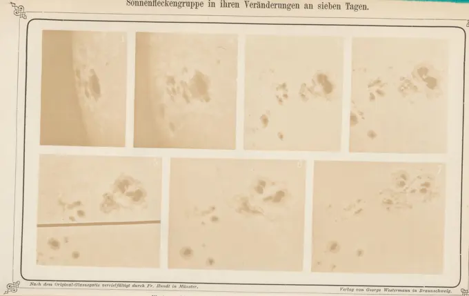
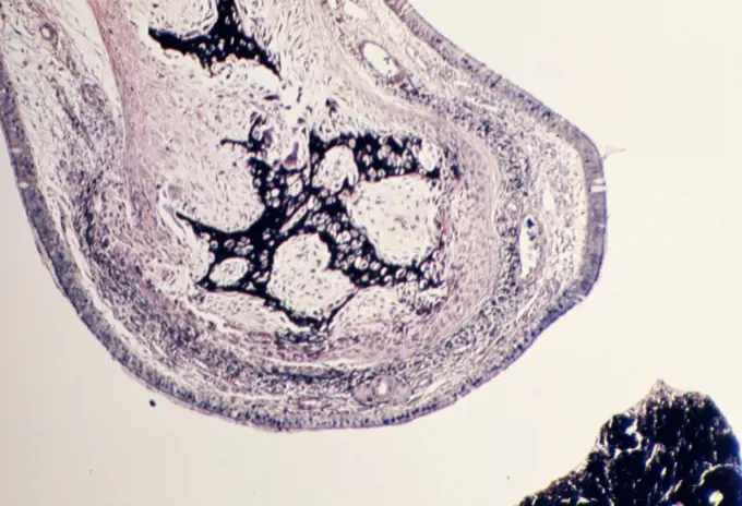
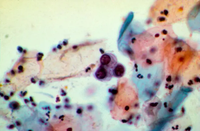
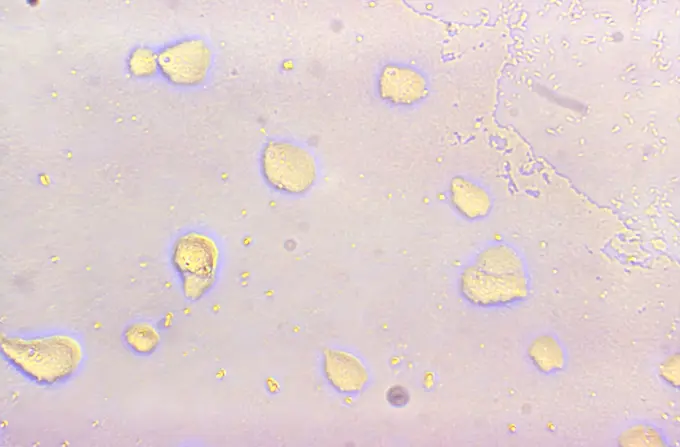
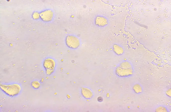
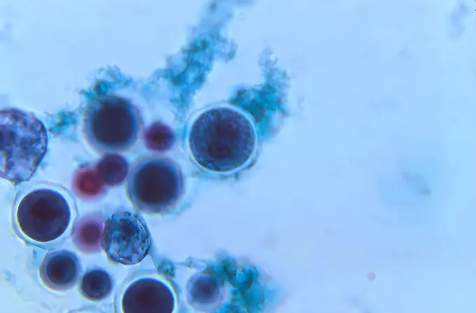
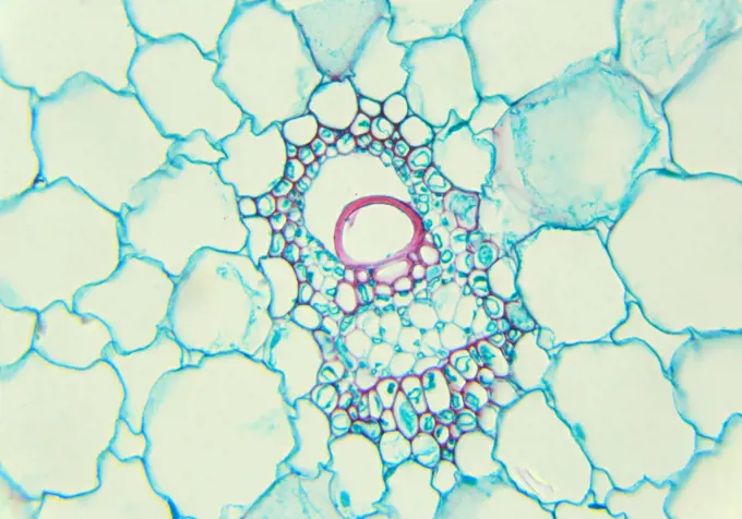
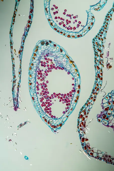
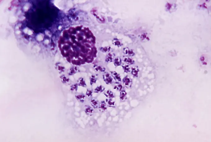
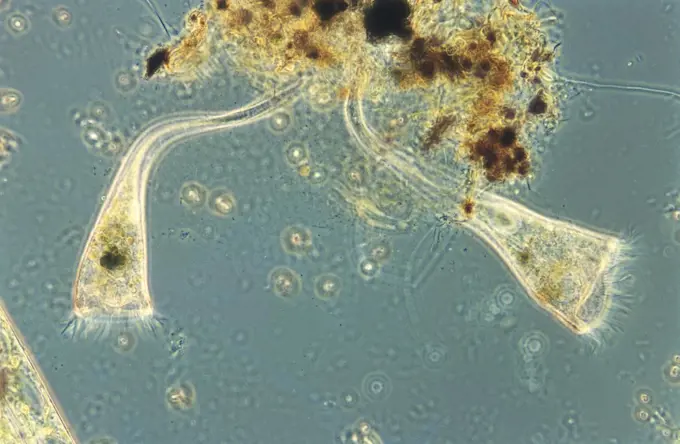
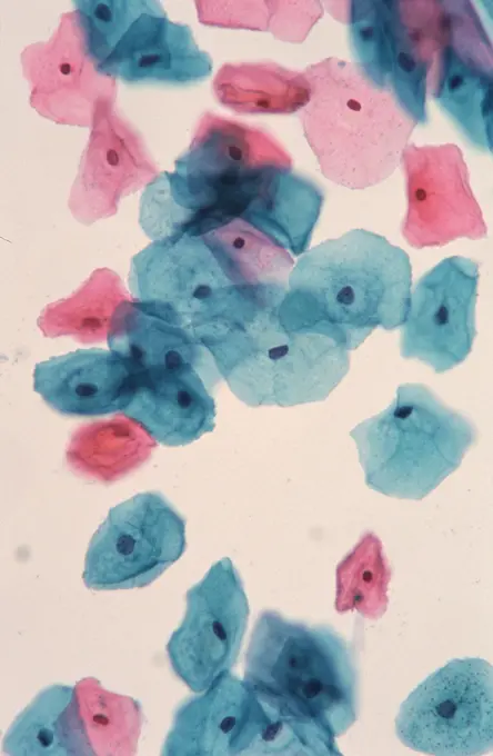
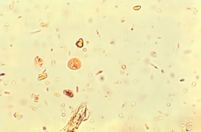
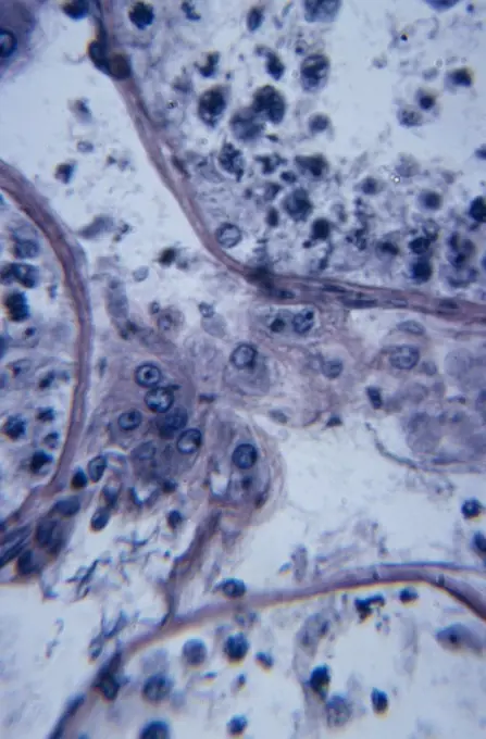
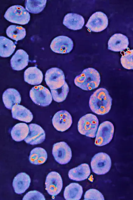
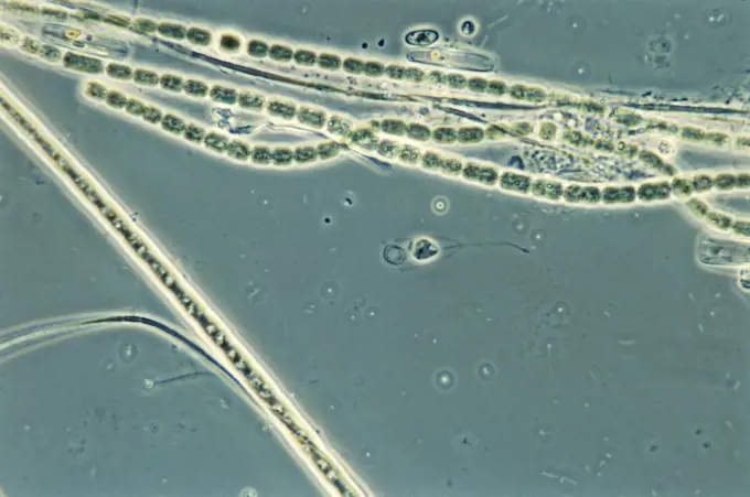
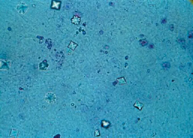
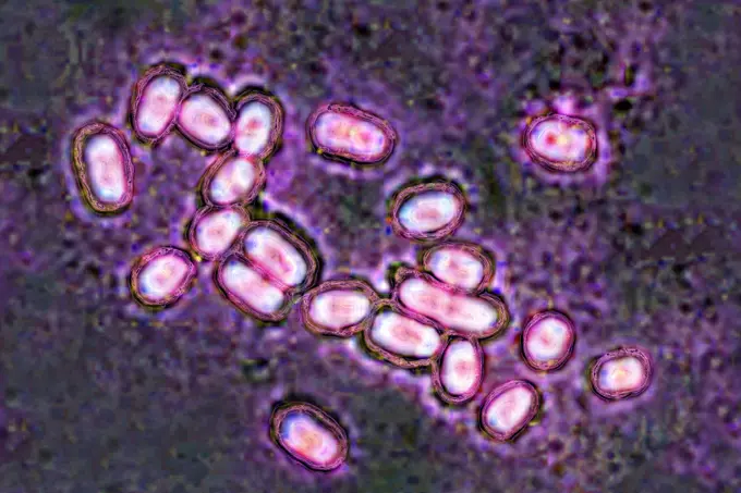
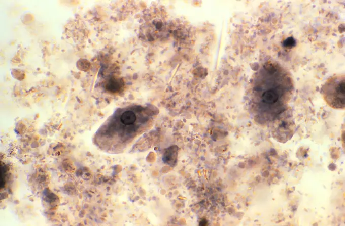
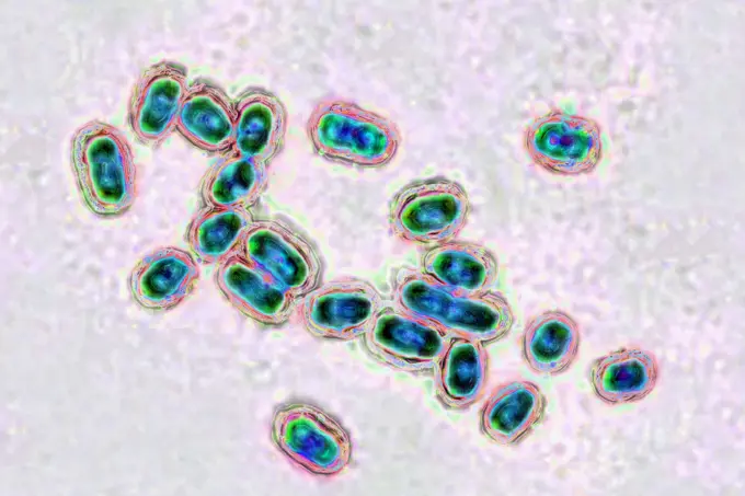
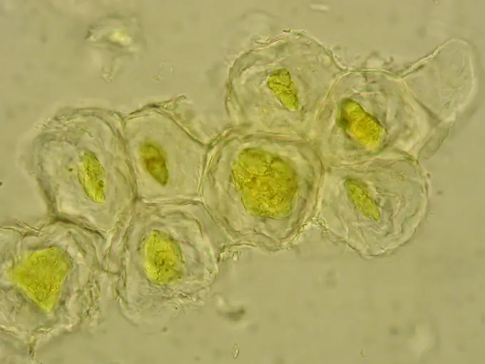
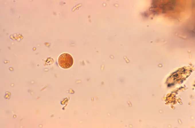
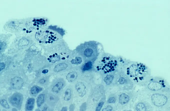
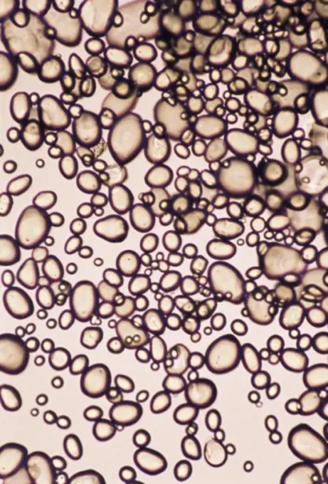
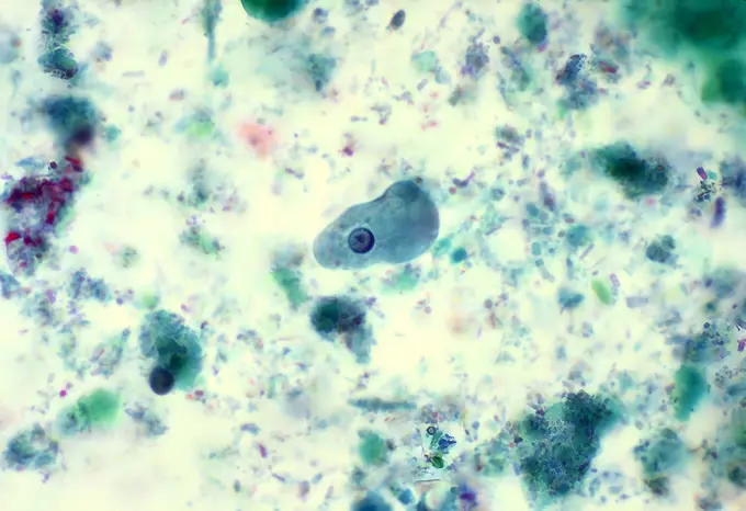
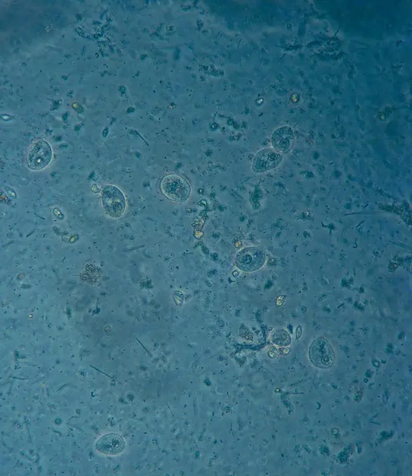
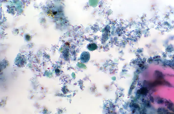
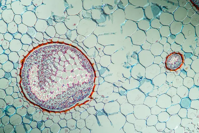
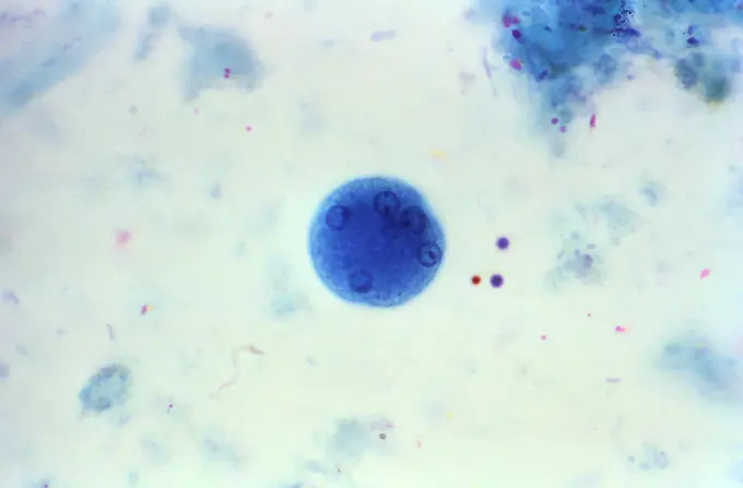
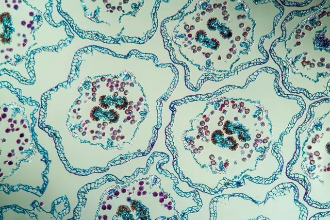
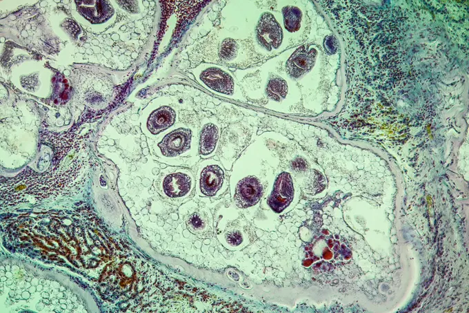
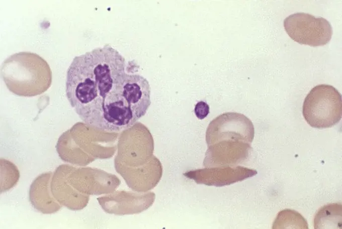
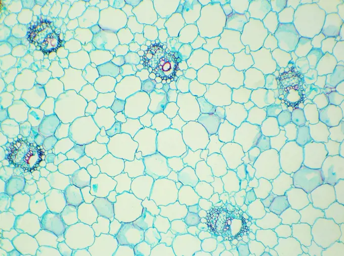
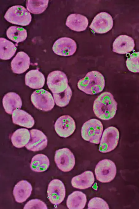
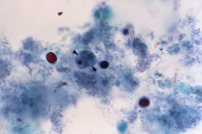
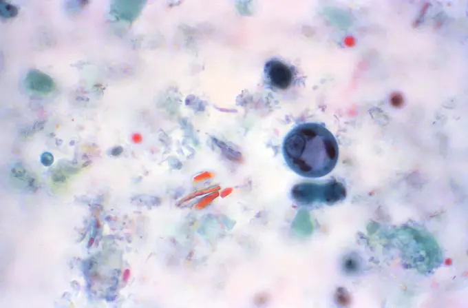
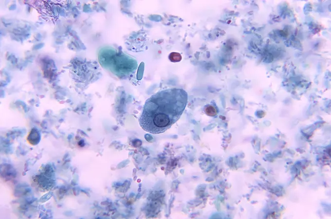
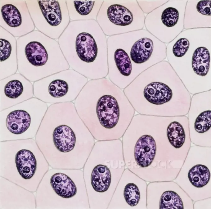
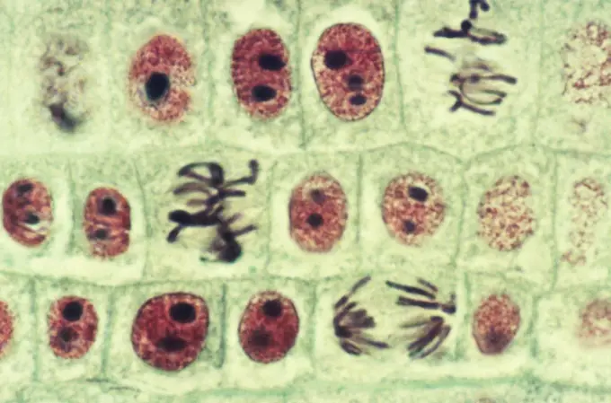
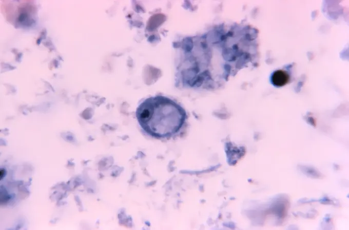
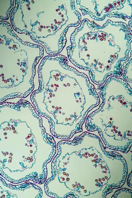
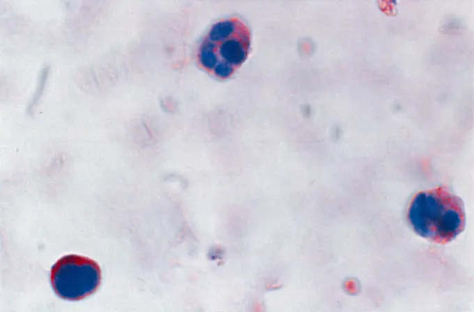
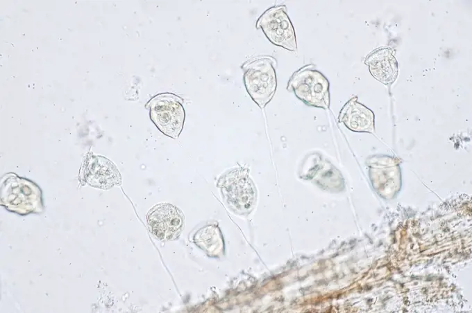
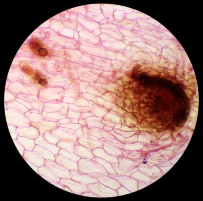
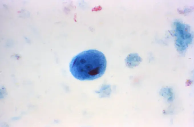
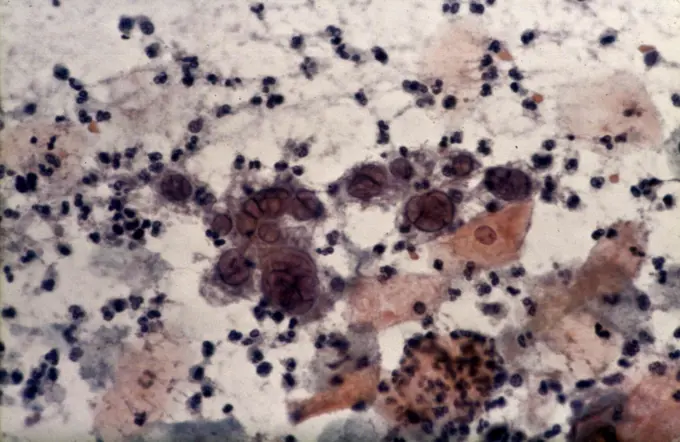
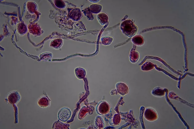
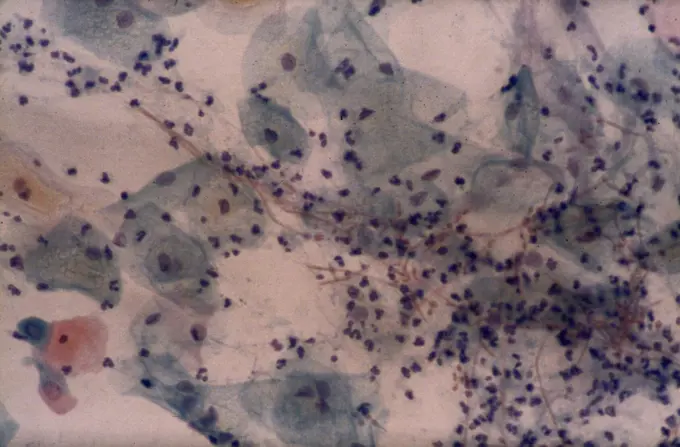
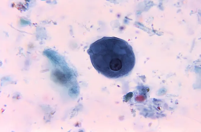
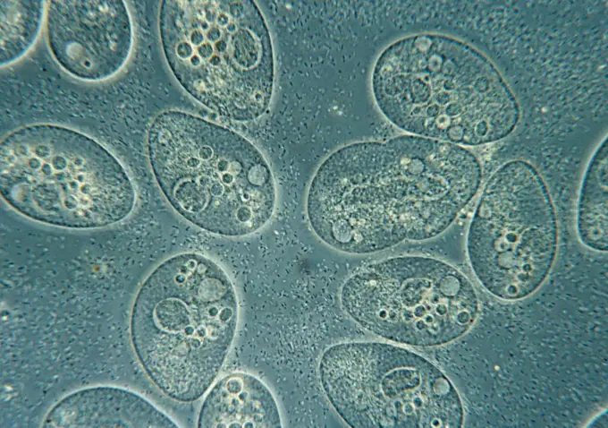
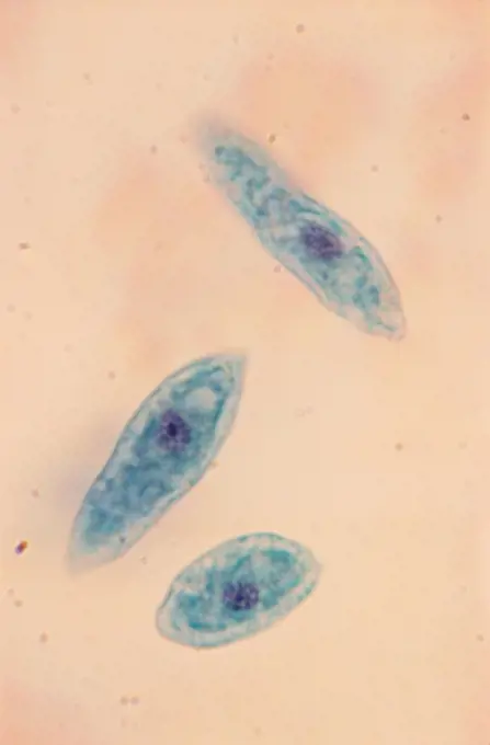
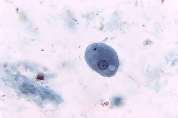
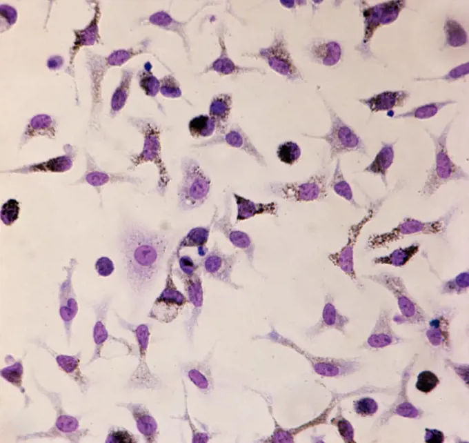
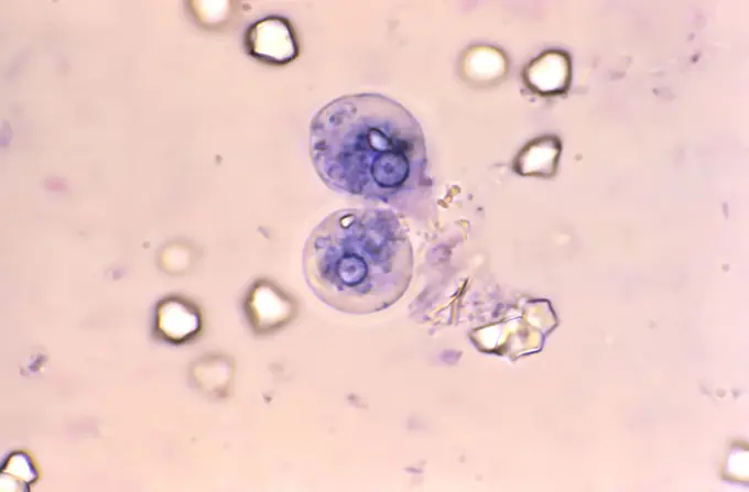
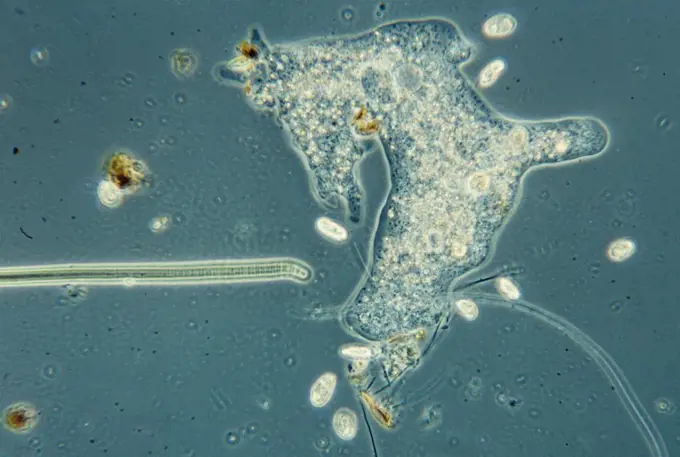
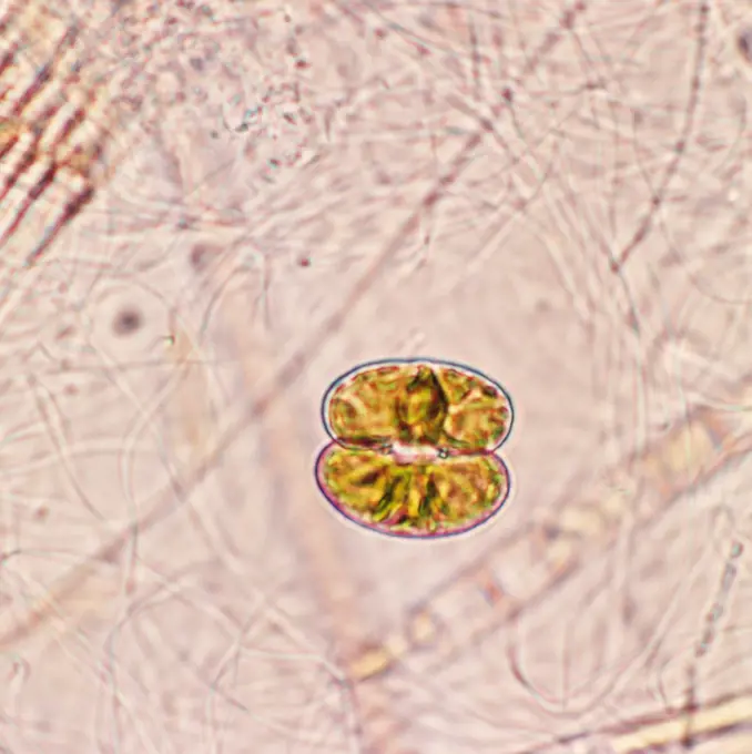
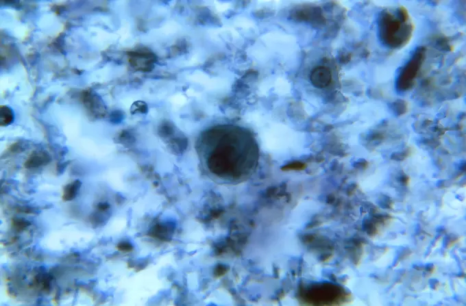
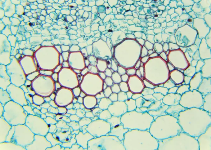
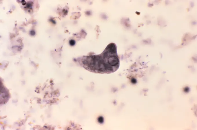
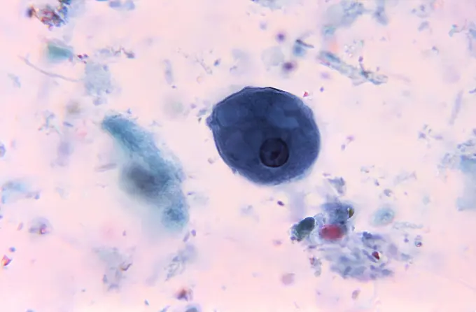
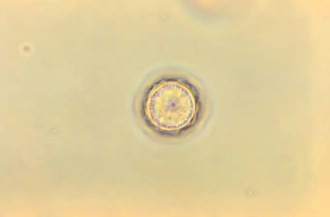
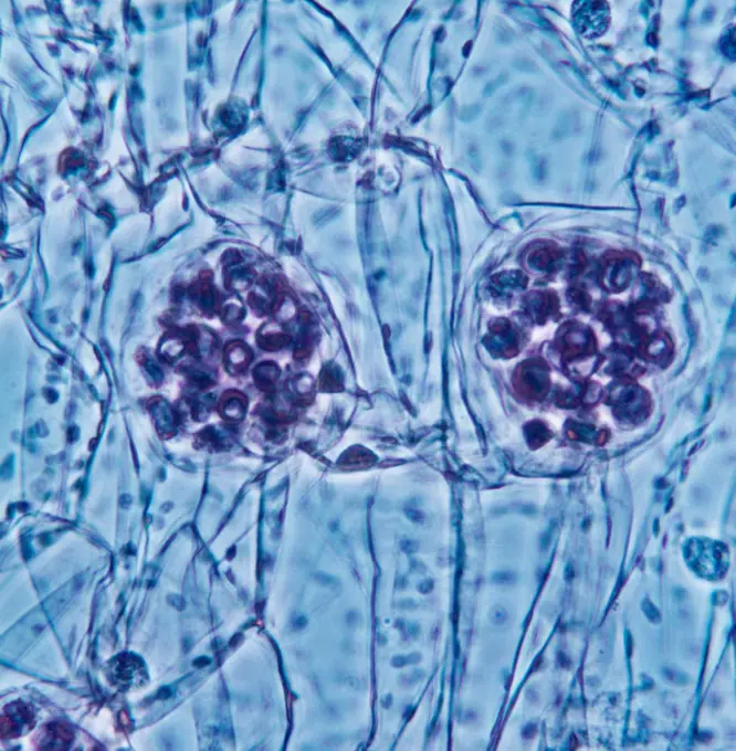
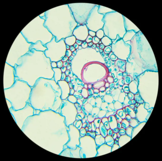
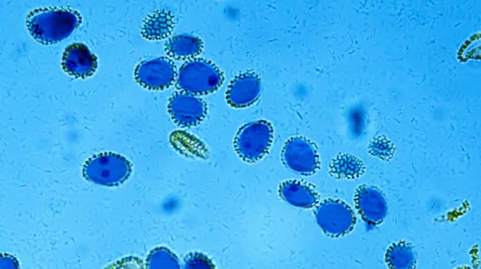
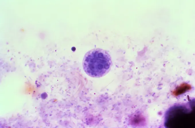
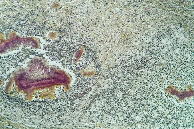
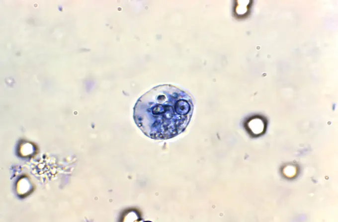
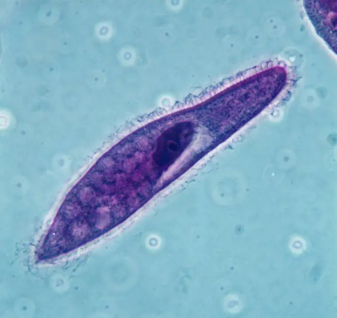
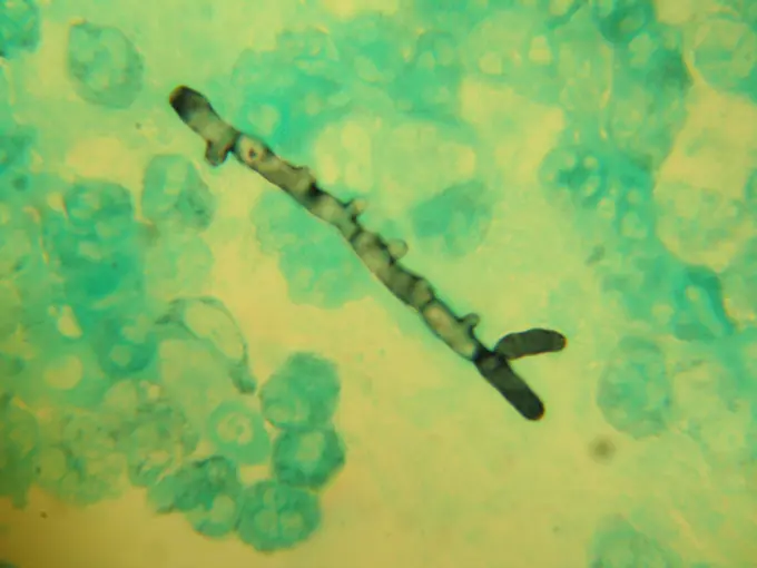
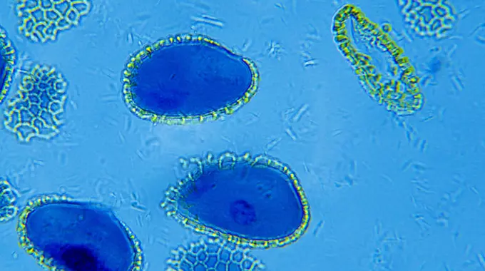
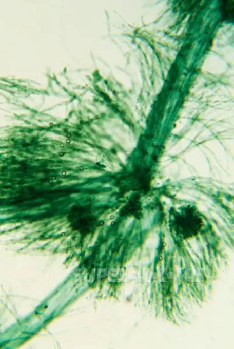
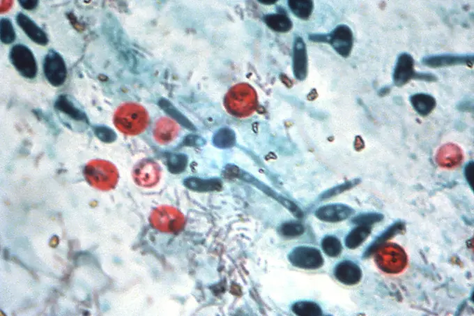
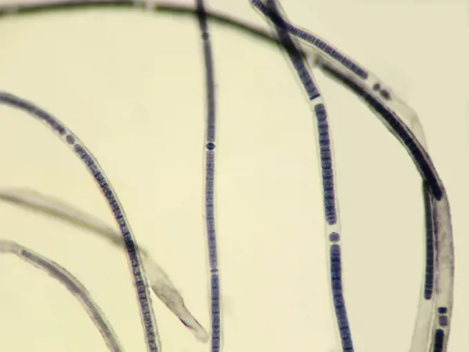
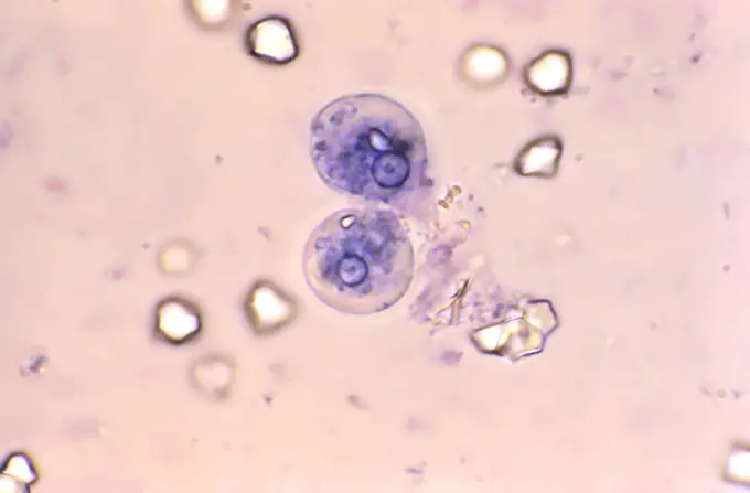
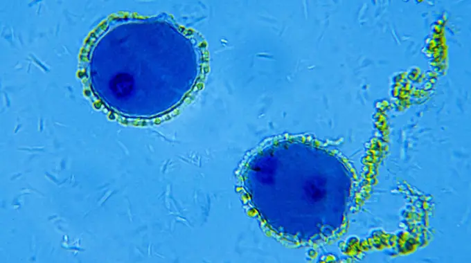
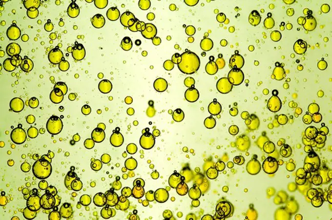
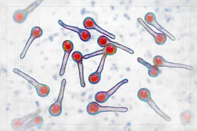
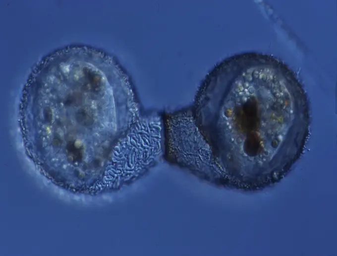
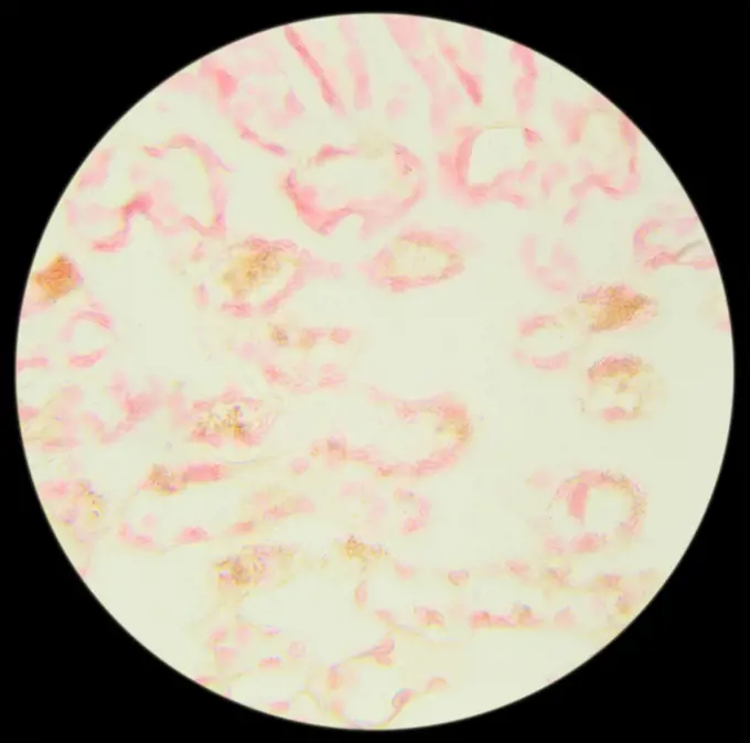
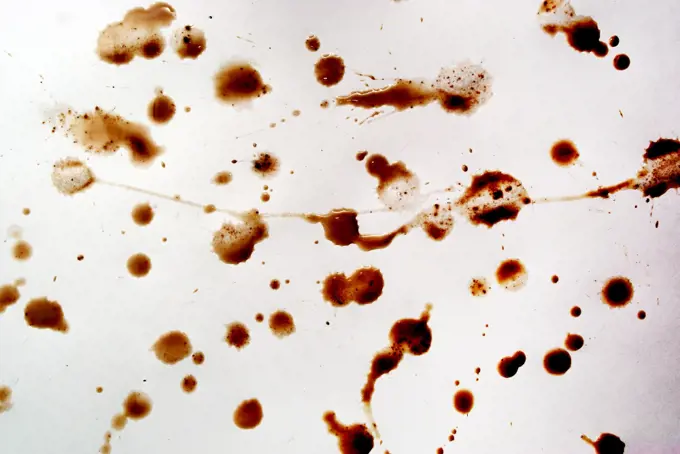
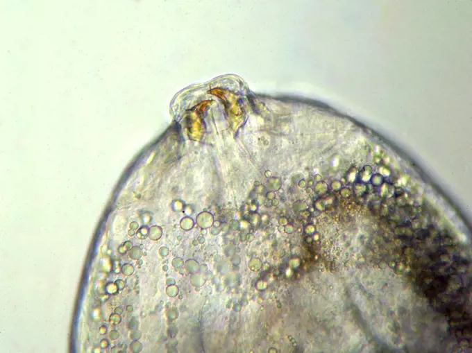
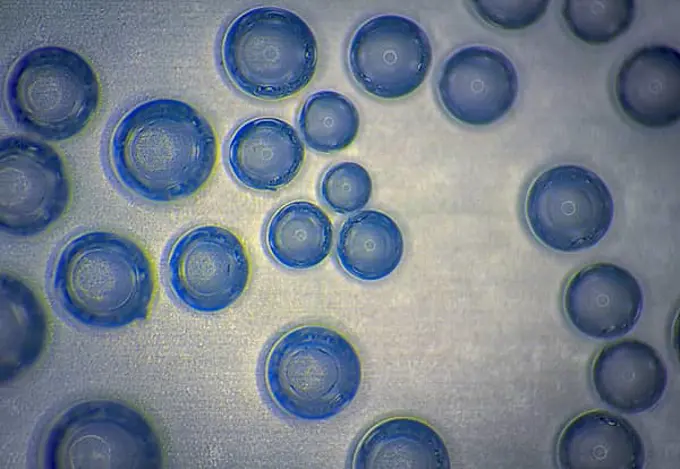
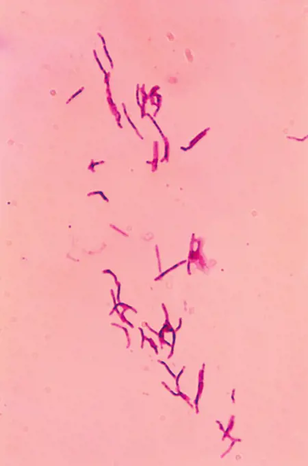
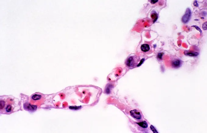
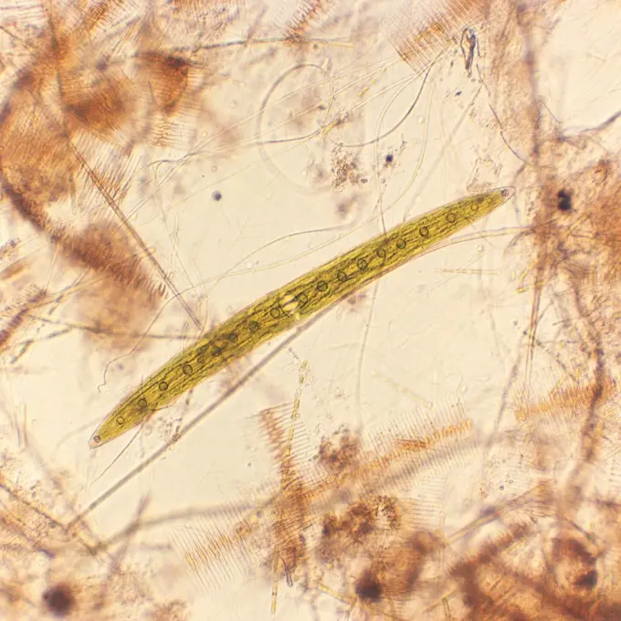
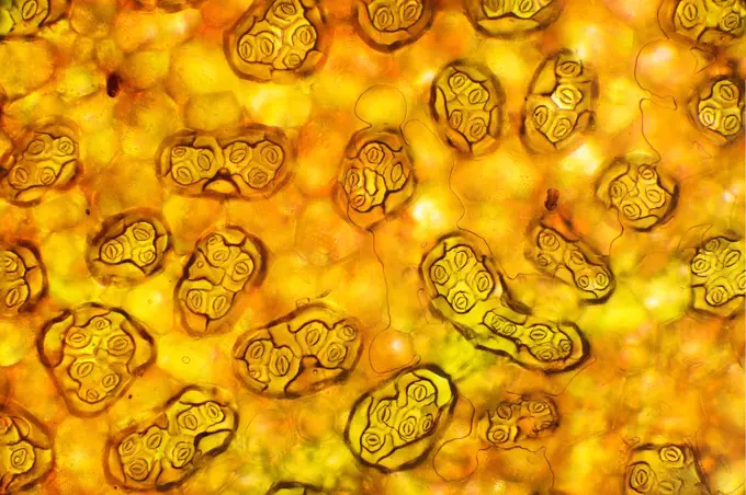
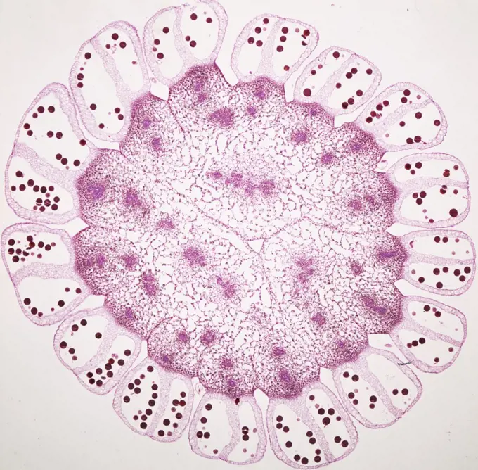
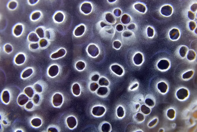
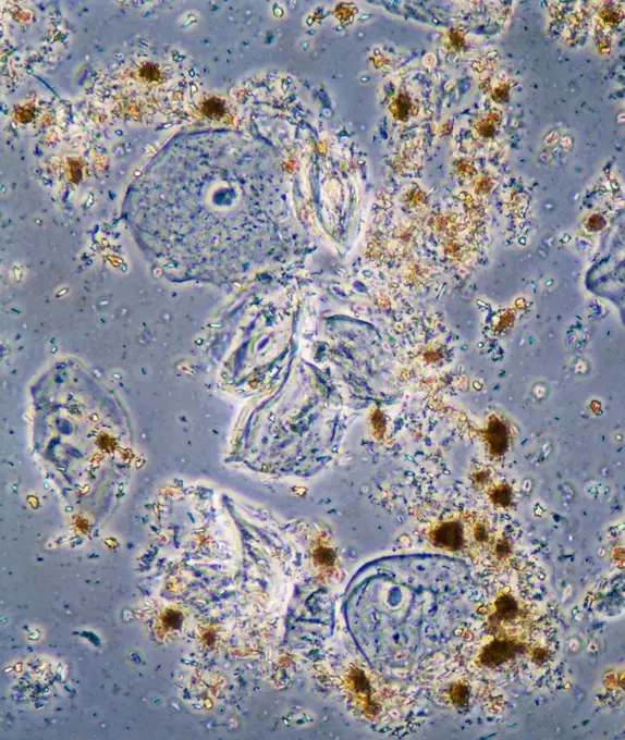
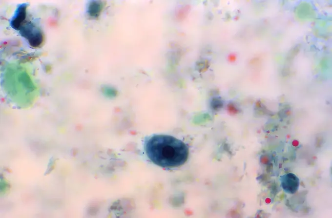
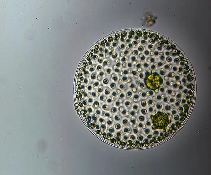
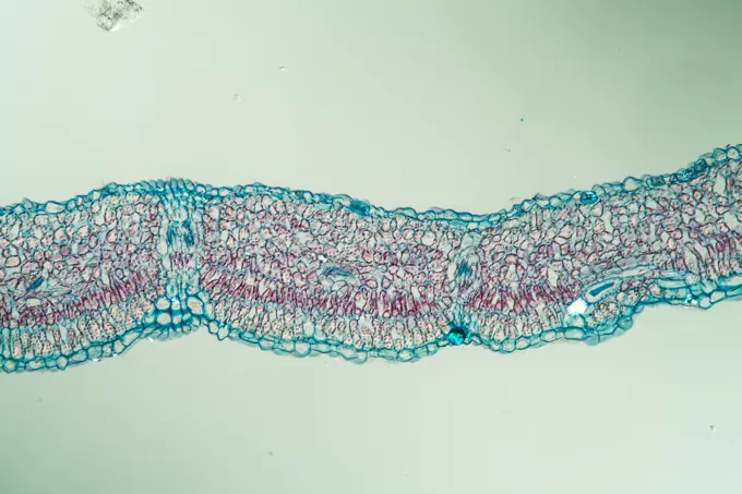
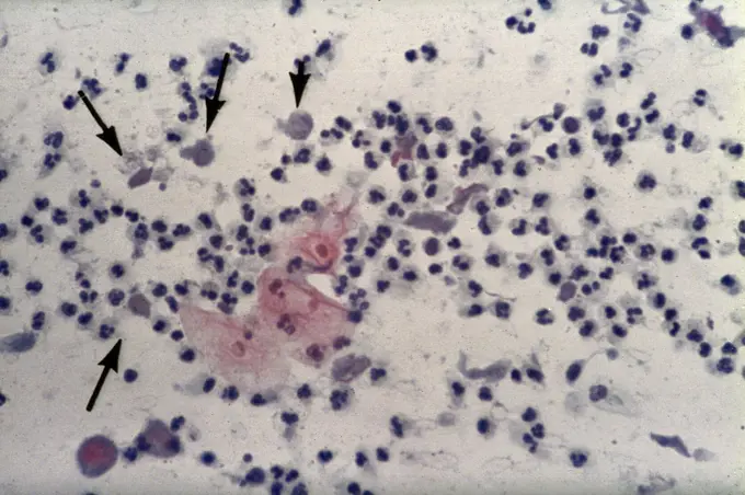
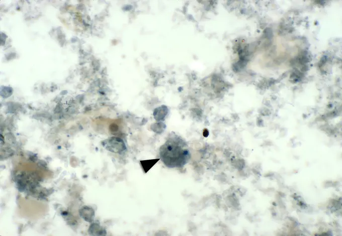
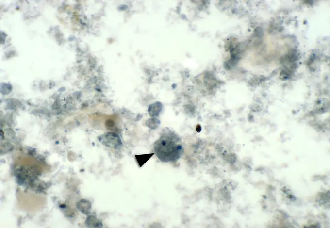
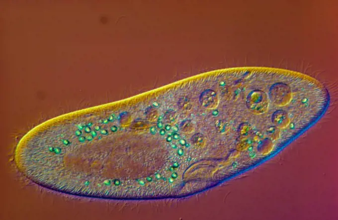
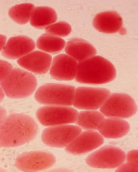
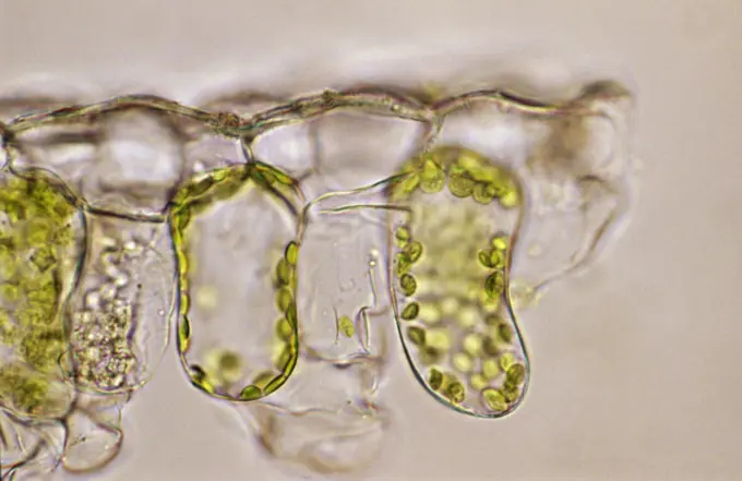
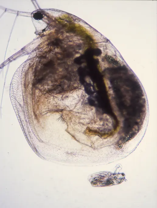
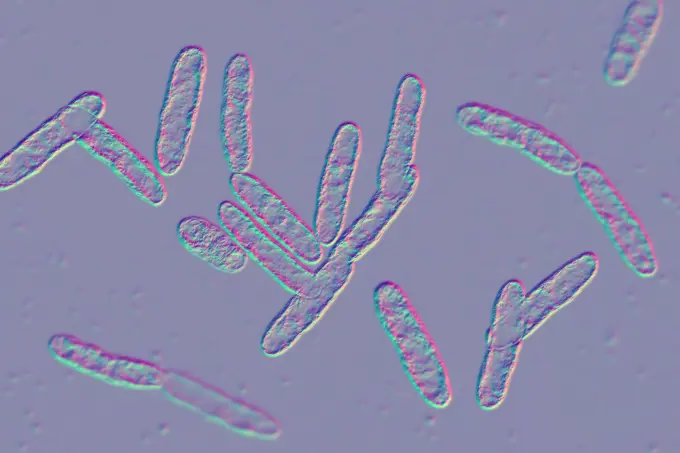
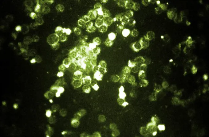
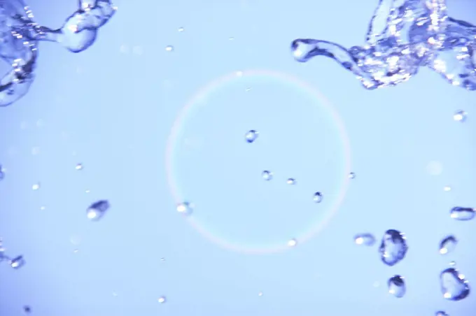
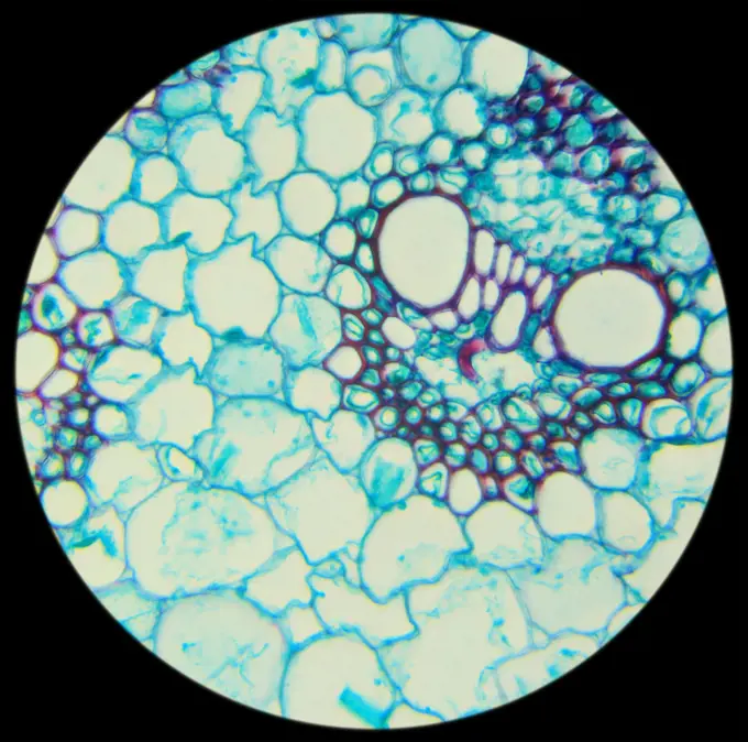
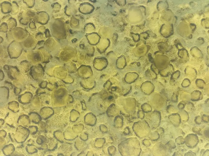
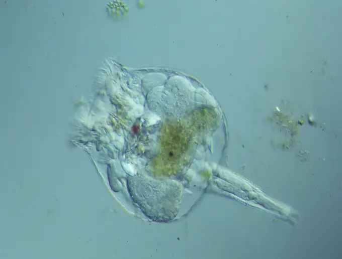
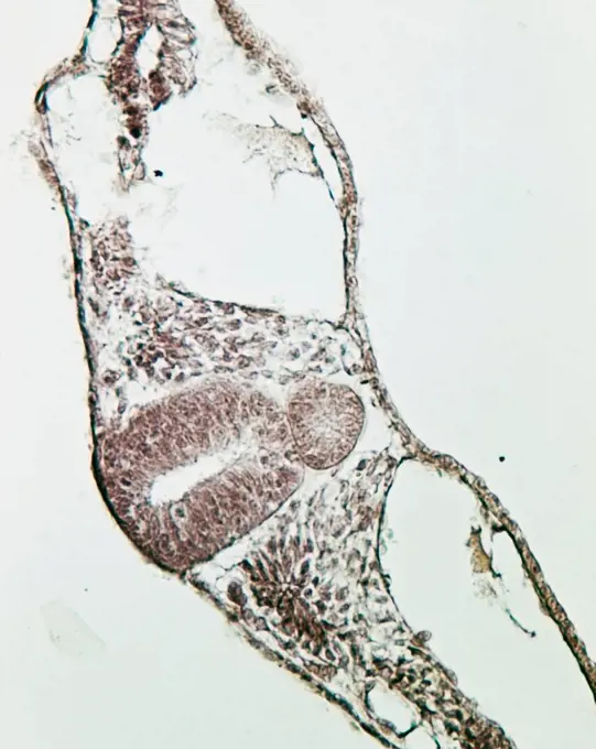
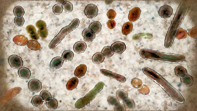
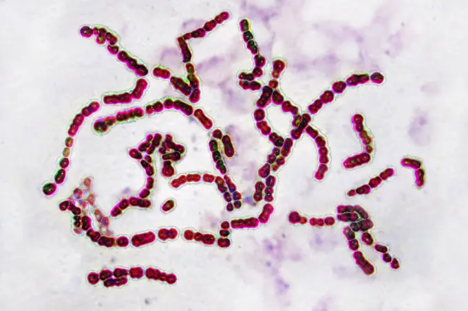
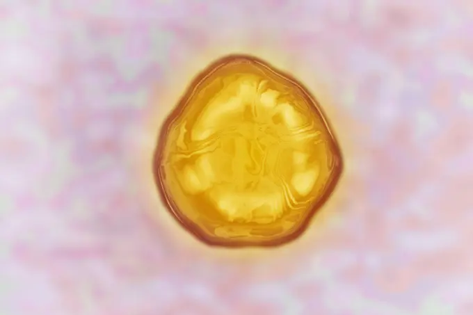
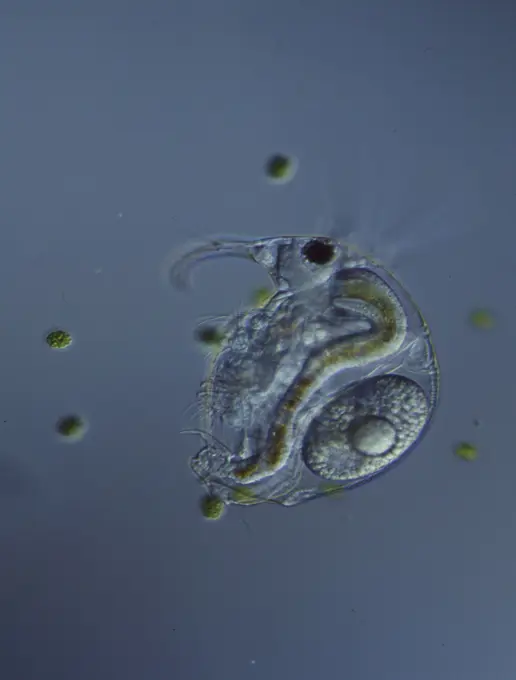
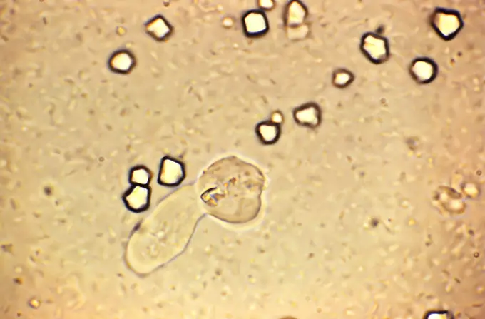
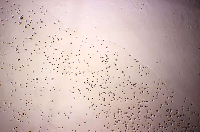
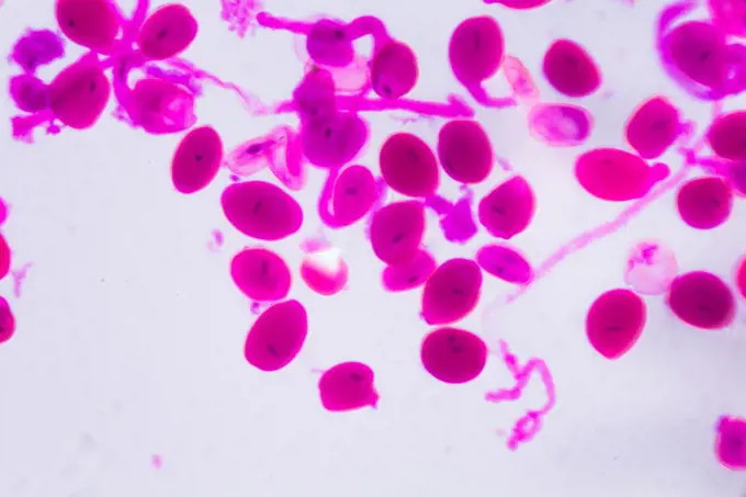
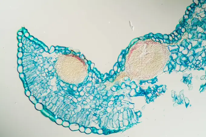
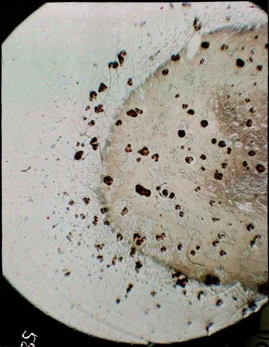
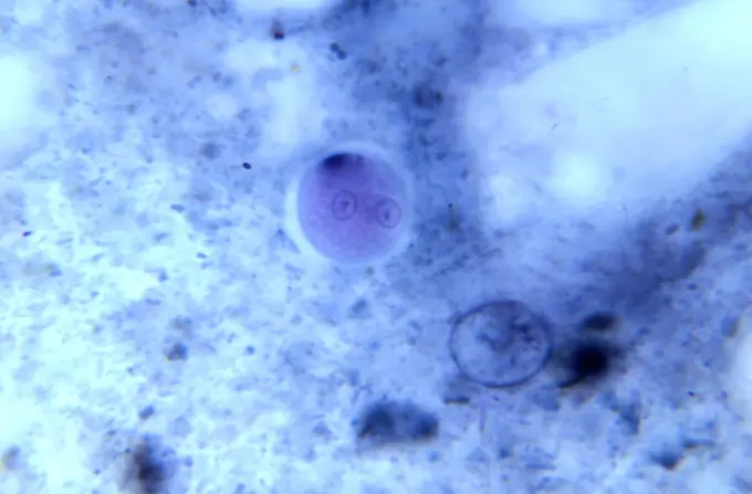
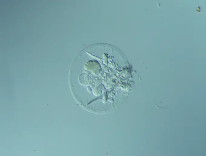
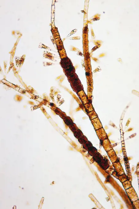
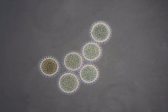
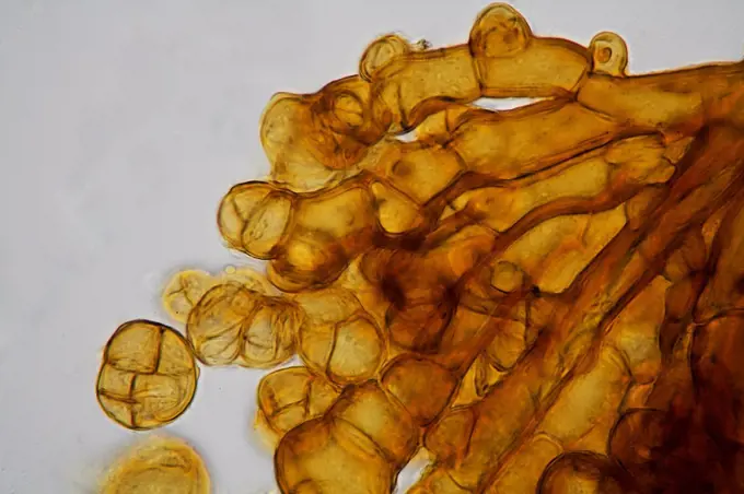
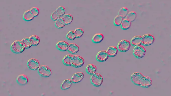
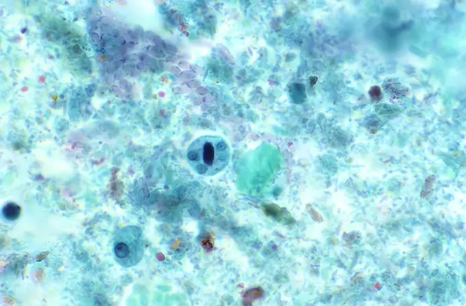
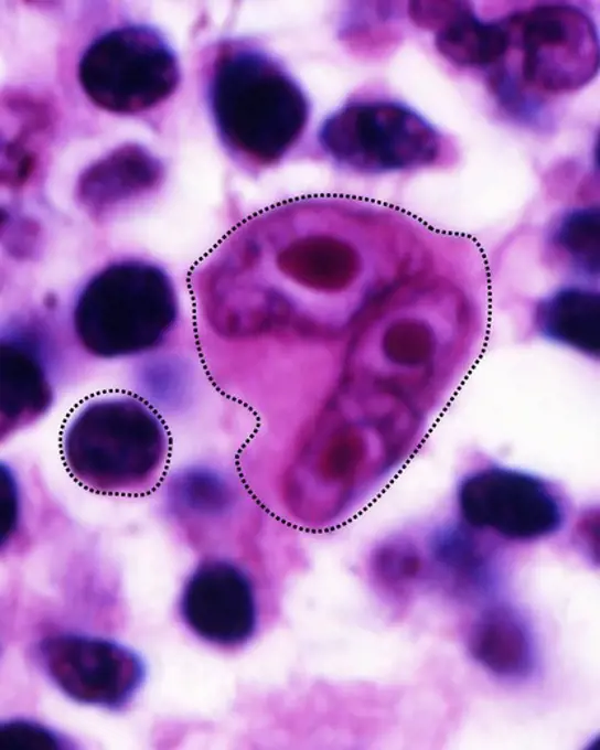
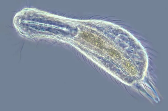
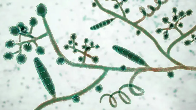
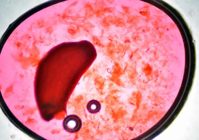
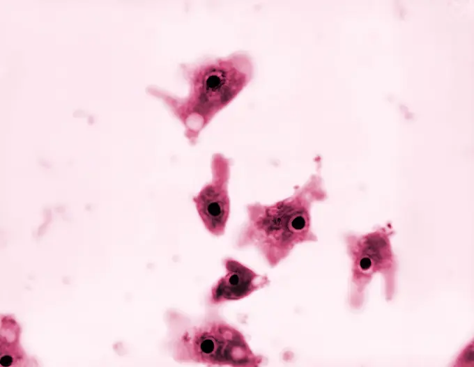
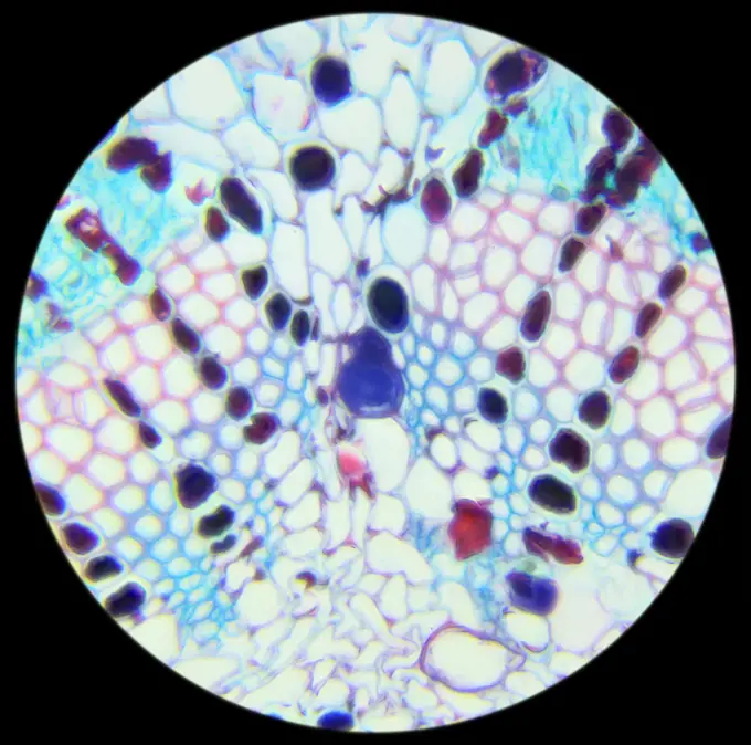
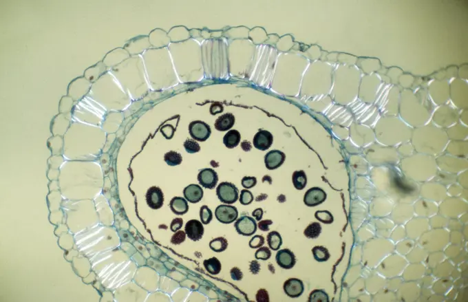
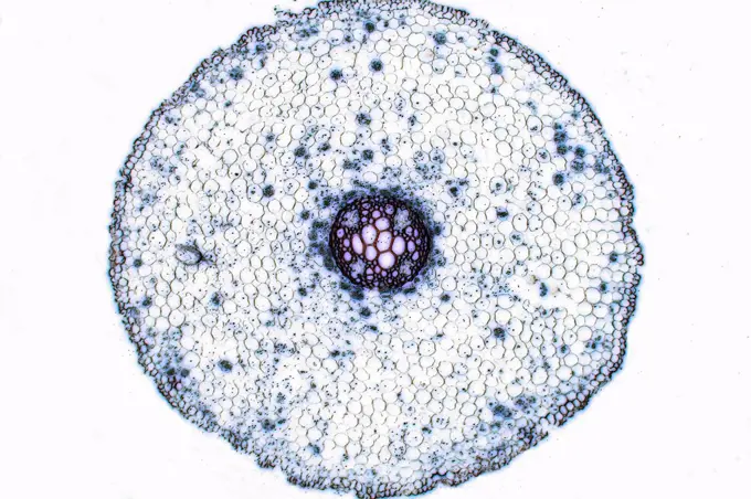
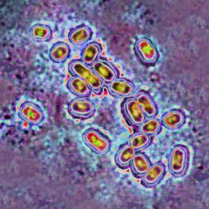
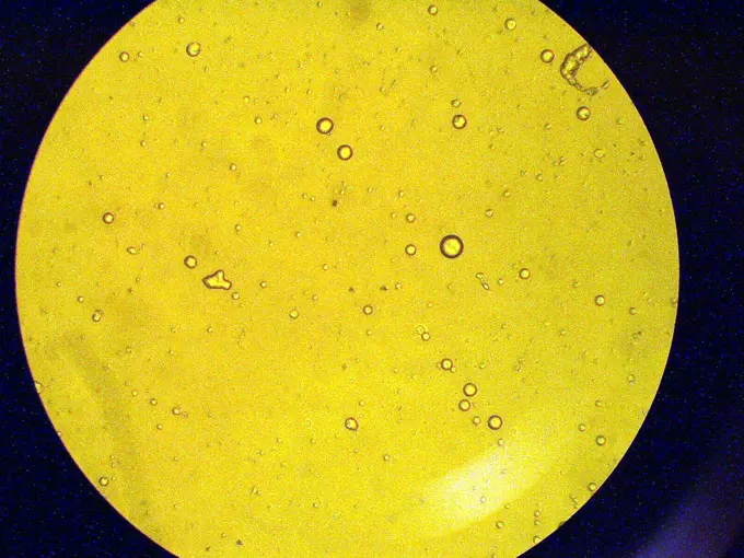
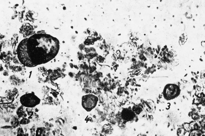
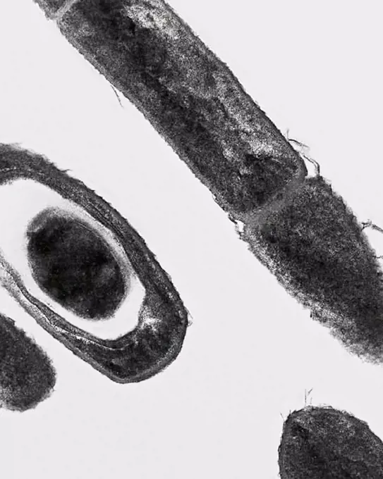
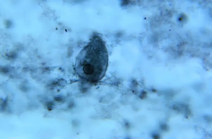
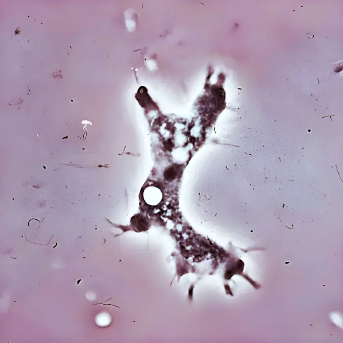
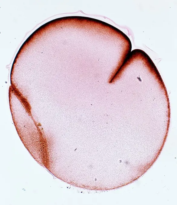
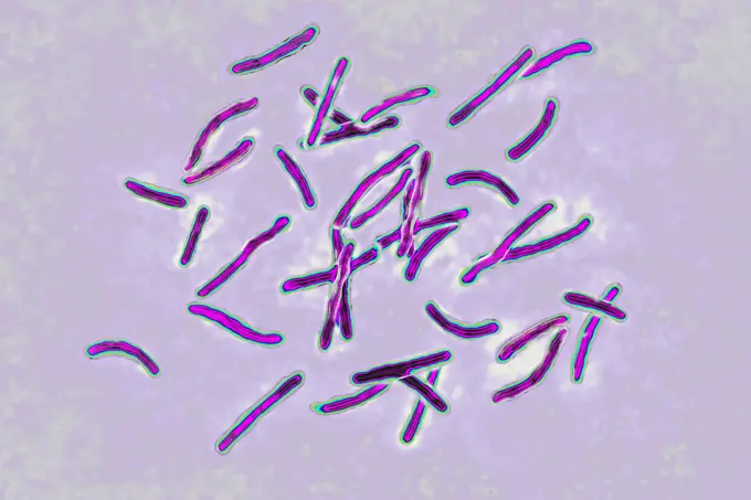
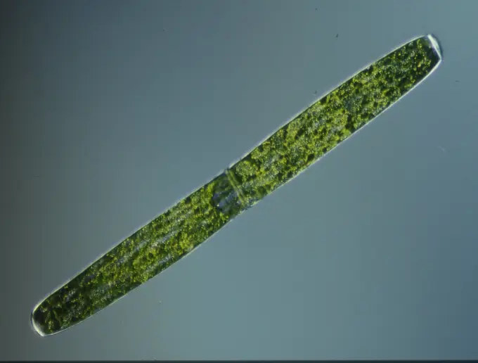
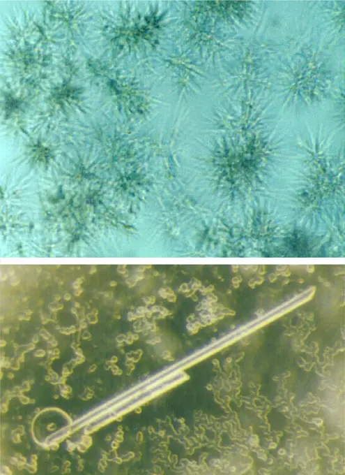
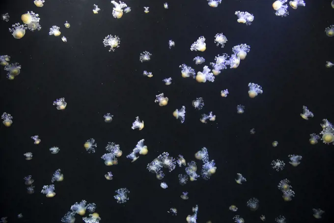
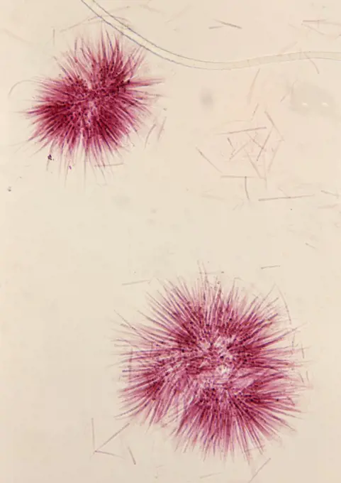
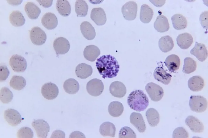
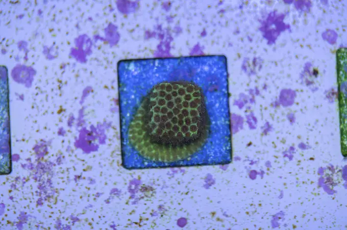
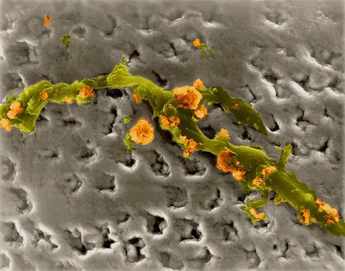
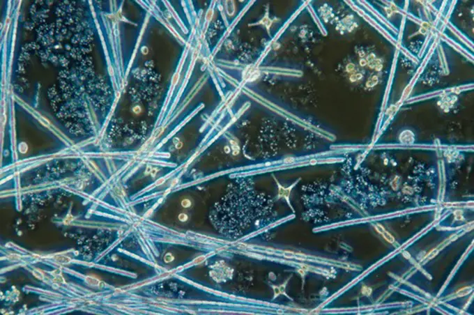
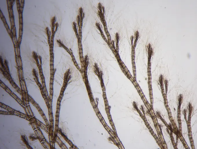
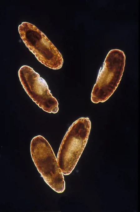
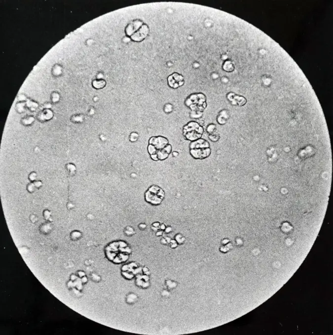
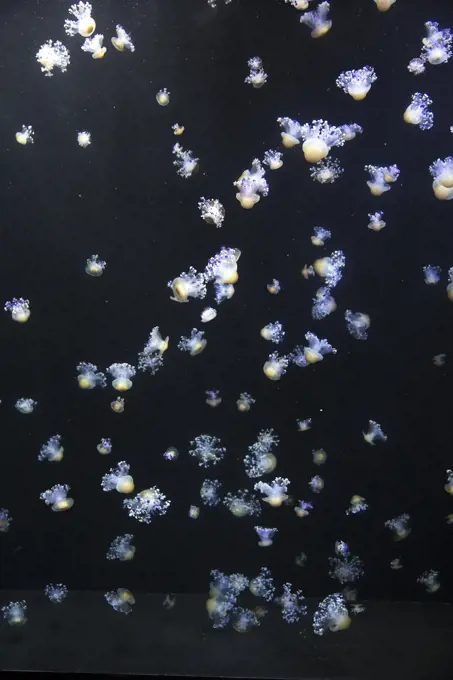
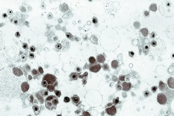
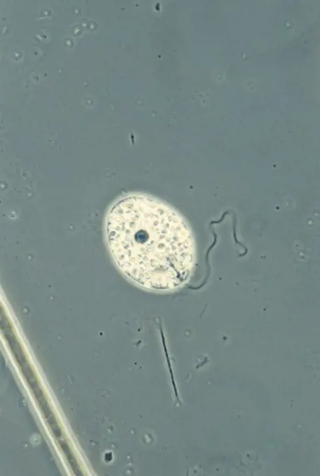
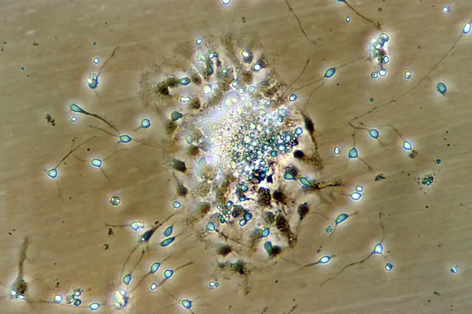
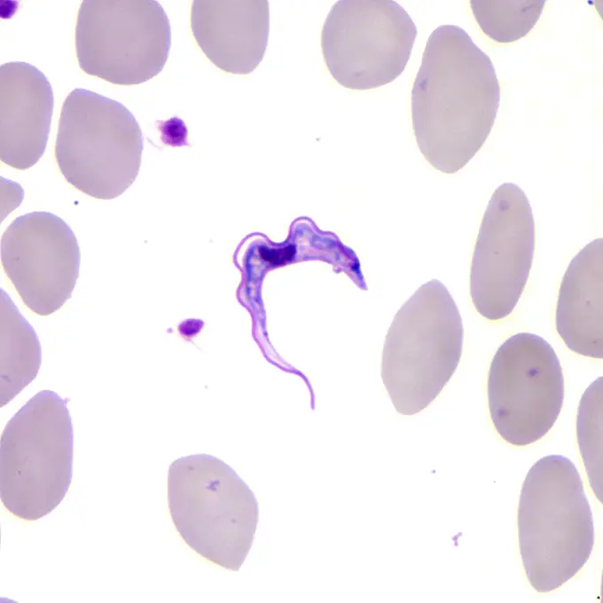
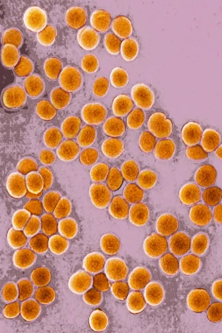
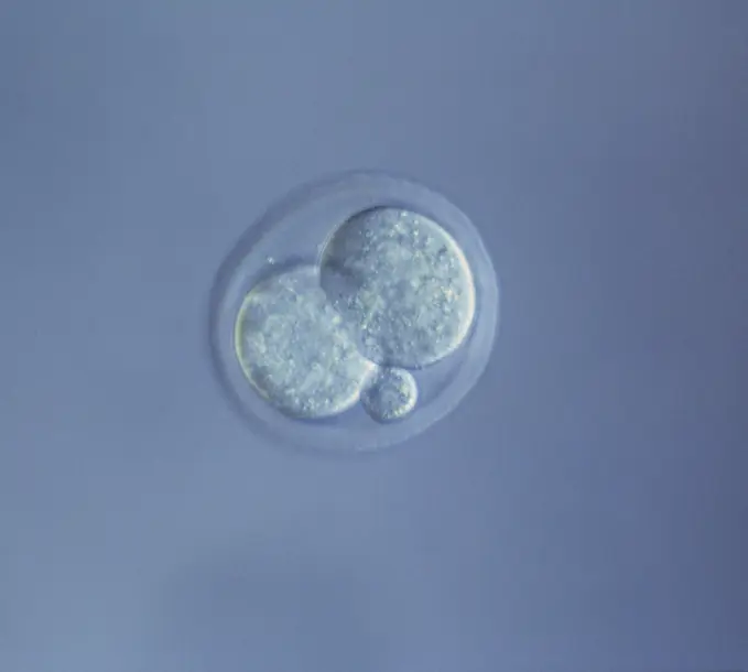
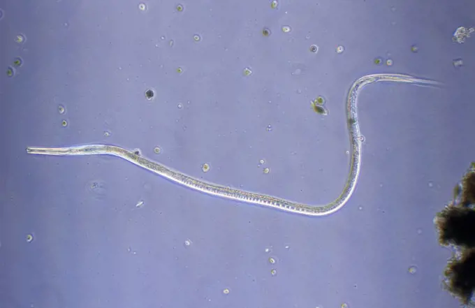
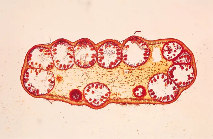
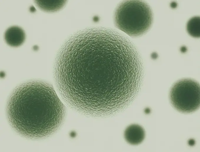
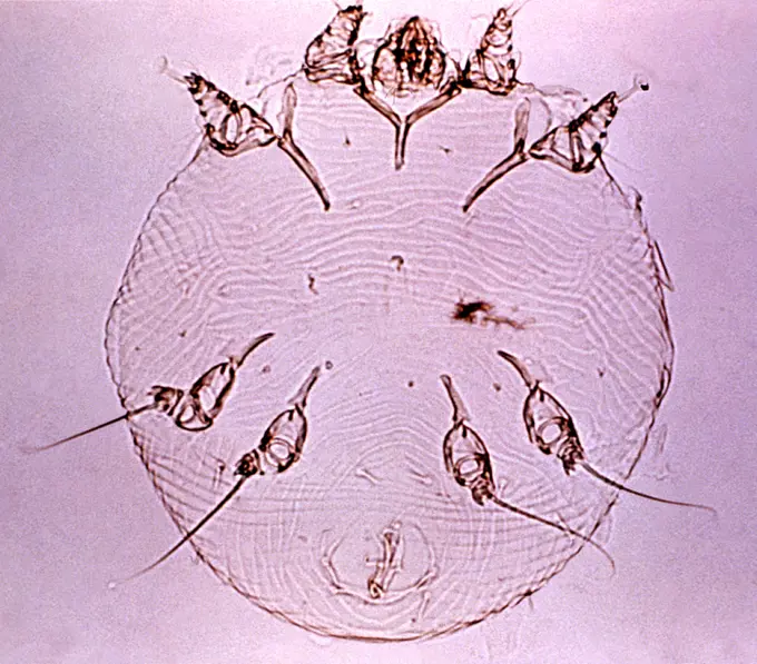
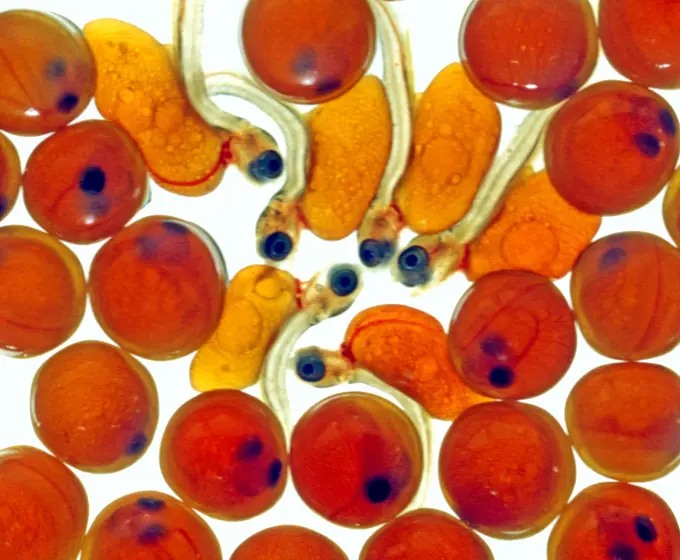
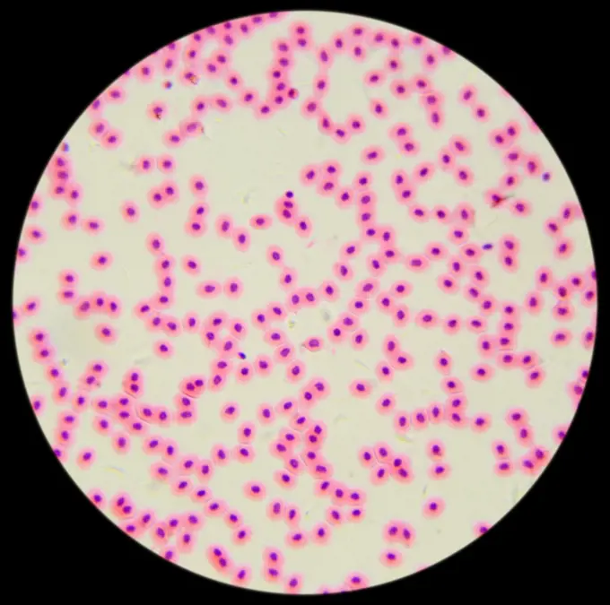
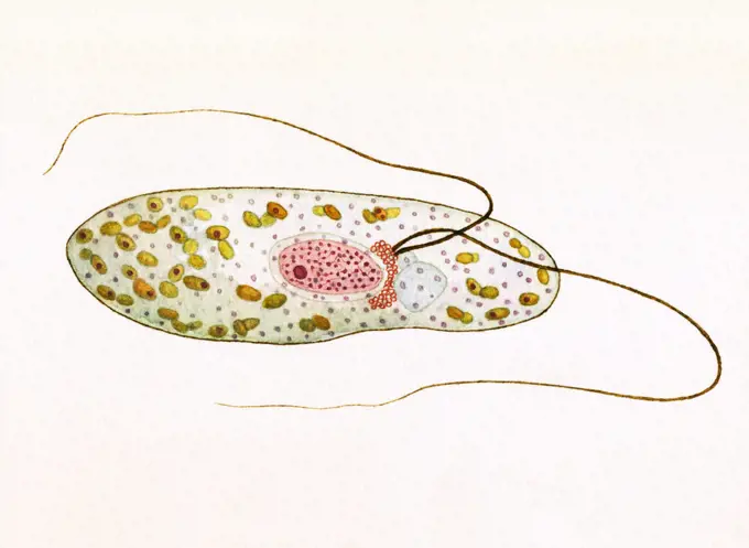
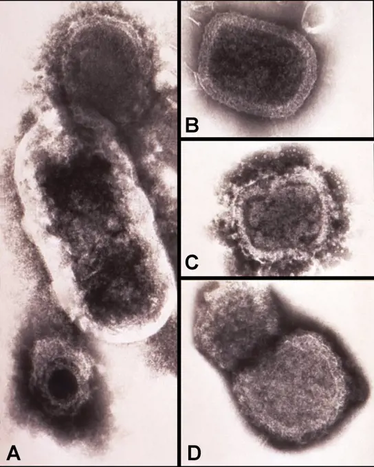
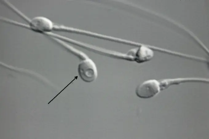
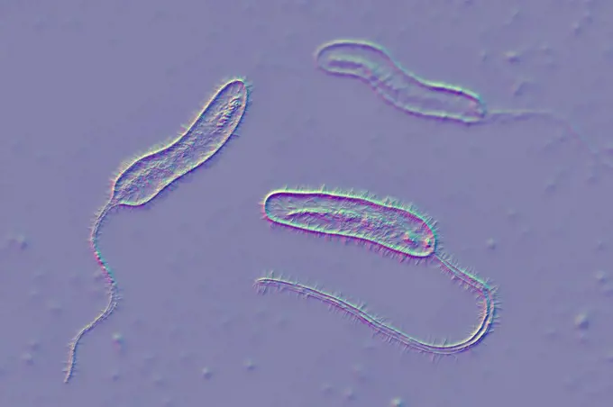
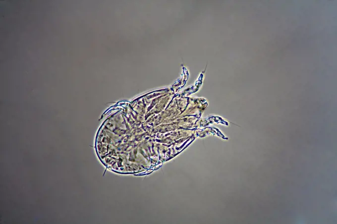
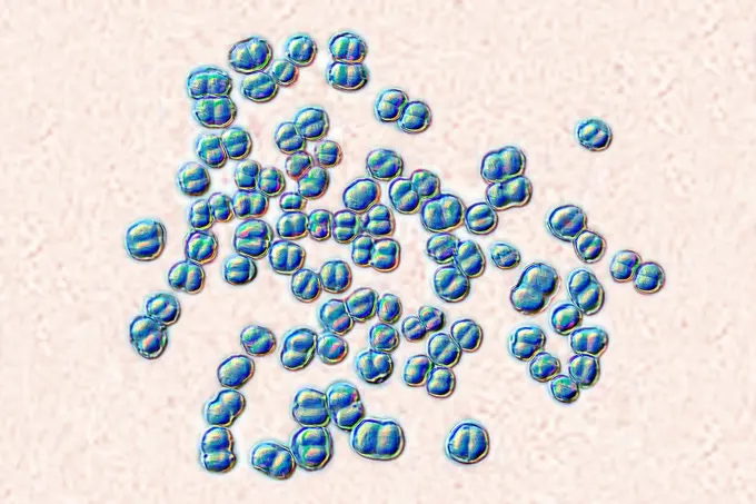
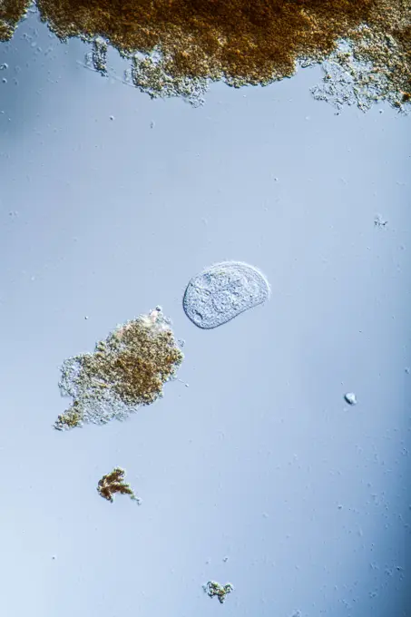
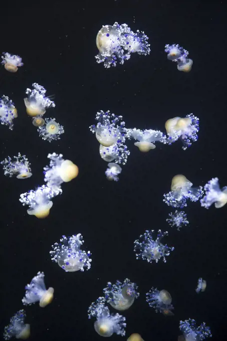
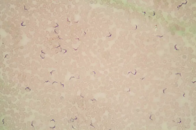
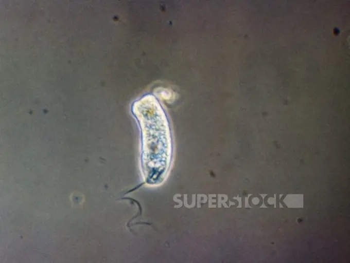
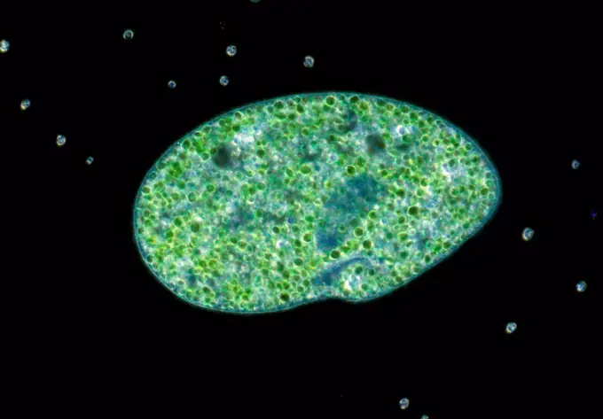
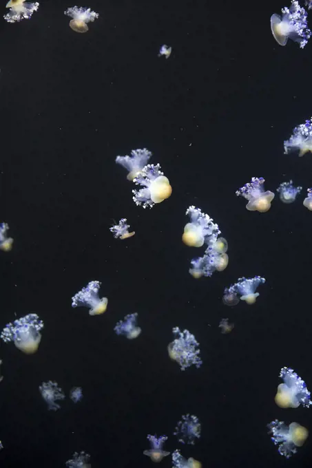
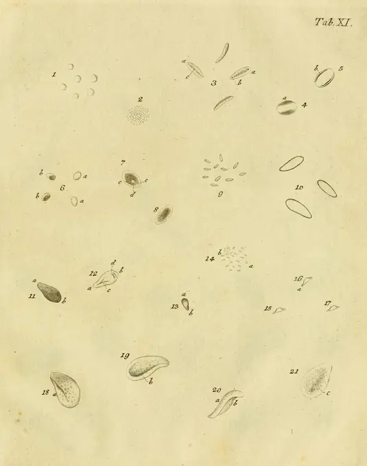
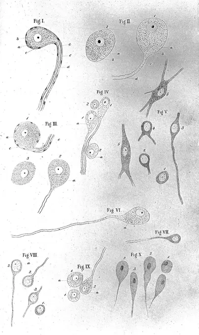
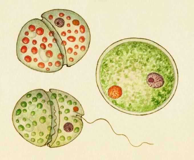
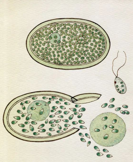
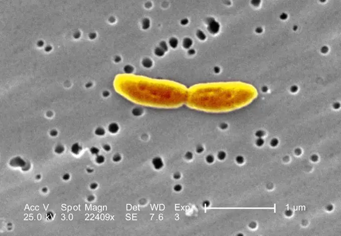
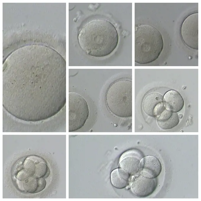
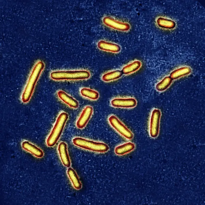
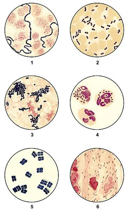
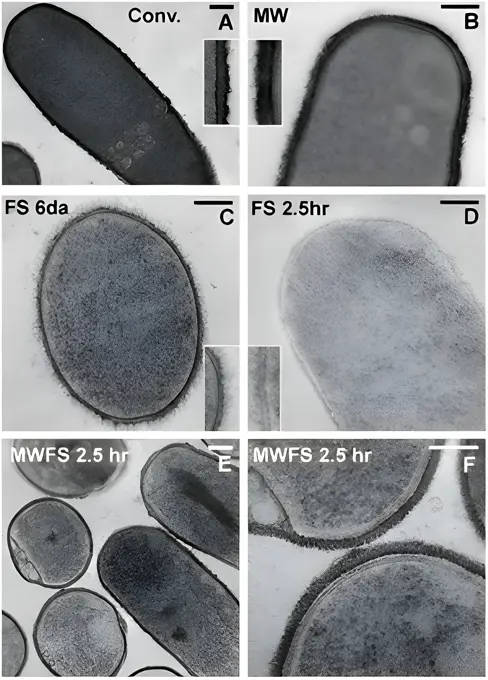
Microscopic Organisms and Tissue Types

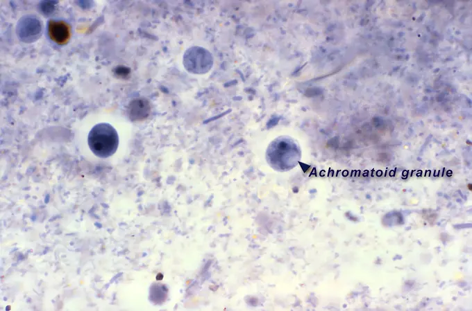
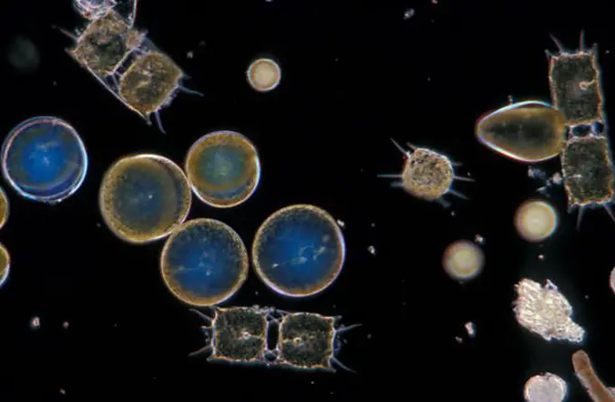
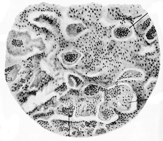
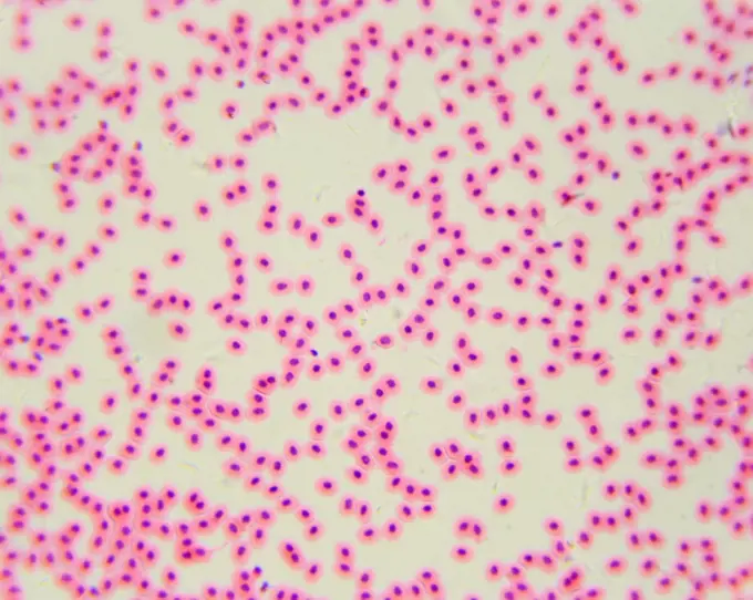
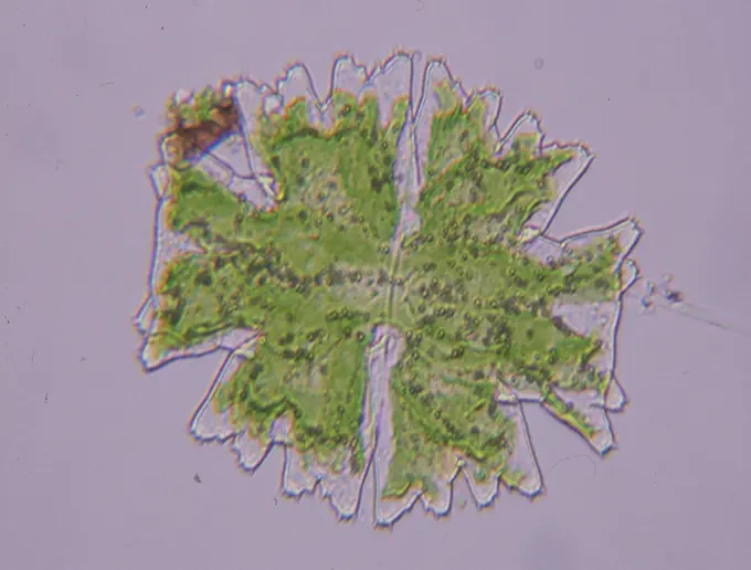
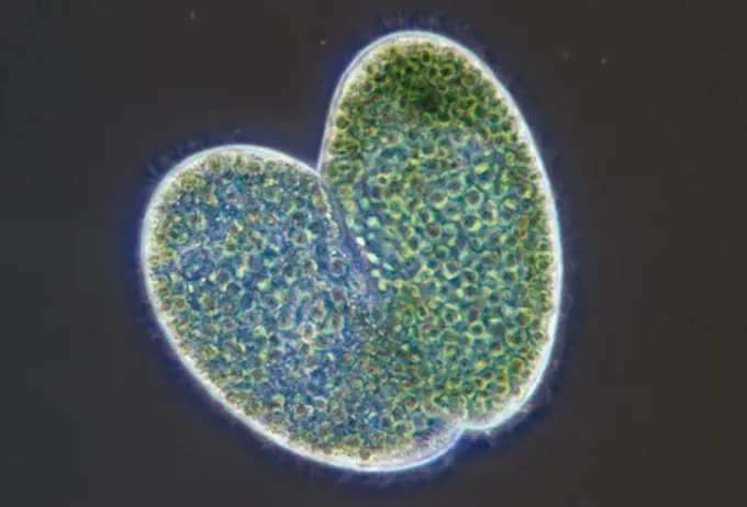
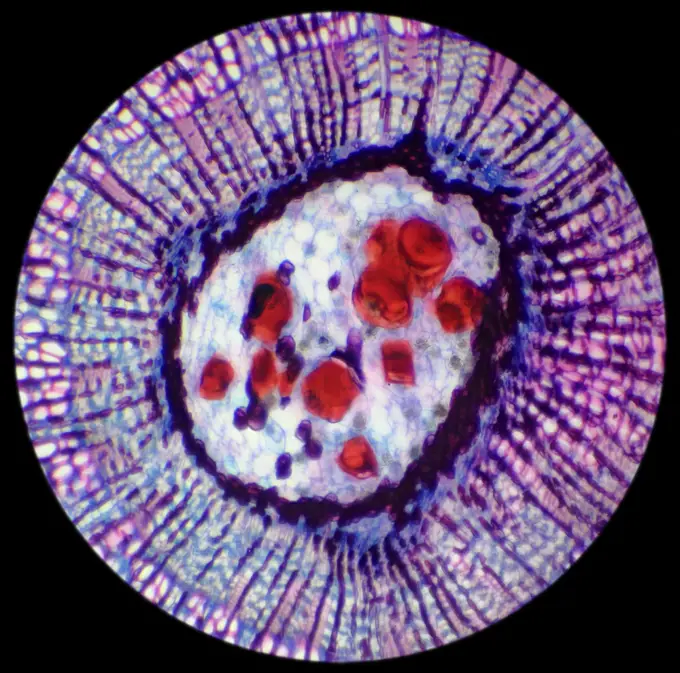
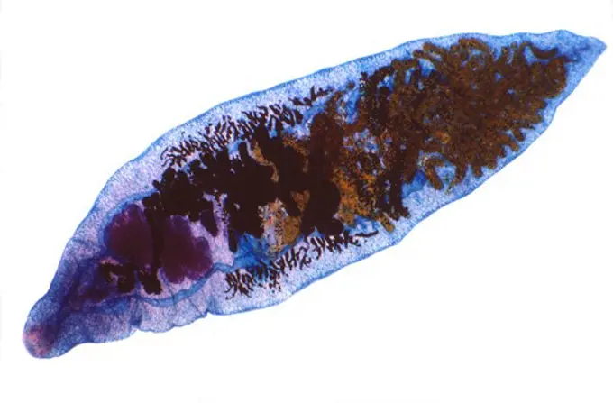
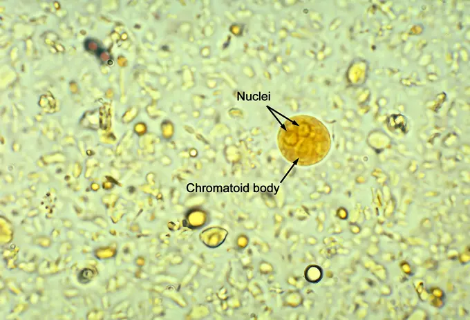
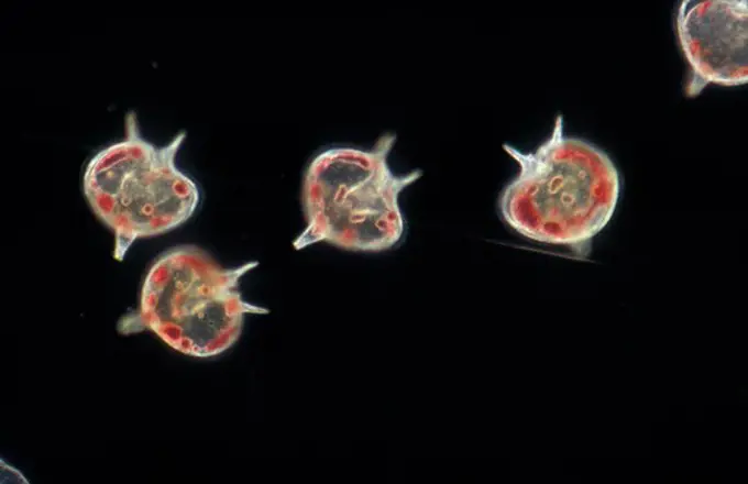
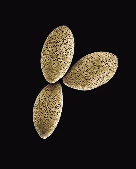
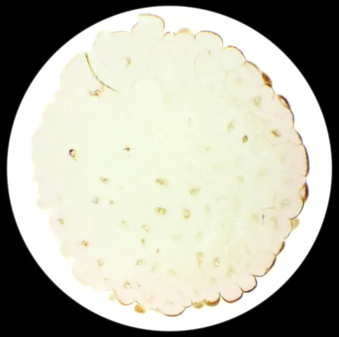
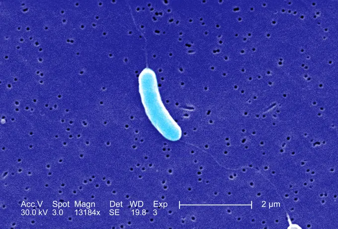
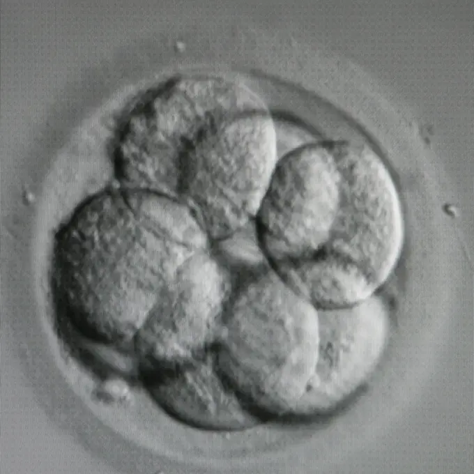
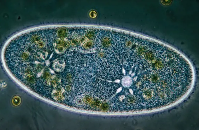
Close-up images of microscopic organisms, including amoebae and cancerous tissue sections, showcasing cellular structures and colors.

Microphotograph of a Section of the respiratory mucosa of a dog, at x100 magnification.

Assets in this Story

1899-85607
Vagina Cancer Histology
1899-61461020
Under a magnification of 250X, this photomicrograph of agar medium contained in a Petri dish, revealed the presence of numerous non-pathogenic Naegleria gruberi amoebae. CDC 1971
824-57659507
Under a magnification of 250X, this photomicrograph of agar medium contained in a Petri dish, revealed the presence of numerous non-pathogenic Naegleria gruberi amoebae. CDC 1971
1899-61460864
This photomicrograph of a trichrome-stained tissue sample revealed the presence of a cyst of a Vermamoeba vermiformis, formerly Hartmannella vermiformis, parasitic amoeba. This cyst morphology was atypical, as usually V. vermiformis contains only one nucleus, CDC/Dr George Healy 1973
1525-19842611
Stalk of a cereal cross-section under the microscope (Corn Stem C.S.), 400x
6188-55644741
Haseltimes hazel casuals bud crosss queps 100x copyright: xzooniups/nr.xnorborxlngxlzex 137569999919
824-63194926
This Giemsa-stained micrograph shows a Trypanosoma cruzi protozoan parasite during its leishmanial stage of development. T. cruzi, causes Chagas disease, a zoonotic disease that can be transmitted to humans by blood-sucking triatomine bugs. Here, the protozoa were cultured in a laboratory setting using a HeLa cell, S-3 culture.
1788-111173160
Two Protozoan, genus Stentor, seen under a microscope, at x68x2 magnification.
1788-111173562
Microphotograph of a Section of the respiratory mucosa of a dog, at x100 magnification.
4269-25179
Histology. Light microscopy of cervical smear showing normal epithelial cells.
824-57659425
This photomicrograph of an iodine-stained specimen revealed an Entamoeba hartmanni cyst, in which you can see four nuclei, along with a number of glycogen vacuoles and indiscernible rounded chromatoid bodies. CDC/Dr Mae Melvin 1977
1788-111173720
Histological slide of human testis seen under a microscope, at x600 magnification.
1899-53511539
Plasmodium falciparum is a malaria-causing parasite or malaria, transmitted by the bite of female anopheles which releases merozoites into the blood which enter red blood cells. Optical microscope view.
1788-111174213
Bicosoeca vacillans, Protozoan, and Chlorophyceae filaments seen under a microscope, at x210 magnification.
6145-44587637
Renal stones are never convenient, but they are a particular concern for astronauts who have limited access to treatment during flight. Researchers are examining how earthbound preventions for renal stone formation work in flight, ensuring missions are not ended prematurely due to this medical condition. The micrograph shows calcium oxalate crystals in urine. These small crystals can develop to form renal stones. Principal Investigator Dr. Peggy Whitson, NASA Johnson Space Center, Houston, TX.
1899-61461215
Pneumococcus (Streptococcus pneumoniae), pneumococcus is a bacteria involved in pneumonia, bronchopneumonia, purulent pleurisy, bacterial meningitis, otitis, sinusitis, and conjunctivitis. Image produced from optical microscopy
1899-61460904
This photomicrograph of an iron hematoxylin stained specimen revealed the presence of a number of amoeboid trophozoites, Entamoeba histolytica. CDC/Dr Mae Melvin 1977
824-57660031
Pneumococcus (Streptococcus pneumoniae), pneumococcus is a bacteria involved in pneumonia, bronchopneumonia, purulent pleurisy, bacterial meningitis, otitis, sinusitis, and conjunctivitis. Image produced from optical microscopy.
824-63210894
Reportage in the French Arkopharma lab in Nice, France. This pharmaceutical lab is specialized in the phytotherapy, natural medicine and dietary supplement sectors. It is European market leader. Analysing capsules that contain turmeric. Turmeric amyliferous parenchyma and reticulated vessel seen in a microscope enlarged X 400.
824-57659392
This photomicrograph of an iodine-stained specimen revealed an Entamoeba hartmanni cyst, which contained a single nucleus, along with diffusely dispersed glycogen particles. CDC/Dr Mae Melvin 1977
6145-29249415
Nasal mucosa: disseminated microsporidiosis with HIV
2001-106
Potato Starch Through Microscope
1899-61460996
This trichrome-stained slide revealed an Entamoeba histolytica trophozoite, which exhibited what was an atypical nucleus, and its peripheral chromatin was unevenly distributed. CDC/Dr Mae Melvin 1977
824-57659375
This photomicrograph of an iron hematoxylin stained specimen revealed the presence of a number of amoeboid trophozoites, Entamoeba histolytica. CDC/Dr Mae Melvin 1977
1788-111174462
Microphotograph of protozoan infusoria in a drop of water, using the cilia that cover their unicellular bodies to move in the liquid, at x100 magnification.
1899-61460908
This photomicrograph of a trichrome stained specimen, revealed the presence of a nonpathogenic intestinal amoeba, Endolimax nana cyst, within which two nuclei were visible. CDC/ Dr. Mae Melvin 1977
1899-61461008
This photomicrograph shows a number of cysts of the amoebic parasite Iodamoeba butschlii (buetschlii). Note the large, lightly colored glycogen vacuole contained within each cyst. CDC/Dr Mae Melvin 1977
6188-55644152
Farn Farn Sproß quer 100x Copyright: xZoonar.com/Dr.xNorbertxLangex 13693903
824-57659429
This photomicrograph of a trichrome-stained specimen revealed the presence of a mature Entamoeba coli cyst, in which you can see five visible nuclei, each with its eccentrically located karyosome and peripherally located chromatin. CDC/Dr Mae Melvin 1977
6188-55644763
Margarite Margarite Blüte im querschnitt, 100x Copyright: xZoonar.com/Dr.xNorbertxLangex 13756950
6188-55643879
Fuchsbandwurm Fuchsbandwurm worms in inficer liver 100x copyright: xzoonar.com/DR.XNORBERBERTXLANGEX 13660665
4297-1831
Light microscope image of a human blood smear
1525-19842581
Stalk of a cereal cross-section under the microscope (Corn Stem C.S.), 100x
1899-53511537
Plasmodium falciparum is a malaria-causing parasite or malaria, transmitted by the bite of female anopheles which releases merozoites into the blood which enter red blood cells. Optical microscope view.
1899-61460896
This photomicrograph of a trichrome-stained specimen, revealed a single Iodamoeba btschlii ( buetschlii ) parasitic cyst, within which youll note two achromatic glycogen granules (arrows), as well as a single, darkly stained intranuclear karyosome. CDC/ Dr. Mae Melvin 1977
824-57659378
This photomicrograph revealed a spherical, immature cyst of Entamoeba histolytica containing a single eccentrically located nucleus with its centrally located karyosome, a large lightly stained glycogen granule, and four elongated, blunt-tipped chromatoid bodies. CDC/Dr Mae Melvin 1977
1899-61461017
Magnified 1125X, this photomicrograph revealed the presence of a parasitic trophozoite Entamoeba histolytica, with its vacuolated cytoplasm, and single nucleus, which has its own centrally located karyosome. CDC 1971
1788-20761925
Polyhedral cells of animal tissue.
1788-111174032
Section of apex of vegetative stems of Allium roots with cells in the mitosis process.
1788-111174231
Opalina ranarum, Protozoan, observed in contrast phase, at x250 magnification.
4070-V19744585
824-57659358
This photomicrograph depicts a single Iodamoeba butschlii (buetschlii) parasitic cyst in which a large glycogen is well visible. CDC/Dr LLA Moore 1970
6188-55644765
Margarite Margarite Blüte im querschnitt, 100x Copyright: xZoonar.com/Dr.xNorbertxLangex 13756952
6145-29297169
Fragile X diagnostic test - normal
4128-17810092
Vorticella protozoa, light micrograph
1525-20635535
Micrograph of new sporophyte of fern leaf prothallium, background (Fern Prothallium W.M.)

824-57659426
This photomicrograph of a trichrome stained specimen revealed a mature Entamoeba coli cyst. In this focal plane, two nuclei were clearly visible, while four others were more difficult to discern. A splinter-like chromatoid body was also visible. CDC/Dr Mae Melvin 1977
6145-29292778
Herpes simplex virus infection
4413-134994
Microscopic view of Lily pollen with pollen tubes
6145-29256815
Candida albicans
824-57659503
Under 1125X magnification, this photomicrograph revealed the presence of a parasitic trophozoite, Entamoeba coli. The trophozoites of E. coli usually measure 15 m to 50 m. Each trophozoite has a nucleus with a characteristically large, eccentrically located karyosome and coarse, irregular chromatin located at the periphery. The cytoplasm is coarse and vacuolated, and is called dirty cytoplasm. CDC 1971
1788-111174160
Paramecium putrinum, Protozoan biciliate, seen under a microscope.
1788-111172846
Euglene, unicellular algae of the genus Euglena, seen under a microscope with counterstaining.
1899-61460935
This photomicrograph was created from an unstained culture specimen and revealed the presence of a parasitic trophozoite, Entamoeba moshkovskii, which had ingested a rice particle. This trophozoite had extended a single pseudopodium. CDC/Dr Mae Melvin 1977
1899-61461015
Under 1125X magnification, this photomicrograph of a trichrome-stained specimen revealed some of the ultrastructural detail exhibited by a parasitic trophozoite, Entamoeba coli. CDC 1971
824-57659499
This photomicrograph revealed the presence of a trichrome-stained parasitic trophozoite Dientamoeba fragilis in this specimen. CDC 1970
1788-111174036
In vitro culture of microphages (large mononuclear blood cells) of mice, seen with counterstaining.
1899-61460912
Using a methylene blue staining technique, this photomicrograph revealed the presence of two Entamoeba moshkovskii trophozoites, which had been cultured in the laboratory. CDC/Dr Mae Melvin 1977
1788-111173140
Pseudopodia formation of Amoeba (Amoeba proteus), Protozoan, seen under a microscope.
1788-111174144
Green algae with circular outline, genus Cosmarium, at x340 magnification.
1899-61460971
Under 900X magnification, this photomicrograph revealed the presence of a trophozoite of the parasitic organism, Entamoeba histolytica, in this tissue sample which had been harvested from an unknown breed of dog and prepared using Kohn's staining method. CDC/Dr Healy 1964
1525-24947301
Japanese paper
1525-19842588
Sunflower stalk cross-section under the microscope, (Helianthus Stem C.S.), 400x
824-57659416
This photomicrograph of an iron hematoxylin stained specimen revealed the presence of a parasitic trophozoite, Entamoeba histolytica. Note the distinct presence of a single, well-circumscribed nucleus containing peripherally located chromatin and, although difficult to see clearly in this focal plane, a slightly eccentric karyosome. CDC/Dr Mae Melvin 1977
1899-61461016
Under 1125X magnification, this photomicrograph revealed the presence of a parasitic trophozoite, Entamoeba coli. The trophozoites of E. coli usually measure 15 m to 50 m. Each trophozoite has a nucleus with a characteristically large, eccentrically located karyosome and coarse, irregular chromatin located at the periphery. The cytoplasm is coarse and vacuolated, and is called dirty cytoplasm. CDC 1971
1899-61460868
This photomicrograph shows you some of the ultrastructural morphology exhibited by an Acanthamoeba polyphaga parasitic cyst. CDC/Dr George Healy 1973
1788-111174373
Antheridiums and antherozoids of a Dryopteris fern, phase contrast microscopy, at x400 magnification.
1525-19842958
Stalk of a cereal cross-section under the microscope (Corn Stem C.S.), 400x
6188-62327757
Lillium anther Lillium anther under Optical Microscope Copyright: xZoonar.com/AlxCarrerax 21993637
1899-61460952
This photomicrograph of an iron-hematoxylin stained specimen revealed the presence of a mature Entamoeba coli cyst, which contained five visible nuclei, each with its eccentrically located karyosome and peripherally located chromatin. CDC/Dr Mae Melvin 1977
6188-55643843
Mushroom disease in the cattle pine fungal disease in the cattle pine fabric 200x copyright: xzoonar.com/DR.XNORBERBERTXLANGEX 13660578
1899-61460911
Using a methylene blue staining technique, this photomicrograph revealed the presence of a single Entamoeba moshkovskii trophozoite, which had been cultured in the laboratory. CDC/Dr Mae Melvin 1977
1788-111174125
Ciliated infusorians seen under a microscope.
4269-27183
Aspergillus. Light micrograph of a lung smear of a fungal threads of Aspergillus, pathogenic fungus agent of aspergillosis. Magnification x 100. Laboratory of Parasitology and Mycology, Department of Pr Dupouy-Camet, Cochin Hospital, Paris, France.
6188-58513100
Lillium anther Lillium anther under Optical Microscope Copyright: xZoonar.com/AlxCarrerax 21600953
1788-20761694
Batrachospermum, freshwater red algae viewed under a microscope.
6176-59553897
Euglena spec. (Euglena spec.), in differential interference contrast
4269-27802
Cryptosporidium sp. Light micrograph of Cryptosporidium sp, a parasite of the digestive and respiratory tracts, causing gastroenteritis (Cryptosporidiosis).
4298-1026
Filaments of blue algae (Cyanobacteria) Lyngbya, magnification 100x
824-57659383
Using a methylene blue staining technique, this photomicrograph revealed the presence of two Entamoeba moshkovskii trophozoites, which had been cultured in the laboratory. CDC/Dr Mae Melvin 1977
6188-67678406
Lillium-Anthere Lillium anther under Optical Microscope Copyright: xZoonar.com/AlxCarrerax 23137773
1397R-78369
Yellow bubbles in the water
4128-28767113
Tetanus bacteria, illustration
6188-55645499
Schalenamöben in division under the microscope 100x shellsam furniture in division under the microscope 100x copyright: xzoonar.com/dr.xnorberbertxlangex 13880828
824-57659360
This photomicrograph depicts a single Iodamoeba butschlii (buetschlii) parasitic cyst in which the nucleus appears to contain a single large, centrally located karyosome. CDC/Dr LLA Moore 1970
1525-19842937
Slice fern leaf under a microscope, (Fern Leaf Sec.), 400x
1525-20468376
Inkblot on the  paper. Elment of design.
4413-43444
Mouth of an Eucalyptus gall wasp larva
1848-69292091
Harmonic backgrounds (screensaver). Air bubbles in water (bleb) on light, smooth surface. Interior design, design creativity
1788-111172727
Mycobacterium leprae, also known as Hansen's coccus spirilly, bacterium that causes leprosy, seen under a microscope.
6145-29293640
Plasmodium falciparum malaria: lung
1788-111174140
Colonies of algae diatoms and filamentous blue-green algae seen under a microscope.
4201-21587639
Begonia (Begonia sp) stomata in leaf, magnified 120x
1788-111175410
Cross section of anthers of zucchini (Cucurbita pepo) seen under a microscope, at x9 magnification.
4201-22106234
Tunicate, Indonesia
1788-111177016
Epithelial cell of a human tongue in vivo, viewed using a phase contrast microscope at x250 magnification.
824-63179028
Invasive basal cell carcinoma, cancer of the uterus. Microscopy of a histological section x 500.
1899-61460942
This trichrome-stained photomicrograph revealed a parasitic Entamoeba histolytica trophozoite that contained a single round nucleus in which you can see peripherally located chromatin and a centrally located karyosome. Also note the presence of numbers of vacuoles in the cytoplasm of the organisms. CDC/Dr Mae Melvin 1977
1848-51687423
Volvox approx. 200x magnified in transmitted light
1848-51610905
Red algae (Porphyridium cruentum)
6188-55644757
Highspry headsprroom lease literates uttecuting 100x copyright: xzoonar.com/r.xnorrbortxlangex 1375693
6145-29269856
Trichomonas vaginalis
824-57659399
This photomicrograph revealed a good example of a binucleate parasitic trophozoite, Dientamoeba fragilis, stained with iron and hematoxylin, in this specimen (arrowhead). CDC/Dr Mae Melvin 1977
824-63211593
Meningococci (Neisseria Meningitidis), the bacteria in meningitis. Seen under optical microscopy X 1000.
1899-61460925
This photomicrograph revealed a good example of a binucleate parasitic trophozoite, Dientamoeba fragilis, stained with iron and hematoxylin, in this specimen (arrowhead). CDC/Dr Mae Melvin 1977
4141-32764
slipper animalcule paramoecium caudatum showing cilia.
6177-V53863465
1788-111177740
Microphotograph of a microscopic section of adipose tissue, at x400 magnification.
1890-105951
Light Micrograph LM of a plant cell chloroplasts, the site where photosynthesis takes place
6188-55645367
Water flea banted to living young animals 100x water flea banted live young animals 100x copyright: xzoonar.com/dr.xnorbertxlangex 13847646
4128-28767096
Cardiobacterium hominis bacteria, illustration.
4297-1854
RSV is the most common cause of bronchiolitis and pneumonia among infants and children under 1 year of age. The majority of children hospitalized for RSV infection are under 6 months of age
1525-24913082
Fountain
1525-19842908
Stalk of a cereal cross-section under the microscope (Corn Stem C.S.), 400x
1848-20188689
Conspicuous patterns at the bottom of the shallow lagoon Salinas de Cerrillos, aerial view, drone shot, Almería province, Andalusia, Spain, Europe
6188-55645408
Radies under the microscope in the water drop 100x wheel animal under the microscope in the water drop 100x copyright: xzoonar.com/dr.xnorbertxlangex 13852649
1788-111176989
Development stage of the embryo with initial outline of chicken organs (Gallus gallus).
4128-28767078
Bacteria of different shapes, illustration
824-57660052
Group A streptococci pyogenes. It is the cause of skin infections (impetigo), abscesses, bronchopulmonary infections, a form of sore throat which can develop into complications such as acute rheumatic fever. Image produced from optical microscopy.
824-63198885
Hepatitis B virus (HBV). HBV causes a sexually transmitted hepatitis, or through blood, which becomes cirrhosis in 20% of cases, 15-20 years after the beginning of the infection. Image produced using high-dynamic-range imaging (HDRI) from an image taken with transmission electron microscopy. Viral diameter around 50-60 nm.
1525-19842569
Stalk of a cereal cross-section under the microscope (Corn Stem C.S.), 400x
6145-29255513
Lung: miliary tuberculosis
6188-55645410
Water flea under the microscope in the water drop 100x water flea under the microscope in the water drop 100x copyright: xzoonar.com/DR.XNORBERBERTXLANGEX 13852653
824-57659410
This photomicrograph was created from an unstained culture specimen and revealed the presence of a parasitic trophozoite, Entamoeba moshkovskii, displaying a single pseudopodia. CDC/Dr Mae Melvin 1977
1899-61460918
Under 31X magnification, this photomicrograph of an agar medium revealed the presence of numerous non-pathogenic Naegleria gruberi amoebae. CDC 1971
1525-25694037
Hemerocallis citrina mature anther under the microscope - Abstract pink dots on white background.
6188-55644670
Pea grate pea rust rust mushroom on wolf milk, 200x copyright: xzoonar.com/dr.xnorberbertxlangeex 13751947
6145-46761714
Lumière autochrome. Medical photographs.A blood cells 12 days. O-serum widespread. Obj. A 2. Hämolysis.
1899-61460981
Under 900X magnification, this photomicrograph revealed the presence of an Entamoeba coli microorganism at the cyst stage, in a sample prepared using the Kohn stain method. In this focal plane you can see two distinct nuclei, each containing a single centrally located karyosome. CDC/Dr Healy 1964
1848-49561584
Sarcodina, Acanthocystis turfacea, "Green Sun Animalcule" at 200x
1788-111172936
Group of algae colonies, genus Pleodorina with counterstaining.
1525-24328209
Good luck and 3d render of gambling games with big jackpot win. Profitable success bets in card game and roulette. Club casino for entertainment of wealthy clients. Good luck and 3d render of gambling games with big jackpot win. Profitable success bets in card game and roulette. Club casino for entertainment of wealthy clients.. Falling poker gold chips
6188-55645413
Radies under the microscope in the water drop 100x wheel animal under the microscope in the water drop 100x copyright: xzoonar.com/dr.xnorbertxlangex 13852659
1788-111172586
Pylaiella (mung), brown algae, seen under a microscope.
1899-61460890
This photomicrograph depicts a single Iodamoeba butschlii (buetschlii) parasitic cyst in which the nucleus appears to contain a single large, centrally located karyosome. CDC/Dr LLA Moore 1970
4413-109749
Microscopic view of pollen Hollyhock
4413-109845
Microscopic view of Spore and Sporangia Moss Barbula
4128-19056160
Lactococcus bacteria, illustration
1899-61460899
This trichrome-stained photomicrograph revealed some of the ultrastructural features exhibited by a parasitic organism known as Entamoeba histolytica. This mature cyst exhibited an oblong-shaped chromatin body, and in this focal plane, three of the organisms' four nuclei were clearly visible, each with its centrally located karyosome. A nucleus was blurred in this view. CDC/Dr Mae Melvin 1977
4391-101
A photo showing normal lymphocytes compared with a Reed-Sternberg cell, which are large, abnormal lymphocytes that may contain more than one nucleus. These cells are found Hodgkin lymphoma.
6145-46838337
2 Pence of George VI, King of Great Britain from Board ofRevenue. Ruler: George VI, King of Great Britain, British, 1895-1952, ruled 1936-52 Mint: Board of Revenue Artist: Unknown
1899-61460892
This photomicrograph of an iron-hematoxylin stained specimen revealed the presence of two spheroid parasitic cysts, Iodamoeba butschlii (buetschlii). The cyst on the right contained a clearly visible achromatoid granule in its cytoplasm (arrow). CDC/Dr Mae Melvin 1977
1848-51674419
Plankton, phytoplankton, winter, x30, coscinodiscus graniBiddulphia regia
1525-28047120
Giant cell sarcoma, vintage engraved illustration.
1525-19882730
Chicken red blood cells under a microscope (blood smear Chicken), 400x

4141-32838
freshwater gastrotrich a ciliated flatworm
4128-20041274
Athlete's foot fungus, illustration
6188-68368798
Die mikroskopische Welt. Ein Tropfen Marmelade unter dem Mikroskop. The Microscopic World. Drop of jam under the microscope. LicenseRF Copyright: xZoonar.com/DmitrixMarutax 23262509
1899-61460966
Under a magnification of 1750X, this photomicrograph of a nasal swab specimen, revealed the presence of numerous, trophozoite-staged amoebae. The species was not indicated. CDC
1525-20635536
Pine needle cross-section under the microscope, background, (Pinus)
1890-110368
Light Micrograph LM of the transverse section of Dehiscence Lilium Anthers with pollen, magnification x300
4413-109764
Cross section of a primary root of buttercup
1899-61461219
Pneumococcus (Streptococcus pneumoniae), pneumococcus is a bacteria involved in pneumonia, bronchopneumonia, purulent pleurisy, bacterial meningitis, otitis, sinusitis, and conjunctivitis. Image produced from optical microscopy
6145-44595021
KENNEDY SPACE CENTER, FLA. -  Pictured is one of the microcapsules removed from the Commercial ITA Biomedical Experiments payload recovered during the search for Columbia debris.  The drug delivery system and spaceflight hardware was developed jointly by JSC, the Institute for Research Inc. and Instrumentation Technology Associates Inc. to conduct microencapsulation experiments under microgravity conditions.  This microcapsule contains an antibiotic for treating deep resistant pulmonary infections.  Dr. Dennis Morrison, senior biotech project scientist, is principle investigator on microencapsulation and urokinase crystal growth.
6145-29605686
Collection Dutch Heidematschappij. Microfoto Pine tree birch time. undated
4384-181
A transmission electron micrographic (TEM) image of Bacillus anthracis from an anthrax culture, showing cell division (right), and spores (left).
824-57659457
Under 900X magnification, this photomicrograph revealed the presence of a trophozoite of the parasitic organism, Entamoeba histolytica, in this tissue sample which had been harvested from an unknown breed of dog and prepared using Kohn's staining method. CDC/Dr Healy 1964
1899-61460547
Acanthamoeba is a microscopic, free-living amoeba, or amoeba (single-celled living organism), that can cause rare but serious eye, skin, and central nervous system infections. The amoeba is found worldwide in the environment in water and soil. The amoeba can spread to the eyes through the use of contact lenses, cuts or wounds in the skin, or by being inhaled into the lungs. Most people will be exposed to Acanthamoeba in their lifetime, but very few will become ill from this exposure
4201-40805
Tardigrade (Pseudobiotus megalonyx) microscopic image, animal can reach more than one mm in length, lives in fresh and brackish water in Europe
1788-111176130
Microphotograph of the segmentation process of the fertilized egg of a frog, beginning of the second division of the egg.
1899-61461194
Bacillus Koch (Bk) or Mycobacterium tuberculosis, it is responsible for tuberculosis. Tuberculosis is making a comeback due to the appearance of multi-resistant bacterial strains. Pulmonary tuberculosis (phthisis) is the most widespread, but there are bone disorders (Pott's disease, white tumor of the knee), renal, intestinal, genital, meningeal and skin disorders (tuberculomas). Image taken from an X 1000 optical microscopy
6188-55645574
Zieralge unter dem Mikroskop 100x Zieralge unter dem Mikroskop 100x Copyright: xZoonar.com/Dr.xNorbertxLangex 13915965
6145-45241824
This image shows crystals of the protein raf kinase grown on Earth (photo a) and on USML-2 (photo b). The space-grown crystals are an order of magnitude larger. Principal Investigator Dan Carter of New Century Pharmaceuticals
1848-18102794
Perigord truffle, black truffle (Tuber melanosporum) Spores
1525-25873589
bright jellyfish floating in the dark sea water
1788-111172622
Rivularia planctonica, blue-green algae, seen under a microscope.
4384-273
Photomicrograph of Simian Blood Sample
4070-19576544
Coral growing in marine tank where impacts of complex environmental changes are researched. National Sea Simulator, Australian Institute of Marine Science, Cape Ferguson, Queensland, Australia. 2019.
4128R-13619959
Human tooth with accumulation of bacterial plaque (smooth areas) and calcified tartar (rough areas) on enamel surface, coloured scanning electron micr...
4141-32751
phytoplankton mixed species from eutrophic lake, mag.x80 anoptal contrast microscopy
6188-55645417
Sea red algae in the water under the microscope 100x sea red algae in the water under the microscope 100x copyright: xzoonar.com/DR.XNORBERBERTXLANGEX 13852666
4201-66211
Fruit Fly (Drosophila melanogaster) embryos at 12x magnification, Spain
1746-30002811
Starch grains of wrinkled pea. From AD Darbishire, Breeding and the Mendelian Inheritance. London, 1912.
1525-25873590
bright jellyfish floating in the dark sea water
4297-1805
Electron microscope image of cytomegalovirus
6188-55645337
Zieralge Micrasterias im Wassertropfen 100x Zieralge Micrasterias im Wassertropfen 100x Copyright: xZoonar.com/Dr.xNorbertxLangex 13819627
4141-32753
slipper animalcule living ciliates paramaecium bursaria in conjugation, photographed under anoptal phase contrast microscopy
1525-20635509
Cross-section of the stem woody plant under the microscope (Basswood Stem C.S.)
4413-43373
Larvae of Brown Soft Scale under mother's carapace
4141-32746
liver fluke of sheep distomum sp. (possibly d. lanceolatum) small fluke parasitic on sheep
1788-111174201
Protozoan, genus Polytoma, seen under a microscope, at x210 magnification.
6145-45250809
Over a billion of mostly third world people are infected with a roundworm known as ascarids. Ascarids are tiny parasites that infect the intestinal tract of vertebrates. Movement of the larvae into the brain or other parts of the body can prove fatal. Space-based research is providing new hope in combating these parasitic worms. Ascarids are dependent upon a substance known as malic enzyme to regulate certain bodily functions. A new drug designed to interfere with normal functioning of malic enzyme should prove deadly to ascarids. The Center for Macromolecular Crystallography, along with the University of North Texas grew malic enzyme crystals on the USML-1 Spacelab mission. Although these crystals proved to be smaller than ground based ones, they were more perfectly formed, therefore producing better data for drug design.
6145-29245774
Charcot Leyden crystals from an endobronchial lesion
824-63168548
Report at the Rouen University Hospital, PMA (Medically Assisted Procreation) service. Biology laboratory. IVF by ICSI.
4269-25256
Neural stem cell. Cultured neural stem cells (multipotent nerve cells) of transgenic mouse on adhesive substrate. At center, a sphere of glial cells (astrocytes), able to reproduce by mitosis, gives by differenciation nerve cells (neurons), seen on the outside of the sphere.  Light Micrograph.
824-63194920
This Giemsa-stained light photomicrograph revealed the presence of two Trypanosoma brucei parasites, which were found in a blood smear. Humans are the main reservoir for Trypanosoma brucei gambiense, but this species can also be found in animals.
824-63211604
Staphyloccus aureus. Bacteria responsible for food poisoning, localized suppurative infections and some cases of septicemia. Seen under optical microscopy X 1000
4220-21334606
Mouse Embryo at Two Cell Stage & a Polar Body.     Date:
6188-55644860
Spitzwegerich Spitzwegerich Stengel cross -section 100x copyright: xzoonar.com/dr.xnorbertxlangex 13769912
4201-40350
Nematode (Euplidogaster striatus) worm
1788-111172976
Section of the thallus of bladderwrack, Fucus vesiculosus, Fucaceae, with female conceptacles, seen under a microscope.
4128R-13682530
Viruses, illustration.
4269-27773
Scabies. Micrograph of a Sarcoptes scabiei var. hominis, an ectoparasite acarus responsible for scabies, which burrows under the sensitive sections of skin, causing severe irritation.
6145-54585138
A close view of salmon eggs and developing salmon fry. Subjects: Fishes; Fisheries management; Alaska.
1525-19886833
Chicken red blood cells under a microscope (blood smear Chicken), 400x

6177-V53477232
6145-45506985
Bliżej Kultury
1788-111172814
Vacuolaria virescens, Chloromonadophyceae. Enlarged drawing.
1746-21103924
High magnification of 150,000X, negatively-stained transmission electron micrograph (TEM) revealing some of the ultrastructure morphology exhibited by a number of different microorganisms. Panel 'A' represents a composite micrograph, for comparing the size difference between a poxvirus at the top, a bacillus in the middle and a herpesvirus at the bottom. Panels 'B', 'C' and 'D' are TEMs depicting the sequential degeneration of variola virus patricles.
4128-V58573367
824-63168547
Report at the Rouen University Hospital, PMA (Medically Assisted Procreation) service. Biology laboratory. IVF by ICSI.
4128-20044975
Cholera bacteria, illustration
4413-109880
Microscopic view of the chicken mite
1525-20846633
Swiss Cheese 02
1899-61461208
Meningococcus or meningococcal disease or Neisseria meningitidis, is a bacteria involved in meningitis. These bacteria are the cause of mild pharyngitis and only one person in approximately 400 becomes the victim of a serious meningococcal infection, most often in the form of acute purulent meningitis. Image produced from optical microscopy
6188-55561711
Ciliaten auf Futtersuche Ciliaten auf Futtersuche Copyright: xZoonar.com/Dr.xLangex 13174906
1525-22717524
bright jellyfish floating in the dark sea water
6188-55643647
Schlafkrankheit Schlafkrankheit Trypanosomen unter dem Mikroskop im Blut 400x Copyright: xZoonar.com/Dr.xNorbertxLangex 13630470
1788-20761710
Euglena unicellular alga viewed under a microscope, Euglenaceae.
4128R-13620000
Darkfield light micrograph of Paramecium bursaria, a ciliate protozoan, that contains endosymbiotic green algae (Chlorella sp.). Paramecium are found ...
6177-V53848781
1525-22717530
bright jellyfish floating in the dark sea water
4443-73161323
Animalcula, Infusoria, Fluvia, Tilia, Et, Marina, Hauniae, Typis, N. Mölleri, 1786, Infusoria, Animalcules, A meticulously illustrated plate showcasing various botanical structures, featuring a range of seeds and spores. The collection is systematically organized, with intricate details highlighting each specimen. Numbers accompany each illustration, indicating individual species or types. The diversity in shape and size is evident, with circular, oval, and elongated forms, capturing the intricacies of plant reproduction. This educational representation emphasizes the significance of each element in botanical studies, reflecting a historical approach to plant morphology and classification.
6145-52833945
A pencil sketch by Carl von Hasenauer depicting architectural design and details on paper.
824-57659491
This iodine-stained photomicrograph revealed ultrastructural morphology exhibited by a binucleate parasitic amoebic cyst of Entamoeba histolytica, in a concentrated wet mount specimen. Notice the chromatoid body with blunt and rounded ends, and that only two nuclei are visible in this focal plane. CDC/Dr Mae Melvin 1977
4421-25060
Plankton - Dinoflagellate (Peridinium depressum) x 43
1848-49277173
Blue Liquid falling into Water against White Background
4201-66319
Torch Lily (Kniphofia sp) SEM close-up view of pollen at 1050x magnification
6145-44934449
Earth Observations taken by Expedition 34 crewmember.
6145-29256509
Nerve cells.
4128-20044978
Cholera bacteria, illustration
1788-111172842
Stages of the life cycle of Glenodinium sanguineum, Dinoflagellates. Drawing.
1788-111172770
Biflagellated gametes of Acetabularia, Polyphysaceae. Drawing.
4297-1457
Scanning electron micrograph (SEM) at an extremely high magnification of 44, 818X, of a single dividing Gram-negative Escherichia coli bacterium
1525-19842940
Frog egg under the microscope, background. (Frogg Egg Sec.) 100x

824-63128596
This scanning electron micrograph (SEM) depicts a flagellated Vibrio vulnificus bacterium, Mag. 13184x. Vibrio vulnificus is a bacterium in the same family as those that cause cholera. It normally lives in warm seawater and is part of a group of vibrios that are called halophilic because they require salt. V. vulnificus can cause disease in those who eat contaminated seafood or have an open wound that is exposed to contaminated seawater. Among healthy people, ingestion of V. vulnificus can cause vomiting, diarrhea, and abdominal pain. In immunocompromised persons, particularly those with chronic liver disease, V. vulnificus can infect the bloodstream, causing a severe and life-threatening illness characterized by fever and chills, decreased blood pressure (septic shock), and blistering skin lesions. V. vulnificus bloodstream infections are fatal about 50% of the time. See image n0831905 on the site.
824-63168551
Report at the Rouen University Hospital, PMA (Medically Assisted Procreation) service. Biology laboratory. IVF by ICSI.
4141-32736
slipper animalcule compressed paramecium aurelia magnification x125
1525-20337735
Human IVF
824-57660038
Pseudomonas aeruginosa or pyocyanin bacillus, pathogenic bacteria, very resistant and often responsible for nosocomial infections. It is one of the most difficult bacteria to eliminate clinically. The infections it causes concern: the eyes, burns, surgical wounds, urine, lungs, stomach, intestines, meninges, blood. Image taken from an X 1000 optical microscopy.
1848-61699854
Bacteria smears of germs for microbiological diagnosis, microscope slides, clinical microbiology, state of scientific research around 1915, 1: Streptococci, 2: Pneumococci, 3: Staphylococci, 4: Gonococci, 5: Sarcina, 6: Influenza virus of the influenza epidemic of 1892
1525-20334880
Human IVF
1899-20311106
 Rouen University Hospital, France. Reproductive biology laboratory.
824-63225798
Transmission electron microscope images of Bacillus subtilis, using ambient and cryo-preparative methods. Mid-log phase cultures were prepared by conventional chemical fixation (A), MW-assisted chemical fixation (B), traditional FS (C), or by rapid FS without (D) or with MW irradiation (E-F). Bars: 50 nm...
6145-52893972
Culpepper Compound Microscope Slide, 18th Century. From microscope storage box 1965T5099.1.. Purchased from Edward H Pinto, 1965.
4443-73206796
Hundt, Fr., Rutherford, Lewis M., Sonnenfleckengruppe in ihren Changeungen an sieben Tagen, photograph, foto, foto's, height mm, width mm, photographer, 1870-09 - 1870-09, third quarter 19th century, salted paper print, salt print