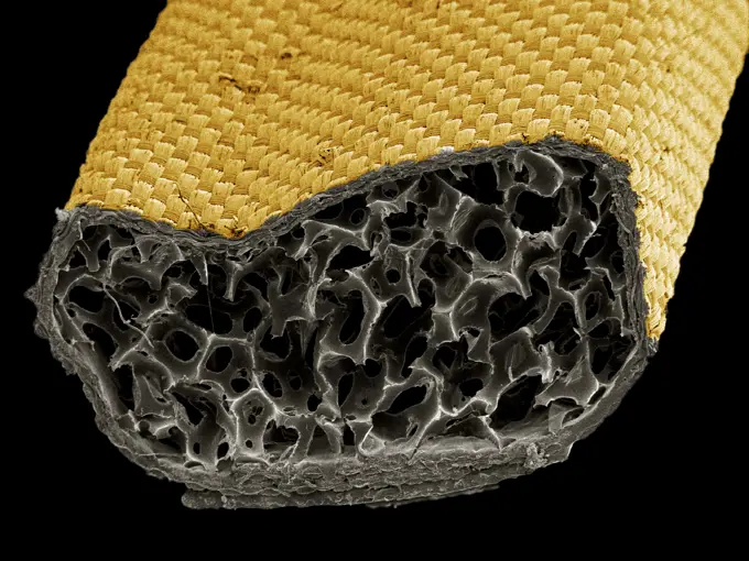
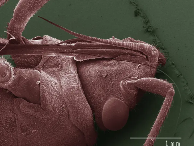
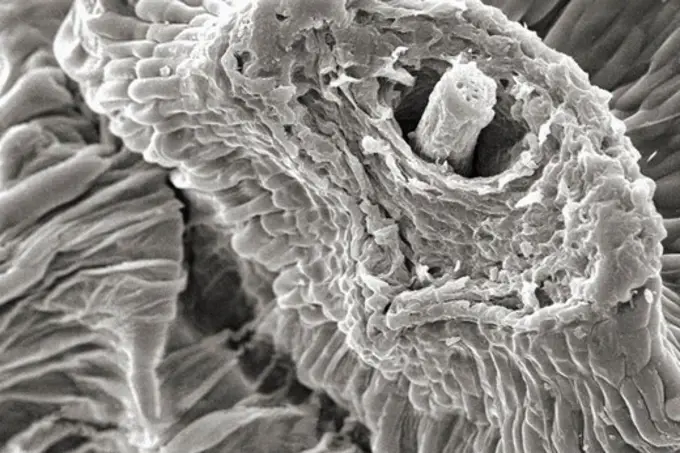
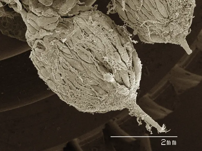
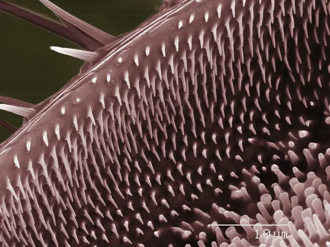
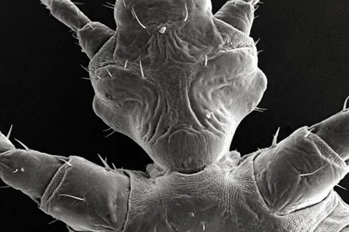
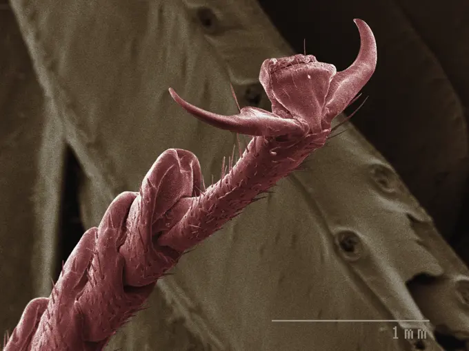
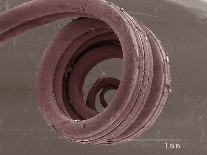
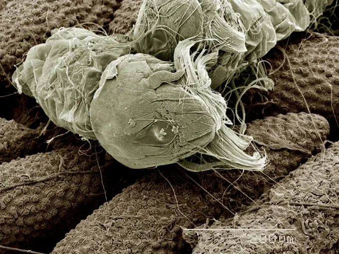
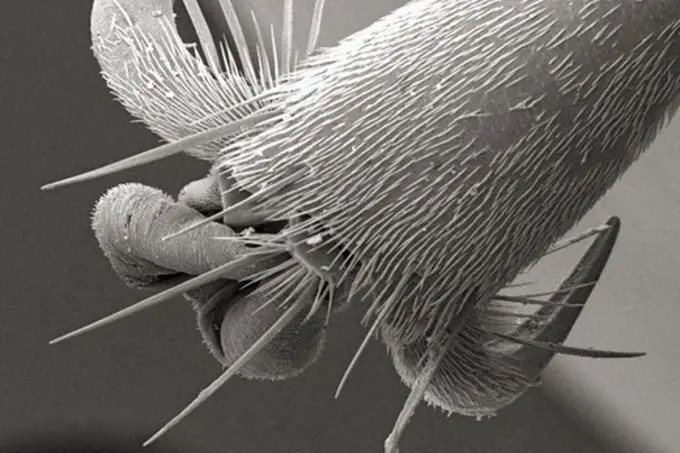
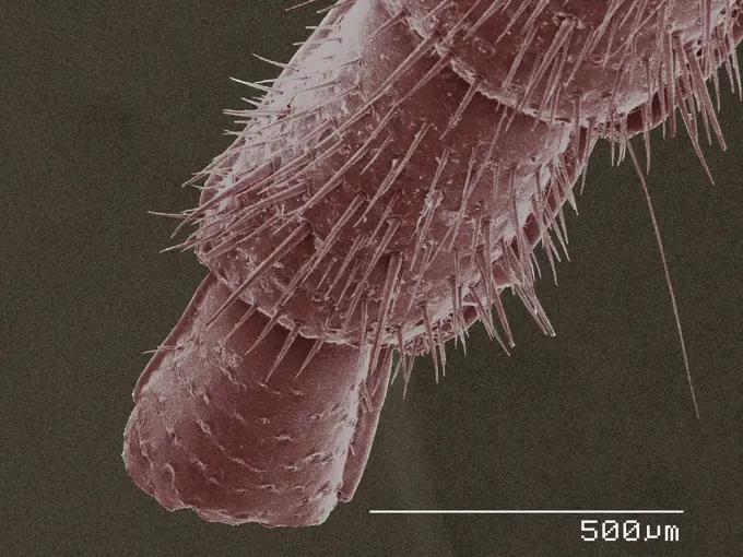
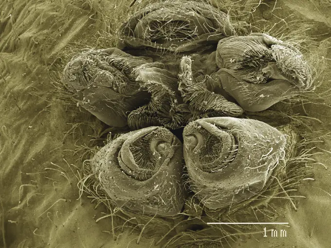
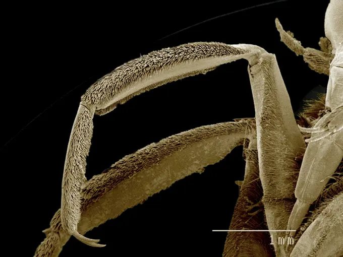
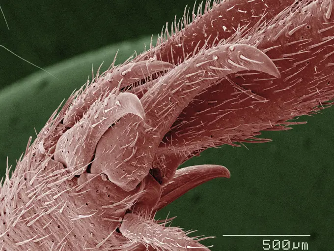
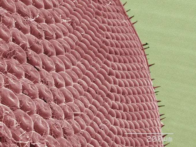
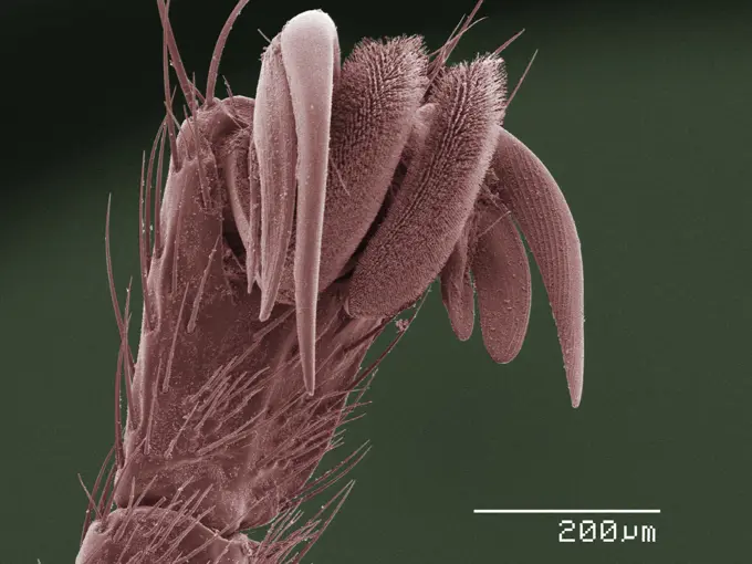
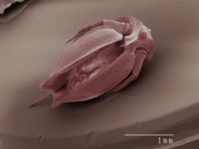
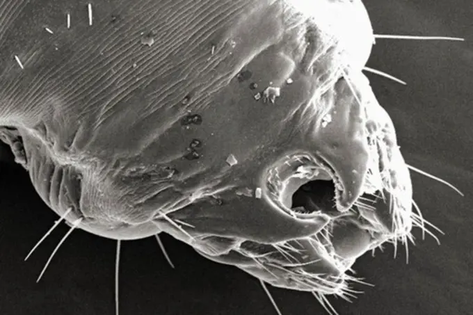
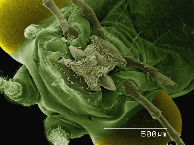
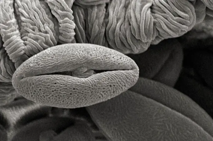
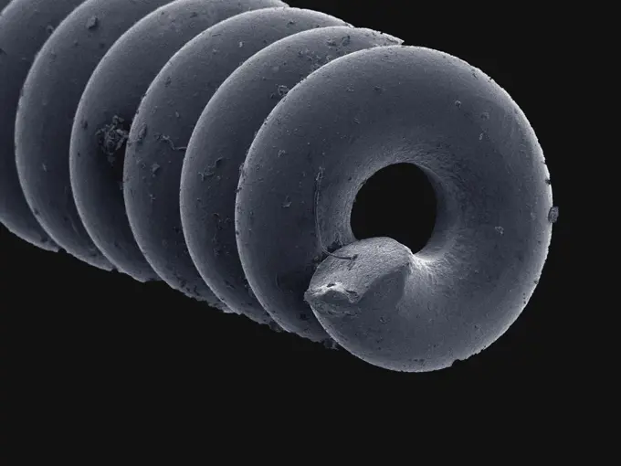
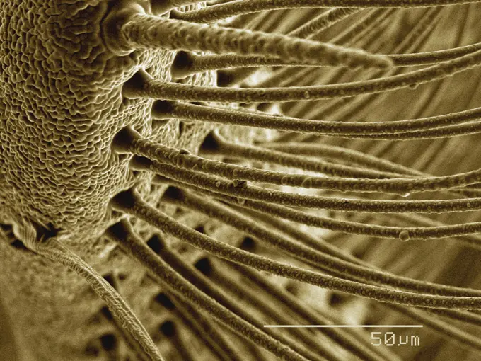
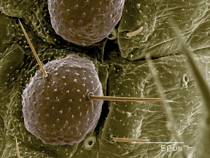
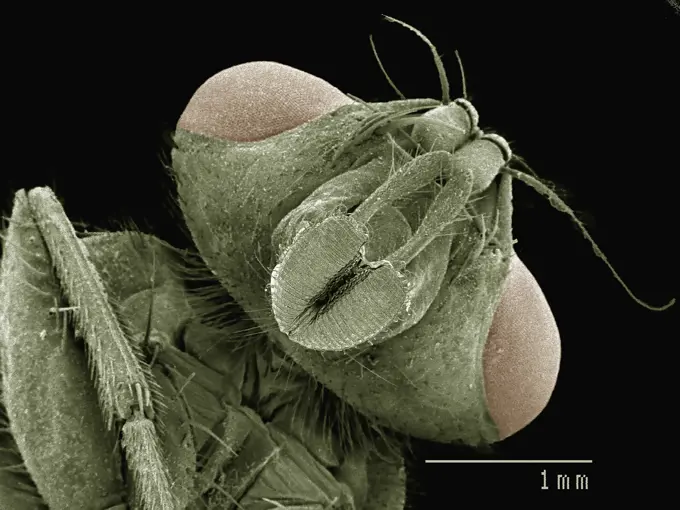
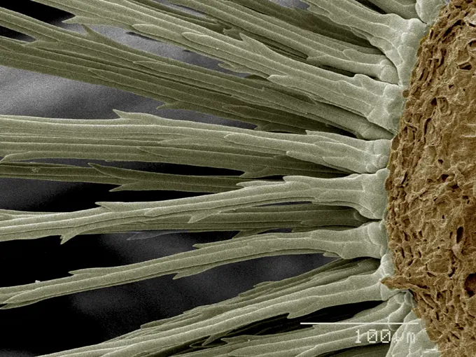
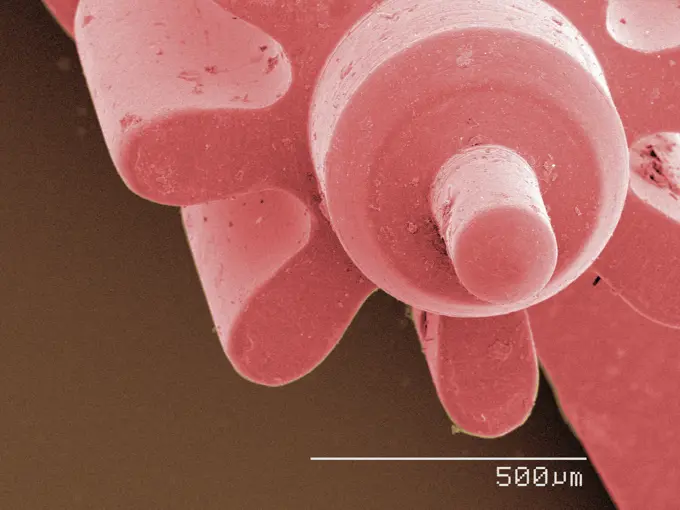
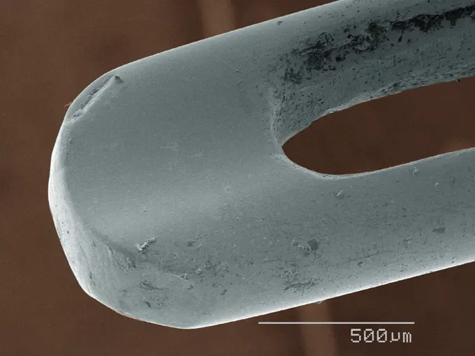
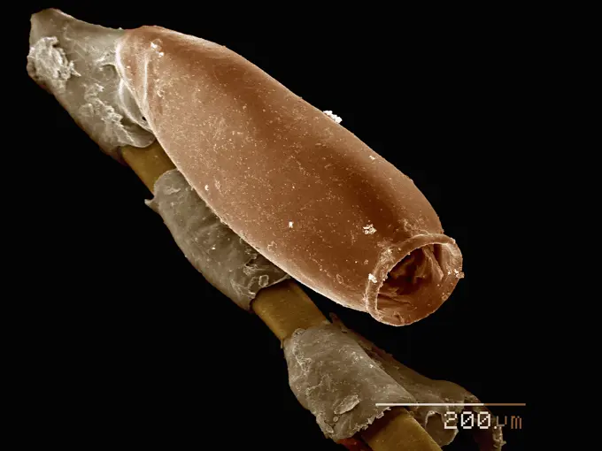
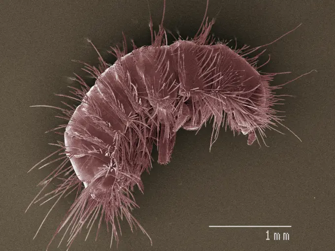
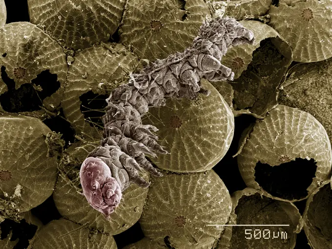
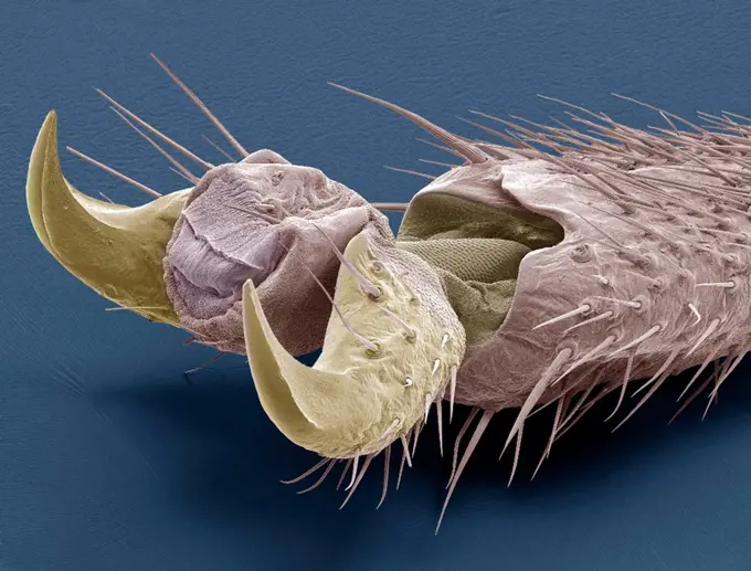
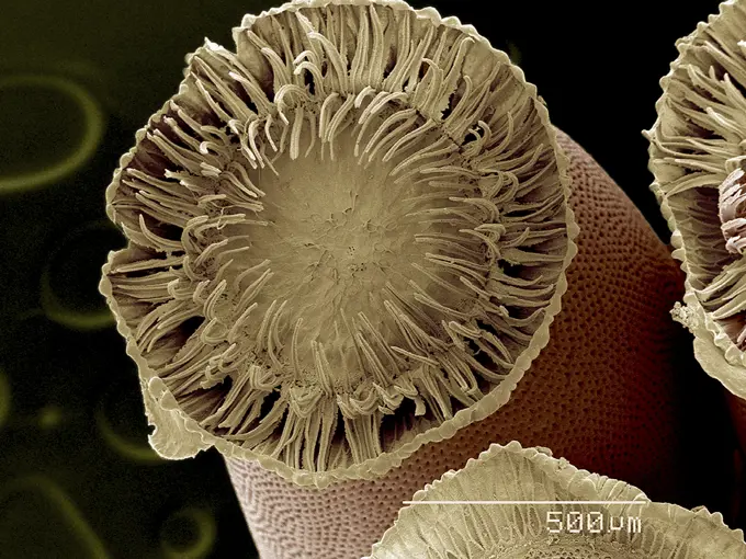
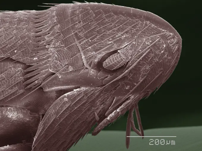
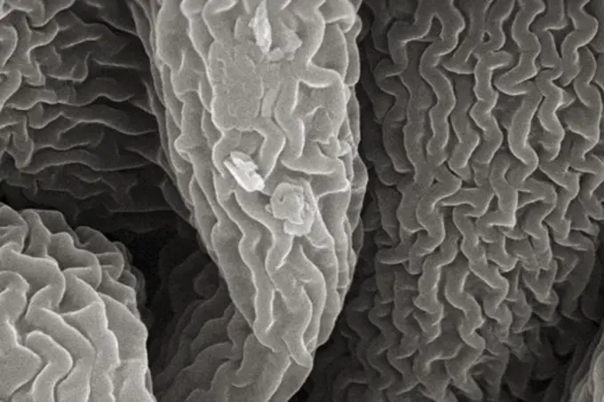
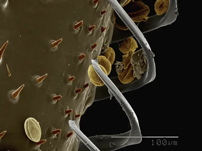
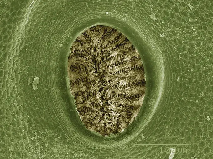
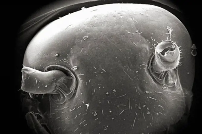
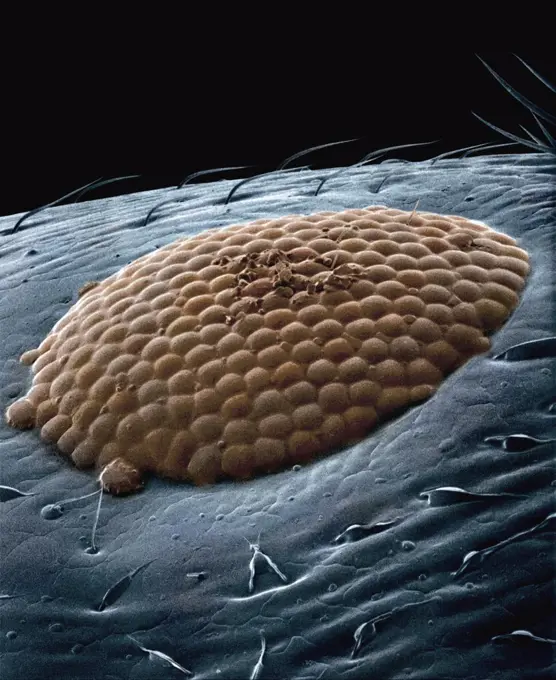
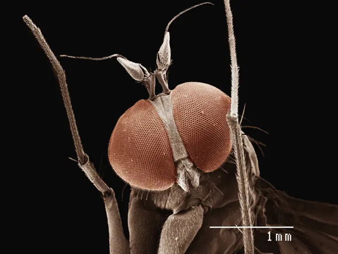
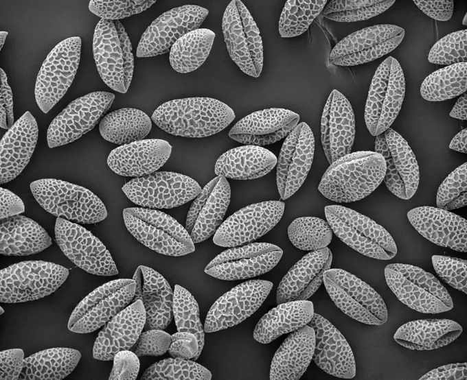
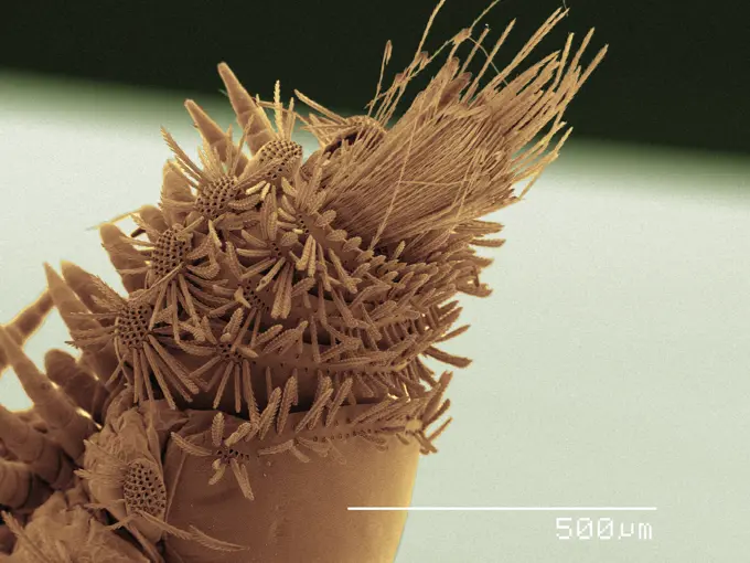
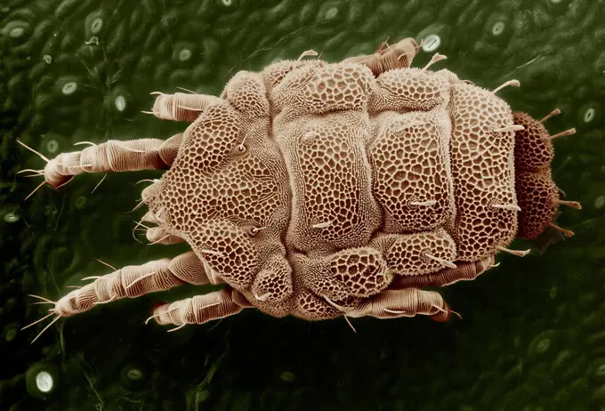
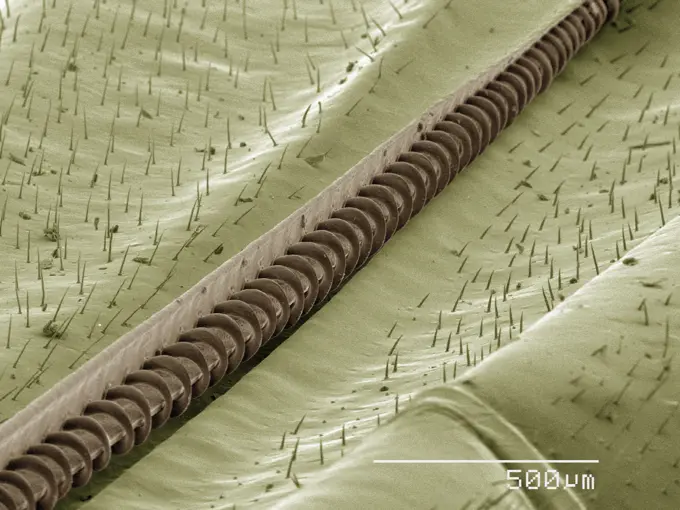
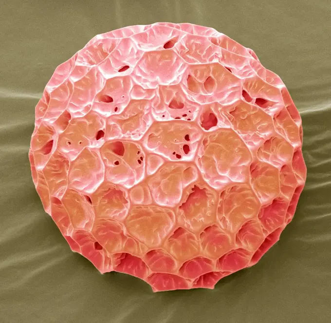
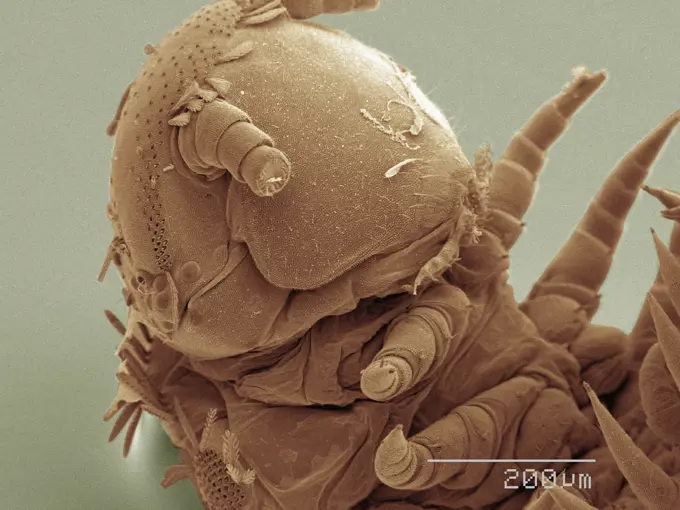
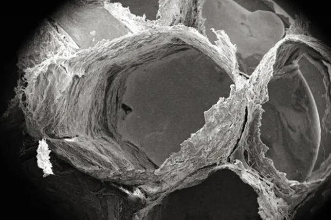
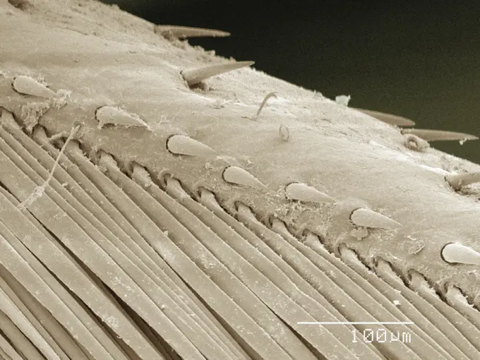
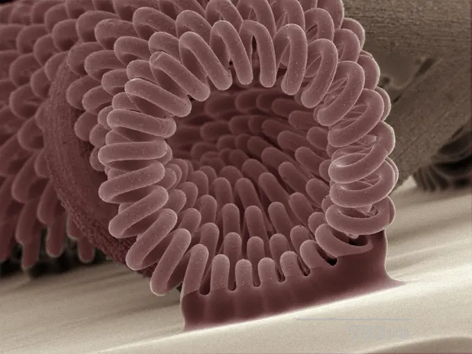
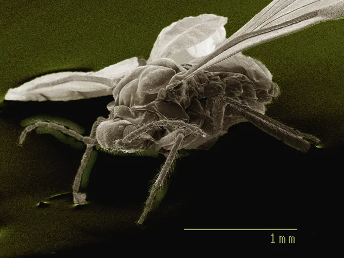
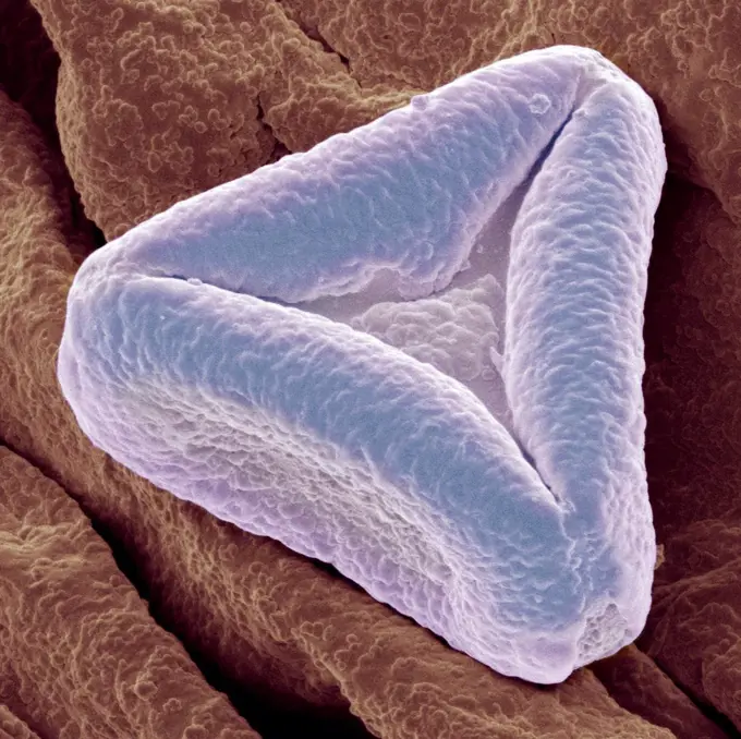
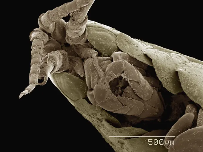
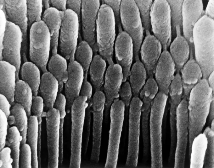
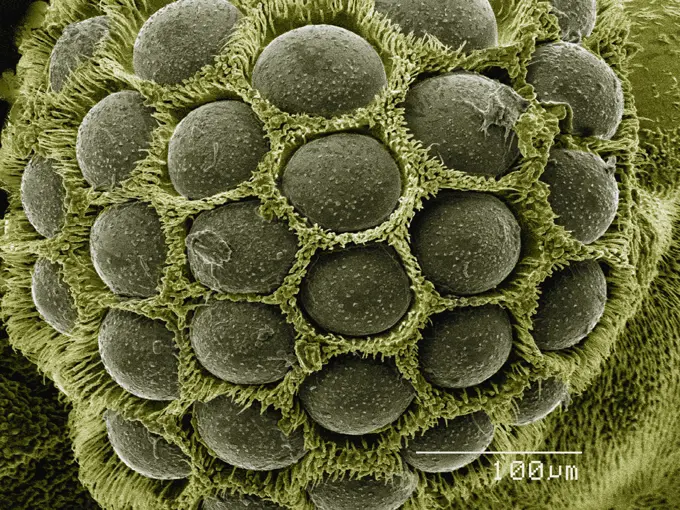
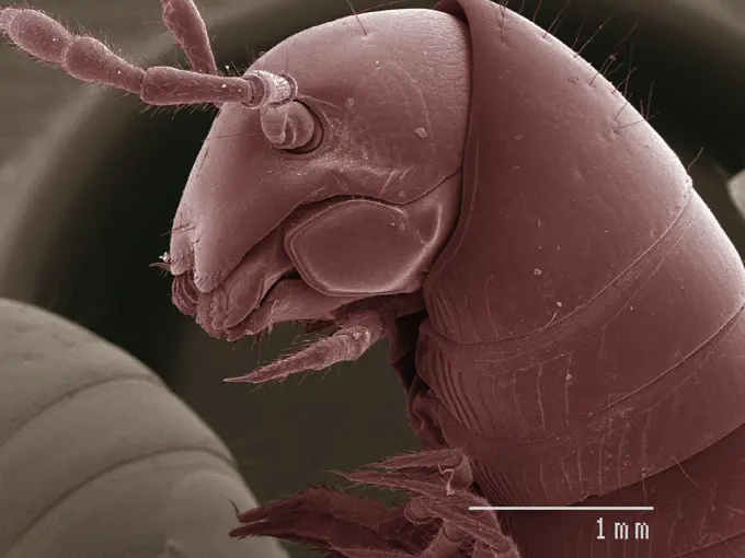
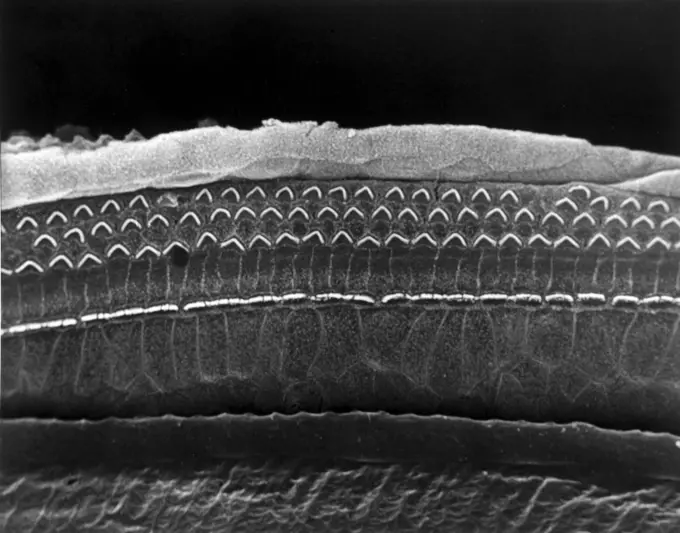
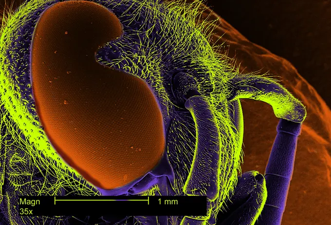
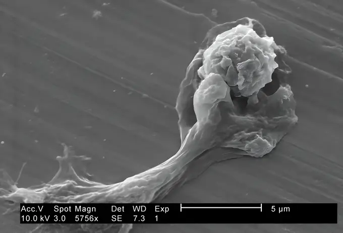
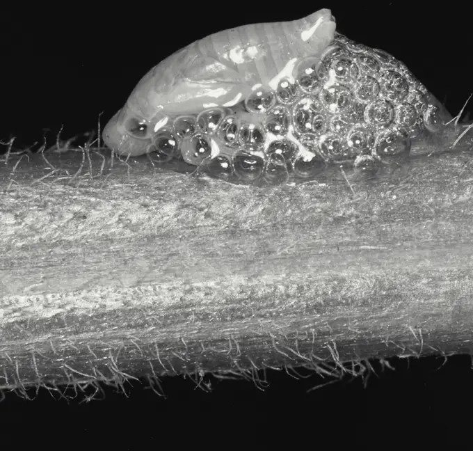
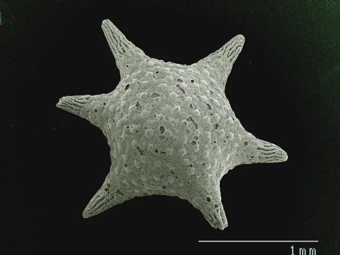
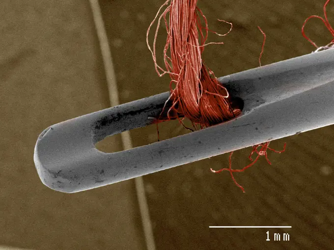
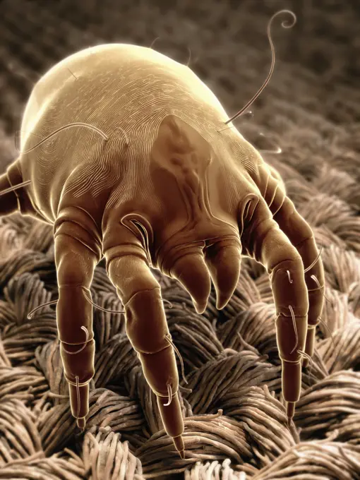
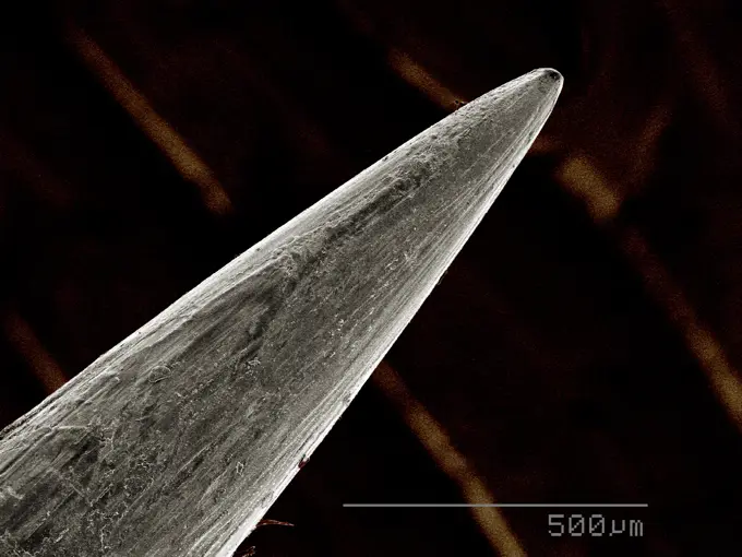
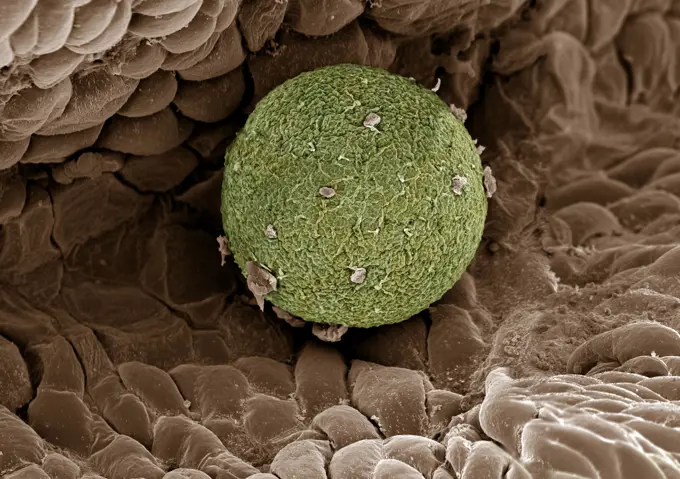
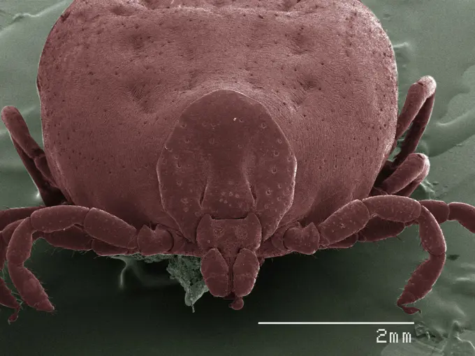
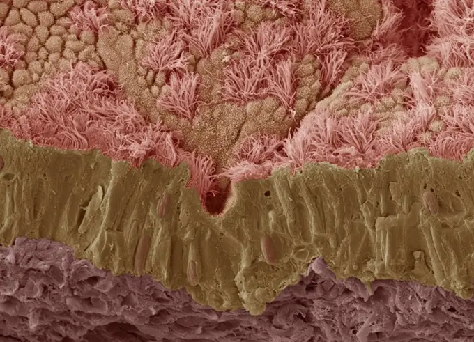
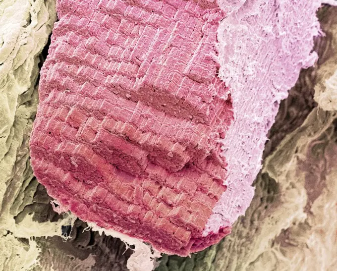
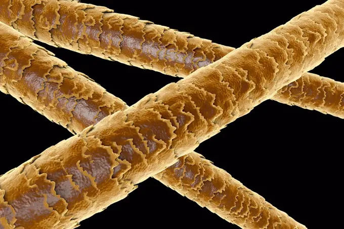
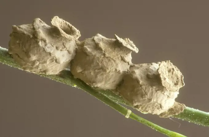
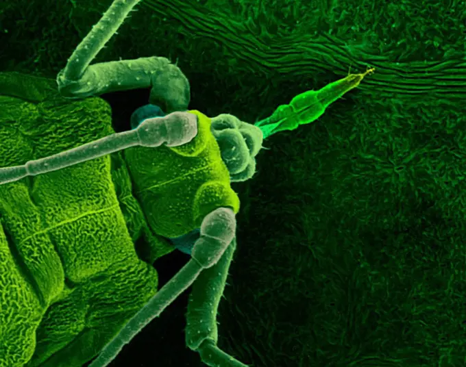
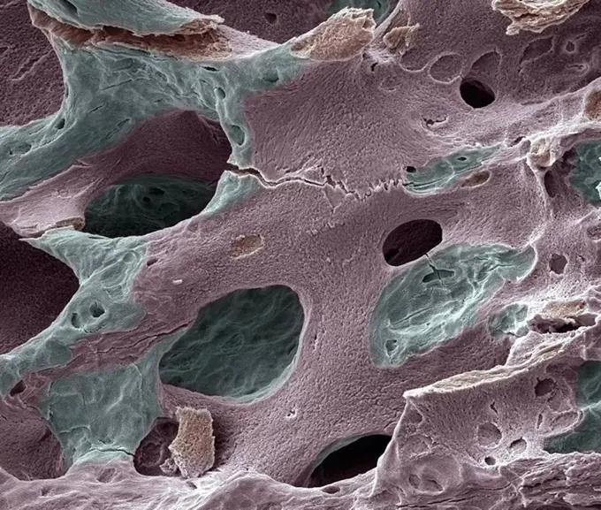
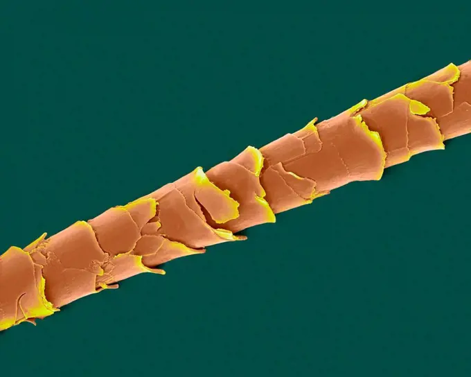
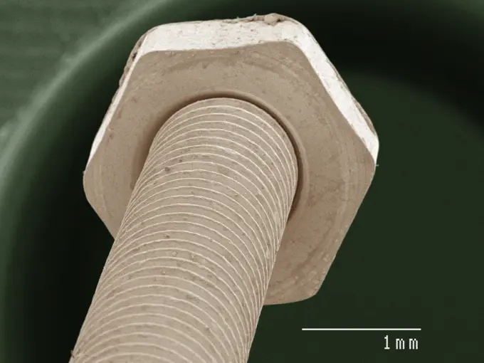
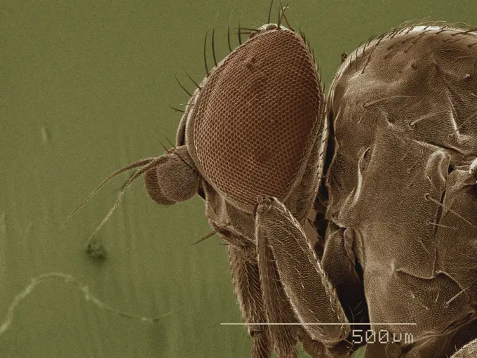
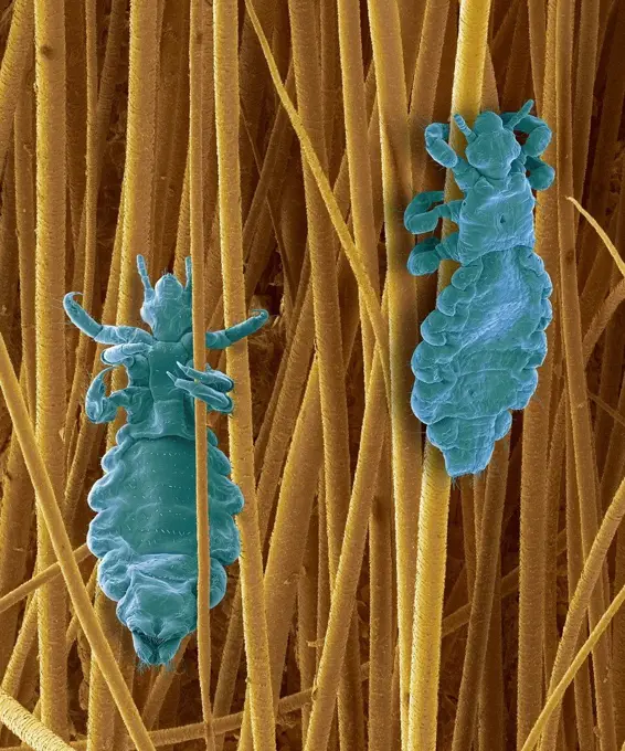
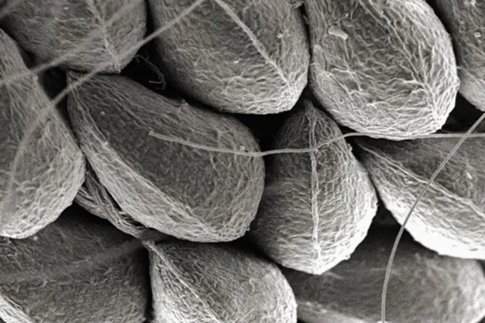
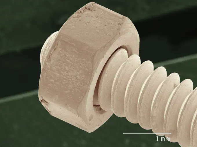
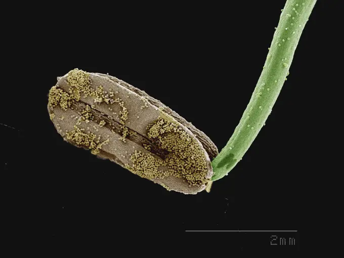
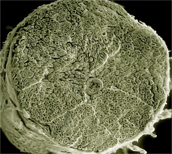
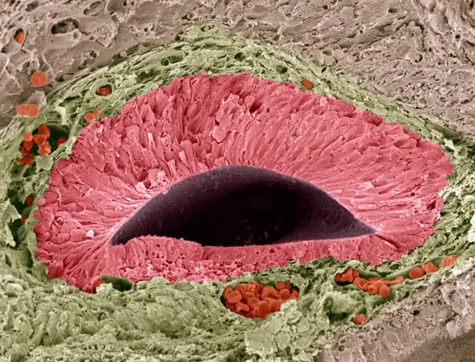
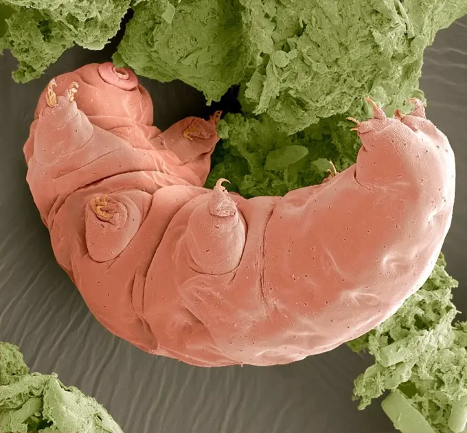
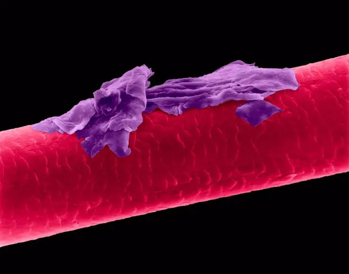
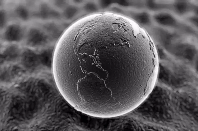
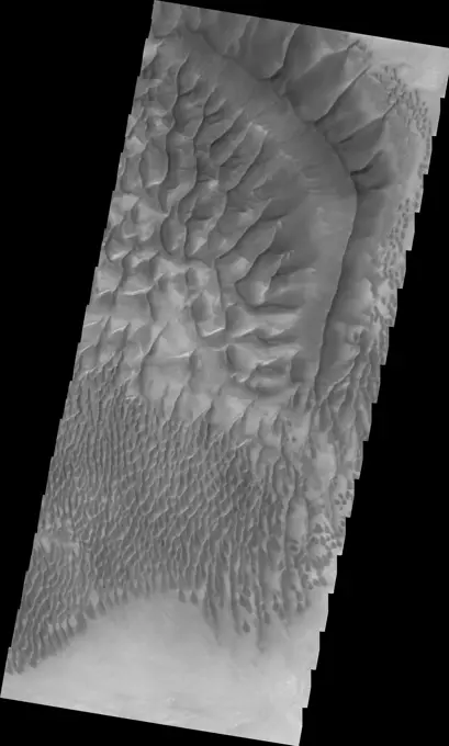
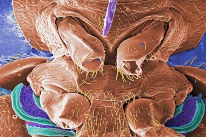
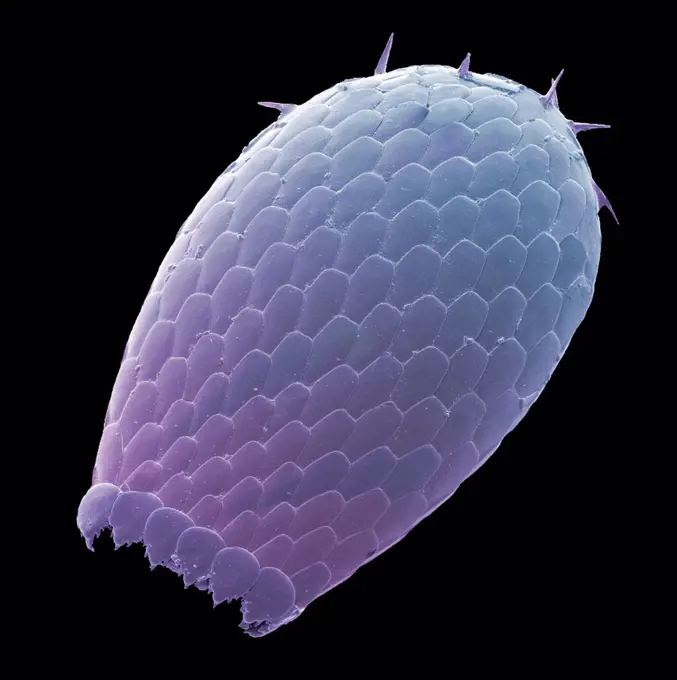
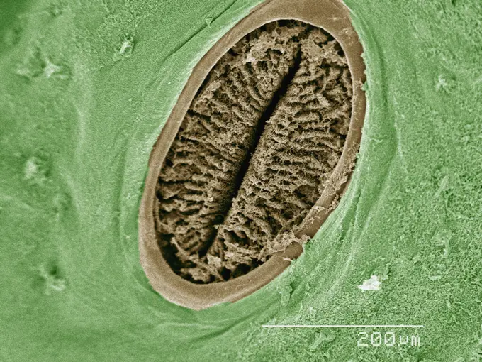
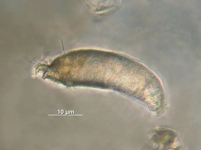
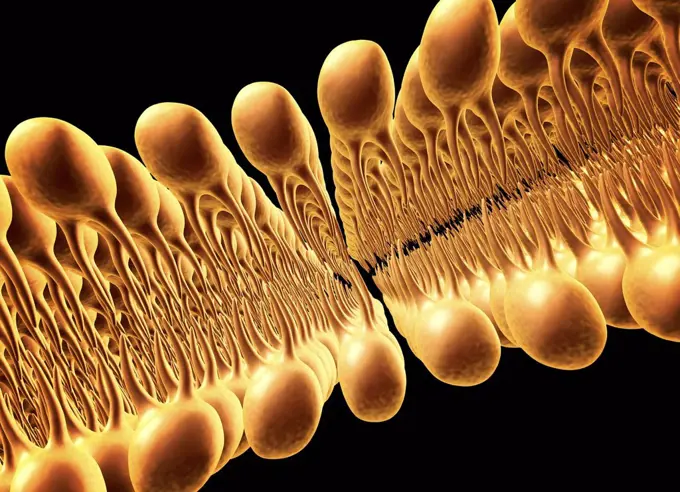
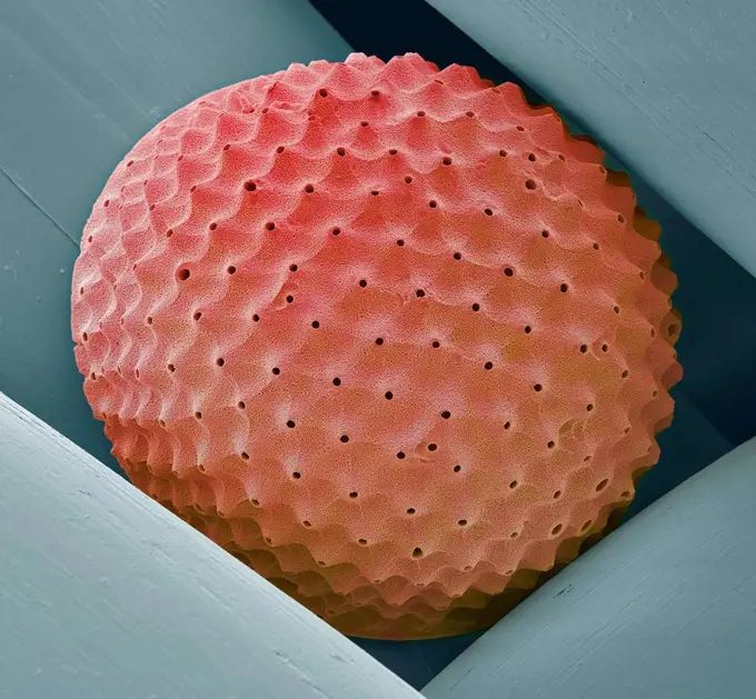
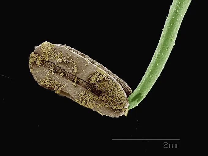
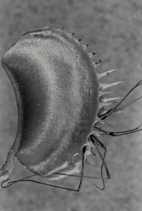
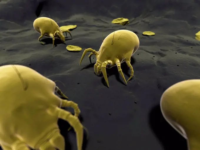
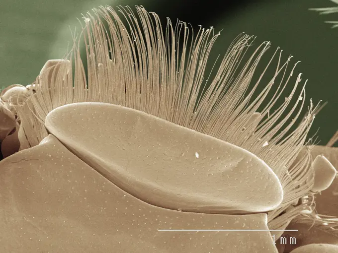
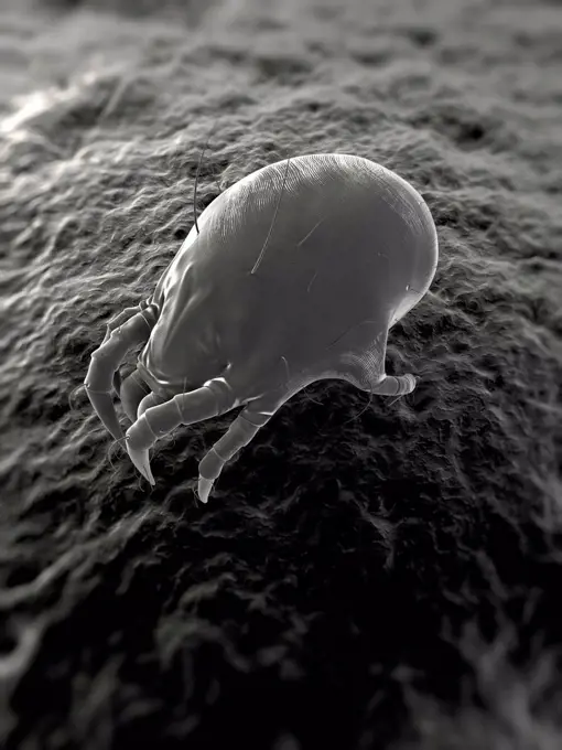
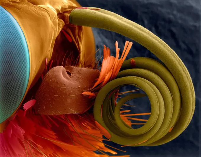
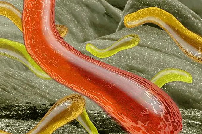
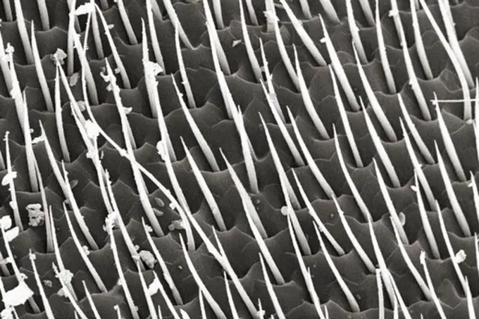
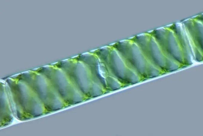
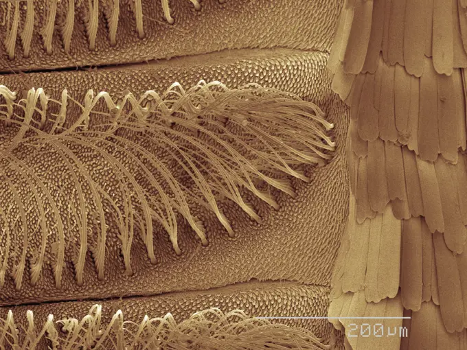
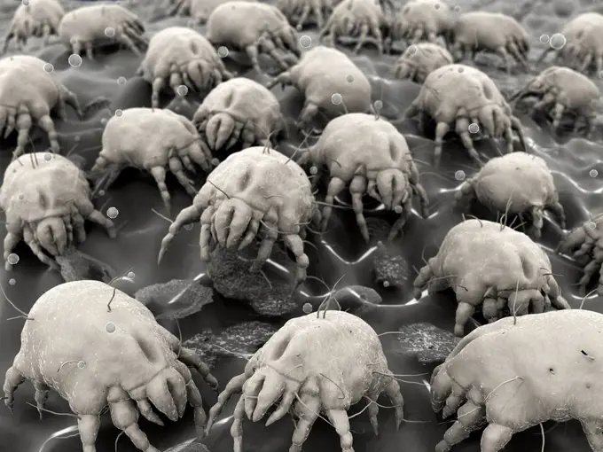
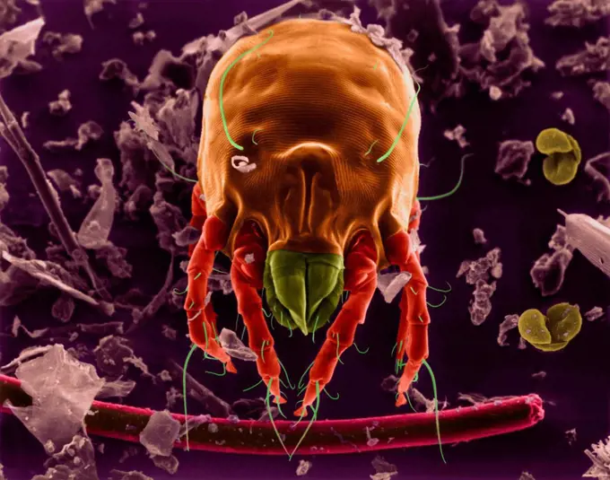
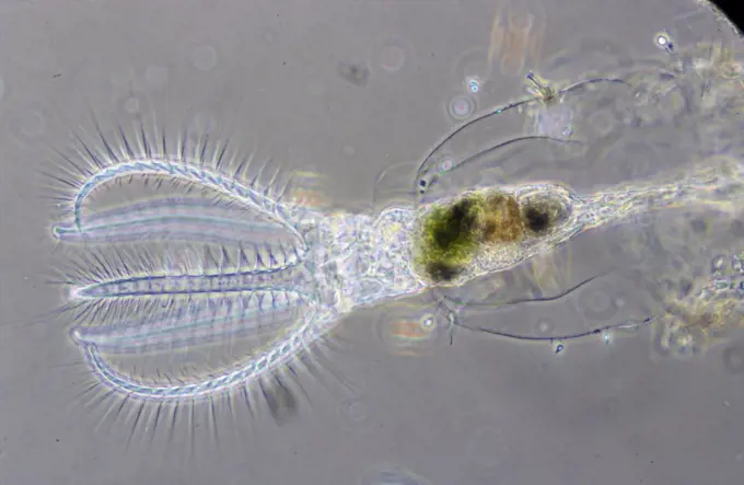
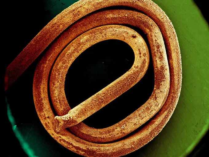
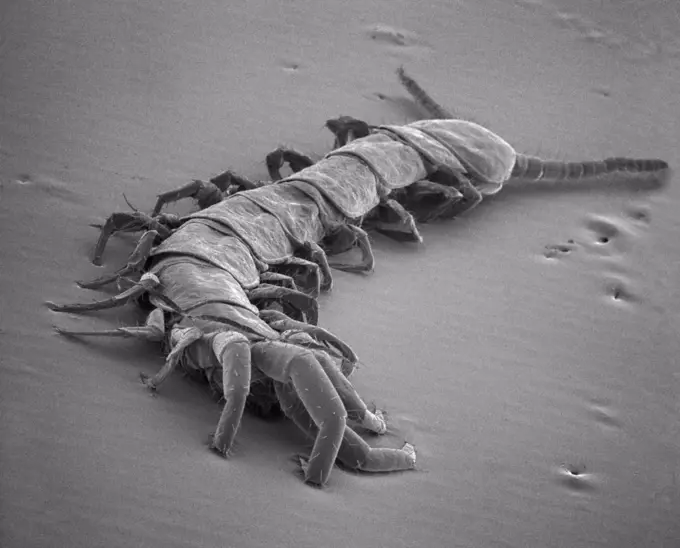
Microscopic Closeups of Structures

Detailed electron microscope images showing intricate structures of insects, plants, and various materials, highlighting their textures and details.

Extreme closeup of an ant captured through an electron microscope.
1439-57940174
Extreme closeup of an ant captured through an electron microscope.
4384-413
Microscopic Detail of Dandelion
1439-57940149
Closeup of intricate textures and structures under the microscope
1439-57940178
Closeup view of microscopic structures in sepia tones.
1439-57940140
Intricate closeup of a cat tongue under microscopic view, revealing the texture and structure
4384-435
Microscopic Detail of Female Body Louse's Exoskeletal Surface
1439-57940195
Closeup view of a microscopic organism.
1439-57940192
Closeup of an insect's limb under the microscope
1439-57940044
Closeup view of an intricate microscopic structure resembling a coiled object
1439-57943334
A closeup of a microscopic landscape resembling rugged terrain
4384-220
Scanning Electron Micrograph of Hornet's Leg
1439-57940198
Extreme closeup of an insect antenna under the microscope
1439-57952384
Intricate closeup of microscopic organisms
1439-57943344
Extreme closeup of an insect leg under a microscope
1439-57940212
A closeup of an ant captured under a scanning electron microscope
1439-57939993
Closeup view of a textured surface, possibly organic, under magnification.
1439-57940046
Up close and personal with a detailed micrograph of a spider.
1439-57940086
Closeup view of a tarantula leg under high magnification.
1439-57940121
Closeup of a bed bug as viewed under a scanning electron microscope with a sepia tone.
4384-433
Microscopic Detail of Female Body Louse
1439-57949059
Magnified view of insect on a plant
4384-397
Fireworks Flower With Pollen
1439-57949063
An upclose, mesmerizing view of a spiral structure with an organic feel
4201-66248
Honey Bee (Apis mellifera) SEM close-up view of the tip of a foot at 105x magnification
1439-57949009
Intricate closeup of a microscopic structure.
1439-57943298
A closeup view of microscopic structures resembling an alien landscape.
1439-57953825
Head of house fly (Muscidae) imaged in a scanning electron microscope
1439-57943332
A closeup exploration of nature's textures under the microscope.
1439-57940197
Intricate microstructure captured in a highresolution microscopic image.
1439-57940219
Closeup view of microstructures under electron microscopy.
4384-236
Scanning Electron Micrograph of Mosquito
4384-259
Scanning Electron Micrograph of Bed Bug's Mouthparts
1439-57939978
Closeup of a microgear mechanism in a vibrant sepia tone
1439-57943295
Intricate textures revealed in a closeup of a plant or biological specimen.
1439-57940058
Extreme closeup of a needle's eye under a microscope
1439-57952402
Closeup of a microorganism under a scanning electron microscope.
1439-57940119
A closeup view of a dust mite under the microscope
1439-57943297
Intricate details of nature captured in a sepiatone micrograph, showcasing the complex textures of leaves and organic structures
1439-57952407
Intricate details of a microscopic organism under high magnification
4413-89053
Detail of an antenna of Honey Bee
1773-100035
SEM image of spider eyes
4128R-13620513
Human hair with dandruff, coloured scanning electron micrograph (SEM). The outer layer of hair (the cuticle) has overlapping scales of keratin. These ...
1439-57953818
Extreme closeup of an insect's head under a microscope
4128R-8950
Stick insect foot, SEM
824-63194414
Under a moderately-high magnification of 8000X, this colorized scanning electron micrograph (SEM) revealed the presence of a grouping of Gram-negative Salmonella typhimurium bacteria that had been isolated from a pure culture.
4413-88889
Glands essential oil on a leaf of Sweet Briar
1439-57949053
Extreme closeup of an insect head under the microscope
1439-57943303
Closeup view of an intricate biological structure with hairlike features
1439-57940106
Intricate closeup of an ant's head under the microscope
4384-390
Microscopic Detail of Crimson Clover Petal
1439-57943320
Closeup of an insect's antennae under the microscope
1439-57952395
Microscopic view of a bee's wing with pollen
1439-57949016
Zooming into nature's microscopic world.
824-63227310
Myelinated axons in a rat vertebral root. Myelin is a type of fat that forms a sheath around and thus insulates the axon to protect it from the loss of the electrical current needed to transmit signals along the axon. The axoplasm inside the axon is shown in pink.
4384-373
Microscopic Detail of a Roach's Head
4201-66336
Harvester Ant (Messor sp) SEM close-up view of eye at 140x magnification
1439-57949051
Closeup of a fly's head with large compound eyes
4201-66257
Trumpet Lily (Lilium longiflorum) SEM close-up view of pollen grains at 105x magnification
1439-57943361
Closeup of arachnid under the microscope with sepia tones
1439-57940031
Intricate microstructures reminiscent of an alien landscape.
1899-18906649
This is an overhead view of the Lorryia formosa mite. Magnified about 200x  Scanning electron microscopy allows mites to be viewed from different angles ca. 28 January 2008.
1439-57940009
Closeup view of microfabric under the microscope
4128R-8941
Phlox pollen, SEM
1439-57940032
Up close and personal with a microscopic marvel.
4384-384
Microscopic Detail of a Wasps' Nest
1439-57940163
Closeup of a textured surface under the microscope
1439-57940125
A closeup view of an ant under a scanning electron microscope.
4128-V58559203
1439-57940027
Microscopic view of a spiral structure
1439-57949078
Closeup of a detailed insect under the microscope
1439-57940216
Closeup of a thorny stem under a microscope in sepia tones.
4128R-9185
Bottlebrush pollen, SEM
1439-57952658
Zoomedin glimpse of a microcosm
6145-29267509
Stereocilia from human cochlea, SEM HP
1439-57948999
Intricate microcosm under the electron microscope
1439-57940075
Closeup view of an ant under an electron microscope
6145-29293865
SEM upper surface of organ of Corti, G pig
824-63127934
An electron micrograph of a wasp's head region and the insect's compound eye, magnified 35x. The wasp's compound eye is actually made up of many identical receptor units called ommatidia. Each receptor contains its own lens and light sensitive cells, and is actually an eye unto itself. See image n1314205 on the site.
824-57659296
Magnified 5756X, this 2002 scanning electron microscope (SEM) image revealed some of the ultrastructural morphology exhibited by two Hartmannella vermiformis amoeba trophozoites. Note that one of the amoebas took on a very distorted shape, having extruded a number of pseudopodia, as it moved across the surface of the coupon. The trophozoite stage of an amoeba's life cycle is its vegetative phase, devoted to feeding, movement, and reproduction. This free-living protozoan moves in response to chemical cues in its environment by extending pseudopodia, or false feet, a number of which are visible in this image. CDC/Dr Rodney Donlan 2002
255-58492114
Vintage Photograph. Spittlebug blowing new spittle.
1439-57949004
Microscopic view of a starshaped diatom
1439-57943365
Extreme closeup of red fibers caught on a metal needle
1849-64860077
Digitally generated image of a mite
1439-57943364
Closeup view of a sharp object tip under high magnification
4220-21334699
Scanning Electron Micrograph (SEM); Human Ovum in Fallopian Tube : This freshly fertilized egg passes down the Fallopian tube on its way to implantation in the wall of the uterus.     Date:
1439-57939991
Closeup of a dust mite under the microscope
4128R-5473
Trachea mucous membrane, SEM
4128R-4463
Skeletal muscle fibre, SEM
4128R-15366447
Human hair, illustration
4269-27258
Anopheles gambiae. Scanning electron micrograph (SEM) of the Anopheles gambiae mosquito's anterior head. Magnification x 114. This mosquito is a vector of the parasite Plasmodium, the agent of malaria.
4179-17882
Mud Chambers of Potter Wasp (Eumenes fraternus) Ontario, Canada
4128R-13620397
Coloured scanning electron micrograph (SEM) of Pea aphid (Acyrthosiphon pisum) on a bean leaf with proboscis penetrating a vein. This insect transmits...
4128-28970690
Cancer cells dividing, illustration
4128R-1469
Osteoporotic bone, SEM
4128R-13620214
Coloured scanning electron micrograph (SEM) Dog hair (shih tzu) with damaged cuticle. Dogs have two types of hair in their coats. The first type is sh...
1439-57940054
Closeup view of a screw and nut under magnification showing detailed threads.
4128-V58557845
1439-57949089
Upclose encounter with an insect under the microscope
4128R-279
Vertebra, SEM
4128-111494395
Coloured scanning electron micrograph (SEM) of Pediculus humanus capitis, human head lice. P. humanus is divided into two subspecies; the head louse P...
4384-429
Microscopic Detail of Wild Strawberry
1439-57940055
Closeup of a micro screw under high magnification
4384-372
Microscopic Detail of a Roach
1439-57949034
Closeup of a flower's stamen covered in pollen against a black background.
824-63227232
Spinal nerves are part of the peripheral nervous system. They run through the spine to carry nerve signals to and from all parts of the body. Spinal nerves allow all the movements we make, from turning our heads to wiggling our toes, control the movements of our internal organs, such as the colon and bladder, as well as allow us to feel the touch and location of our members.
4128R-5639
Foetal sense organ, SEM
4128R-9191
Water bear, SEM
4128R-13619958
Human hair with dandruff, coloured scanning electron micrograph (SEM). The outer layer of hair (the cuticle) has overlapping scales of keratin. These ...
4378-872
The earth as if seen under scanning electron microscope (conceptualization)
6145-45258890
This image shows the central part of the dune field on the floor of Russell Crater. Russell Crater is located in Noachis Terra. A spectacular dune ridge and other dune forms on the crater floor have caused extensive imaging. The Odyssey spacecraft has spent over 15 years in orbit around Mars, circling the planet more than 69000 times. It holds the record for longest working spacecraft at Mars. THEMIS, the IR/VIS camera system, has collected data for the entire mission and provides images covering all seasons and lighting conditions. Over the years many features of interest have received repeated imaging, building up a suite of images covering the entire feature. From the deepest chasma to the tallest volcano, individual dunes inside craters and dune fields that encircle the north pole, channels carved by water and lava, and a variety of other feature, THEMIS has imaged them all. For the next several months the image of the day will focus on the Tharsis volcanoes, the various chasmata o
4384-260
Scanning Electron Micrograph of Bed Bug's Mouthparts
4128R-8942
Euglypha amoeba shell, SEM
1439-57949002
Closeup of a microscopic texture in green hues.
4413-20032306
Colomerus vitis (Pagenstecher, 1857) A species of the family Eriophyidae. These mites are microscopic in size. The one in this photograph measures only 43 µm (1/2 millimetre) and is therefore completely invisible without optical equipment such as a microscope. The Colomier mites live on the underside of the vine leaves where their bites cause a reaction of the plant in the form of blisters due to the formation of numerous filaments. The mites live in the middle of these filaments and are almost invisible. This disease is called grapevine erinosis.
4128R-32072
Phospholipid bilayer. Computer artwork of the phospholipid bilayer that forms the membrane around all living cells. The cell membrane is made of phosp...
4128R-12571802
Radiolarian. Coloured scanning electron micrograph (SEM) of a radiolarian. Radiolarians are amoeboid protozoa that produce intricate mineral skeletons...
1439-57952414
A closeup view of a lily anther coated with pollen grains
255-32335
Vintage Photograph. Close-up view of Venus Fly-trap plant.
4128R-11288968
Dust mite, computer artwork.
4297-1892
Scanning electron microscopic image of a Klebsiella pneumoniae bacterium
4141-32824
freshwater bryozoan plumatella repens portion of a colony
1439-57940101
Close-up view of a microstructure under an electron microscope
4128R-15324
Dust mite. Computer artwork of a house dust mite Dermatophagoides pteronyssinus.
4128R-13620138
Coloured scanning electron micrograph (SEM) of Woodland skipper butterfly compound eye and proboscis (Ochlodes sylvanoides). (Ochlodes sylvanoides). S...
1848-53914854
Bacteria, illustration
4384-351
Honey Bee's Exoskeletal Surface
4141-32837
spirogyra cell
1439-57940005
Closeup of textured microscopic structures in sepia tones
4128R-11291603
Artwork of dust mites.
4128R-13620537
Coloured scanning electron micrograph (SEM) of Dust mite (Dermatophagoides sp.). Millions of dust mites inhabit the home, feeding on dead human skin t...
4201-22092
Rotifer (Rotaria rotatoria) a microscopically small animal with a length of 004 to 240 mm, showing cilia on its trochal discs, waterborne and can inhabit highly acidic environments
1439-57939996
Closeup view of microscopic structures with a scale indicator
1773-95772
Science: SEM of chemically deposited Antimony on metallic substrate
4201-66381
Stone Centipede (Lithobius sp) SEM close-up view at 21x magnification
1439-57952403
A closeup view of microchip circuitry with vivid green highlights
1848-49235415
Hollow stem at insect hotel for solitary bees occupied by spider
4128R-13620137
Coloured scanning electron micrograph (SEM) of Woodland skipper butterfly compound eye and proboscis (Ochlodes sylvanoides). (Ochlodes sylvanoides). S...
1439-57943353
Closeup of a crosssection of a yellow fibrous material against a black background.