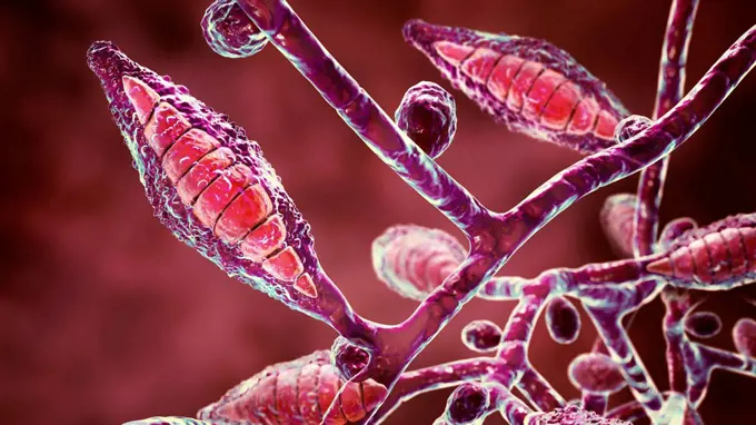
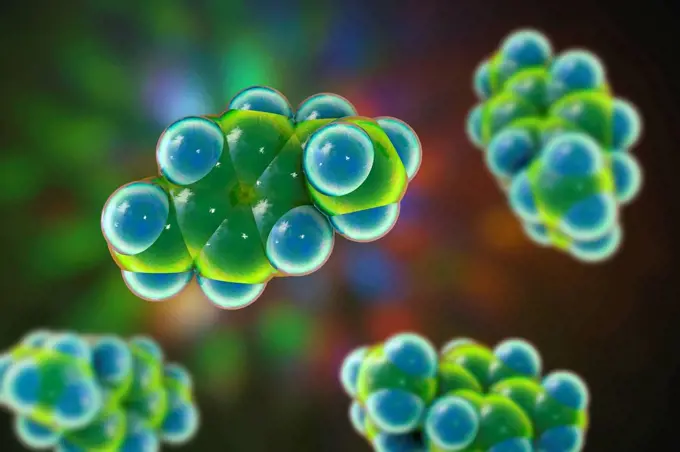
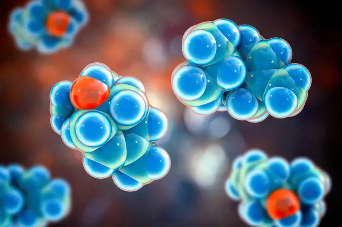
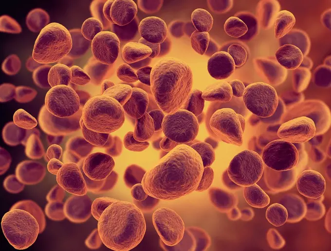
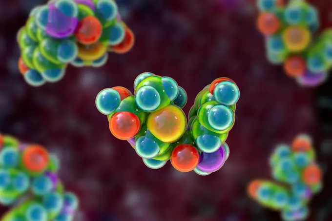
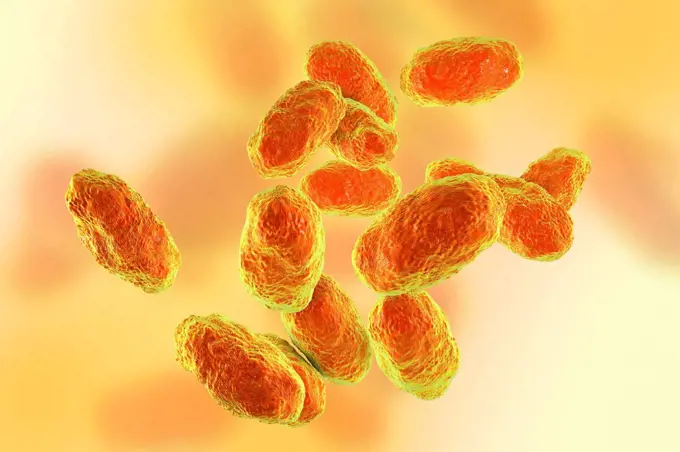
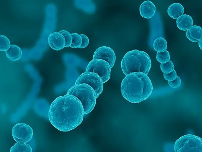
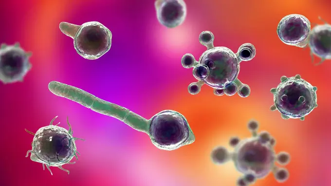
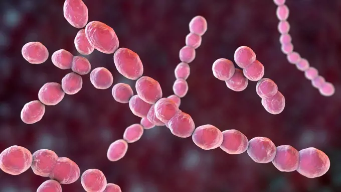
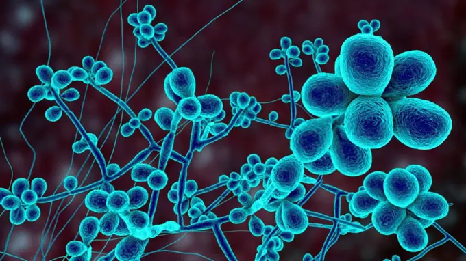
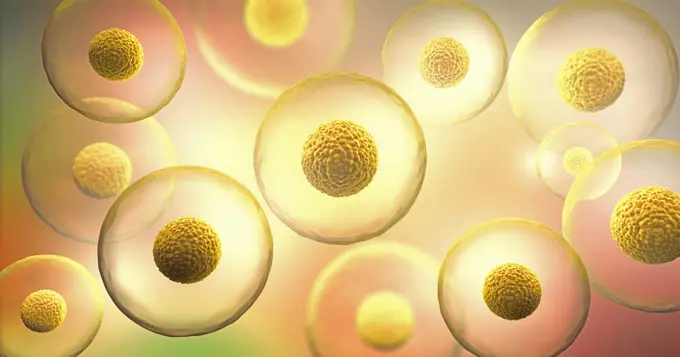
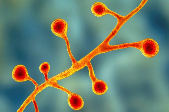
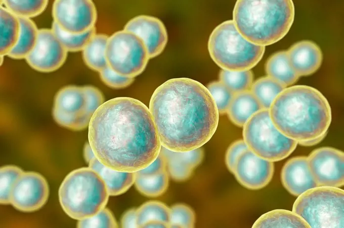
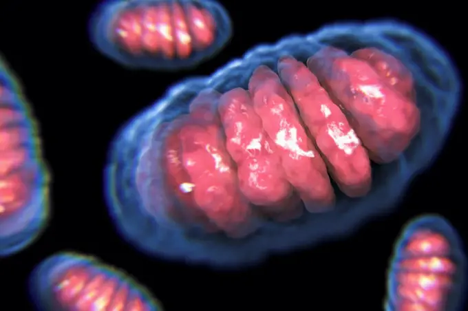
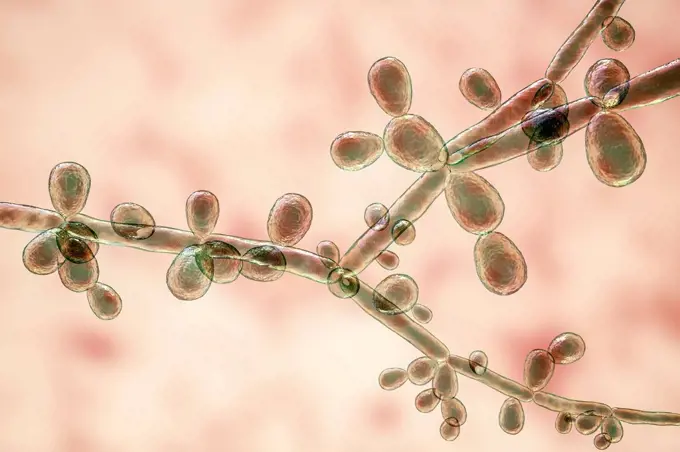
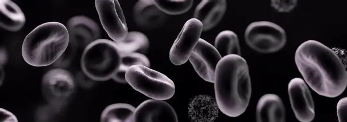
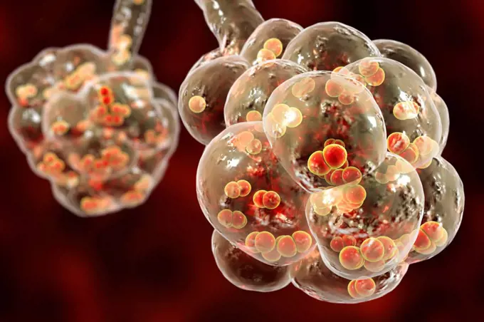
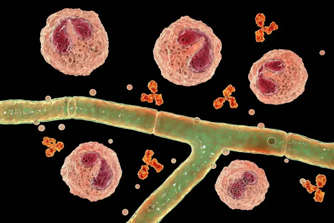
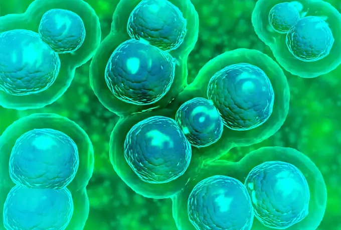
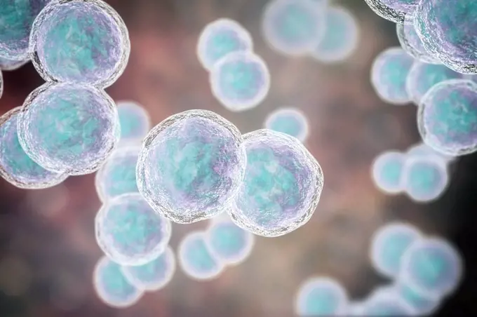
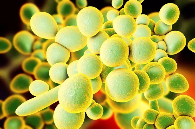
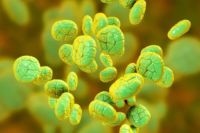
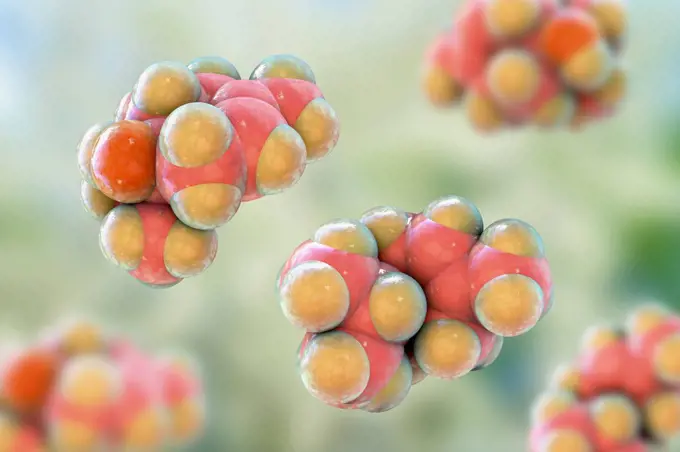
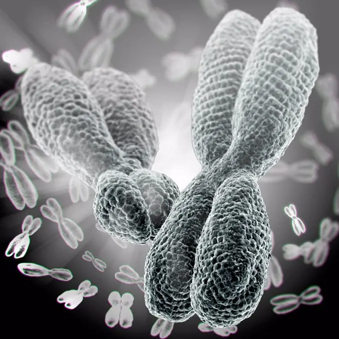
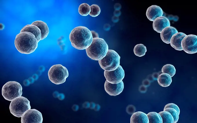
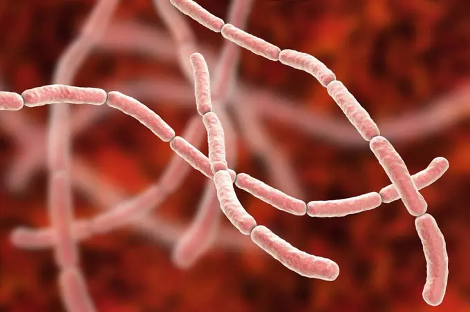
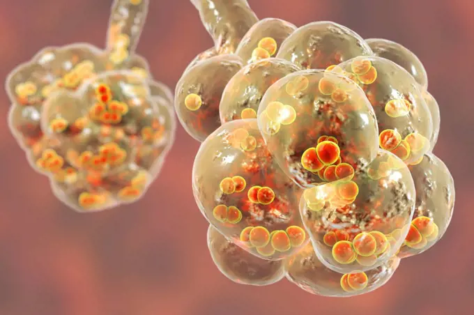
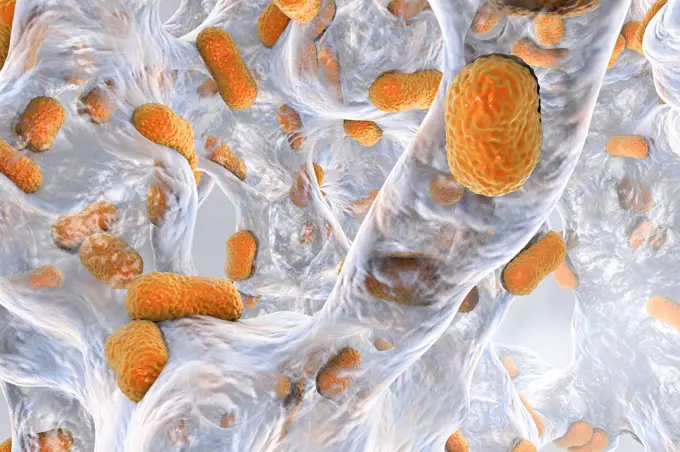
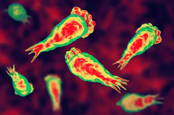
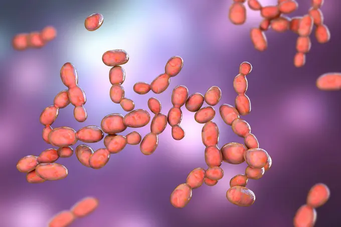
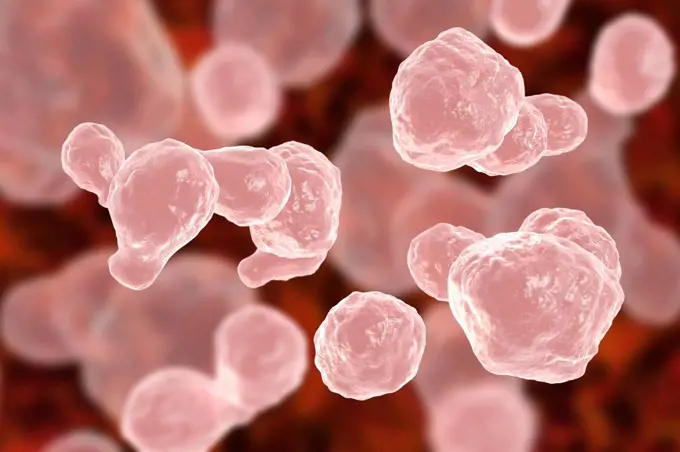
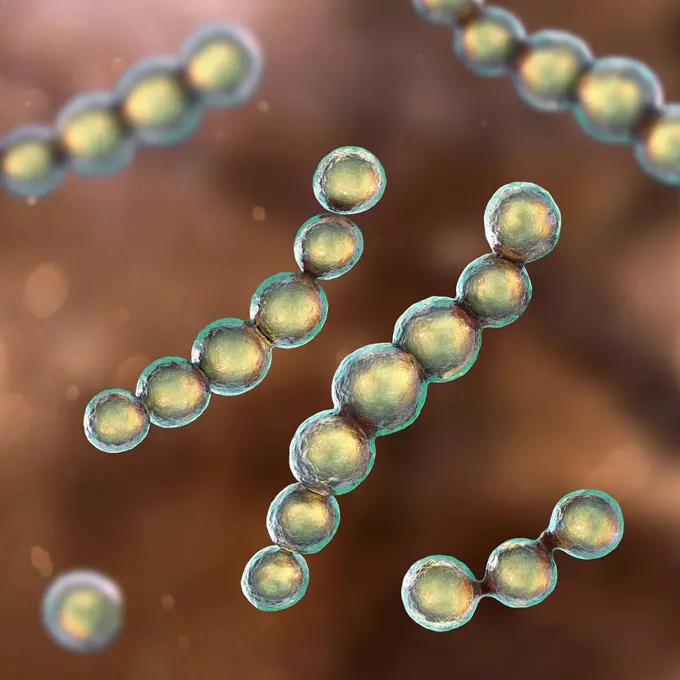
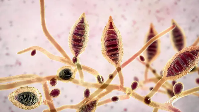
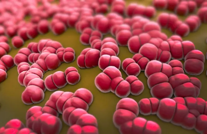
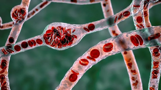
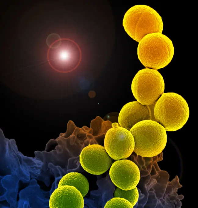
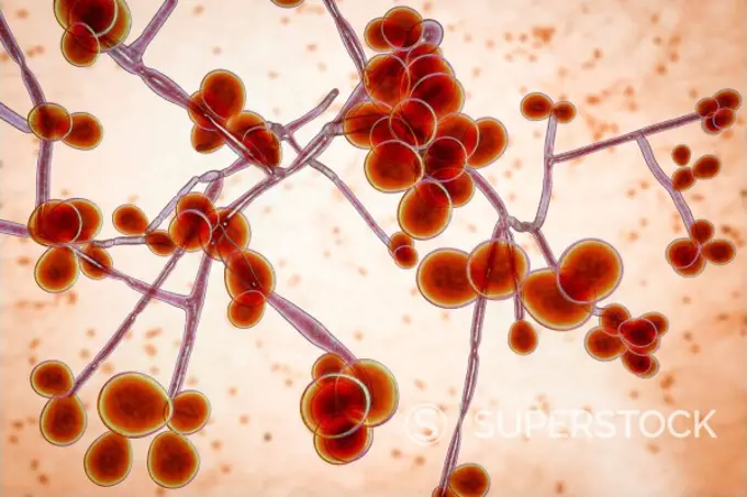
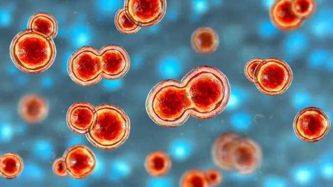
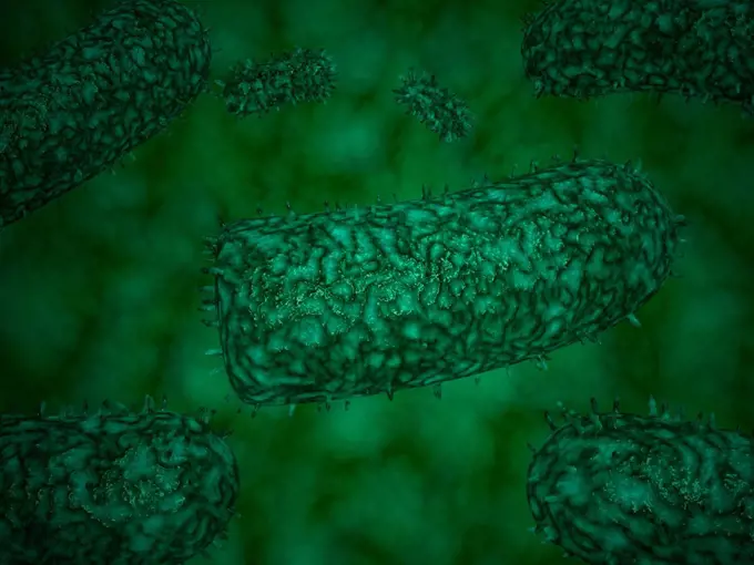
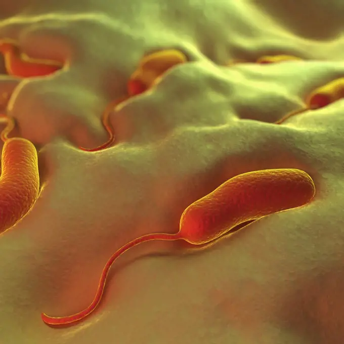
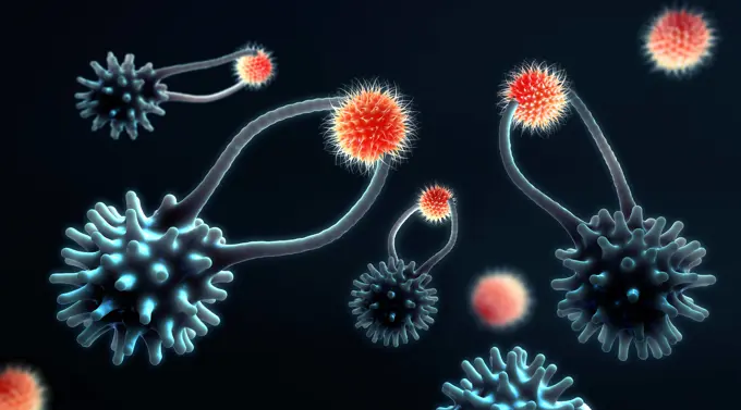
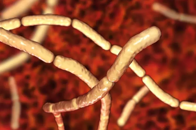
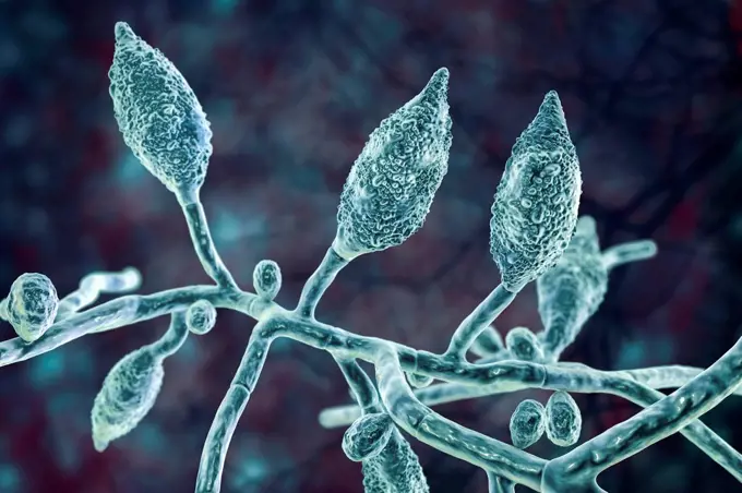
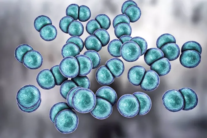
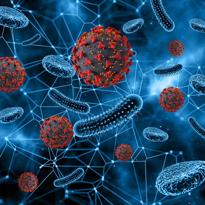
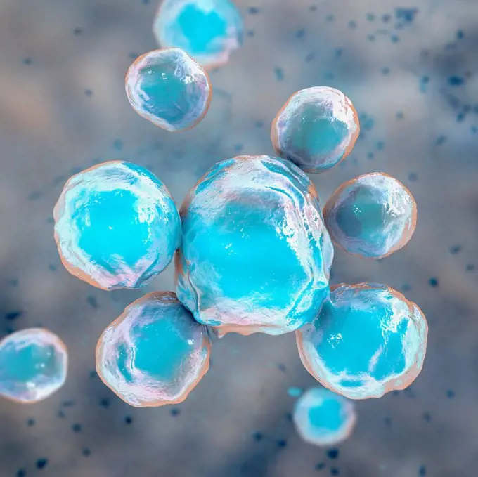
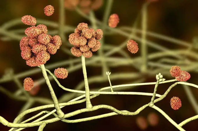
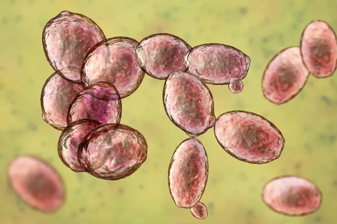
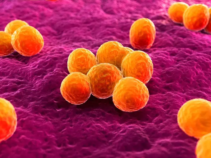
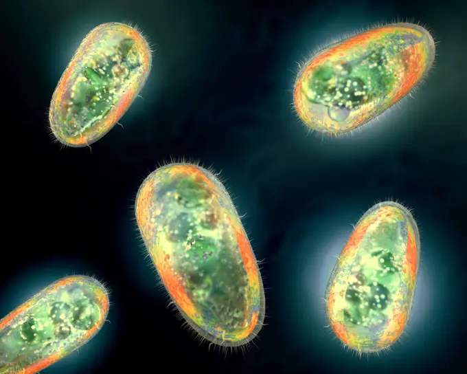
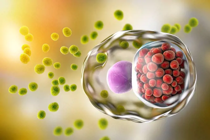
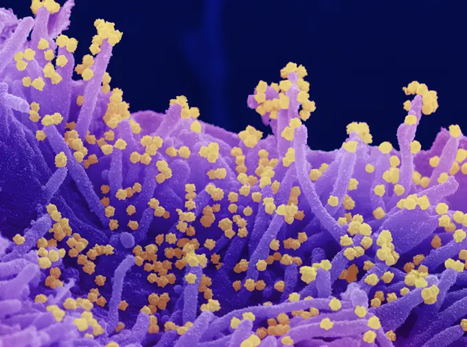
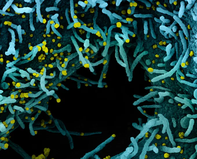
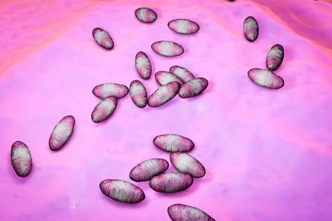
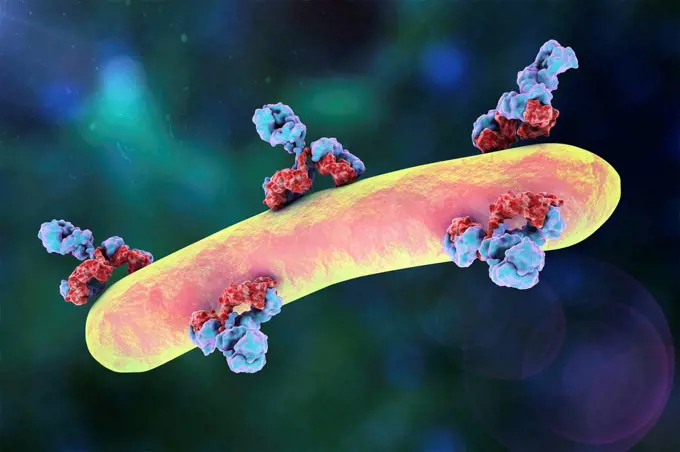
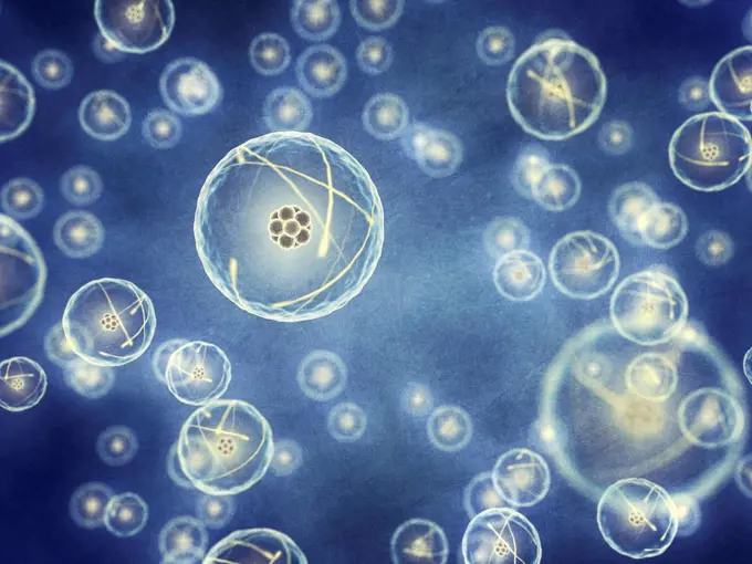
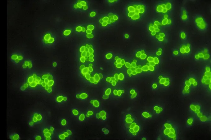
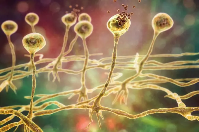
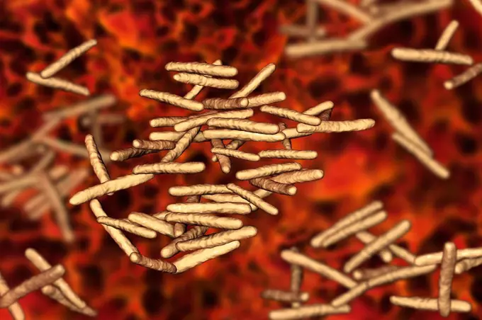
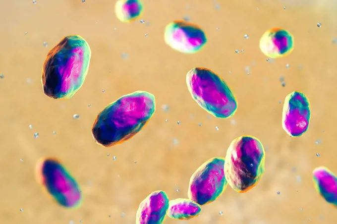
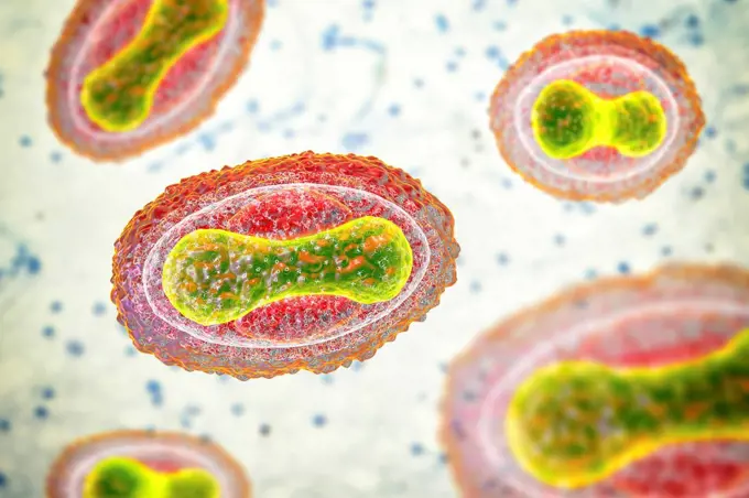
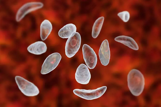
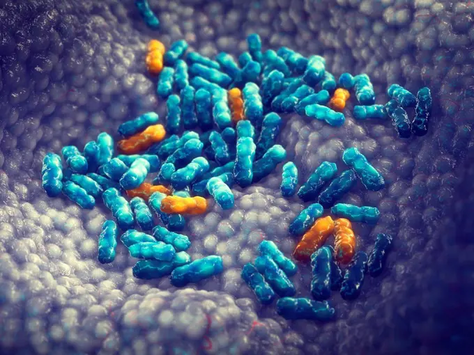
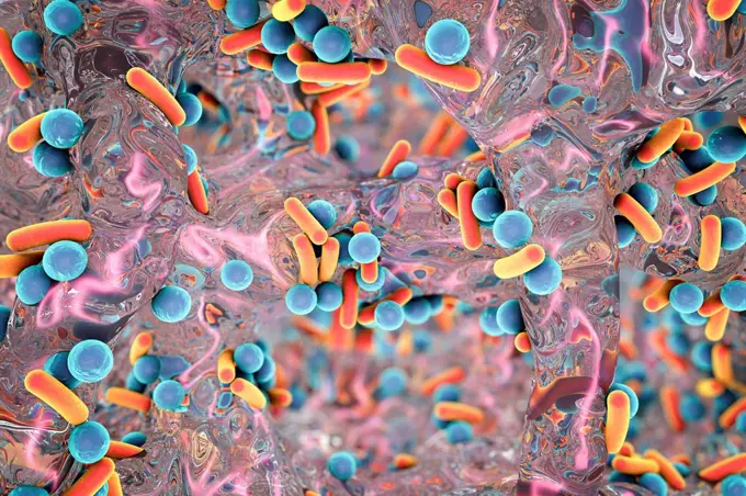
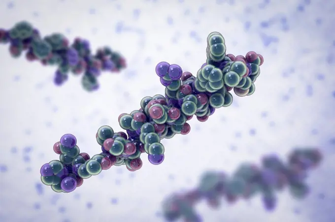
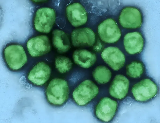
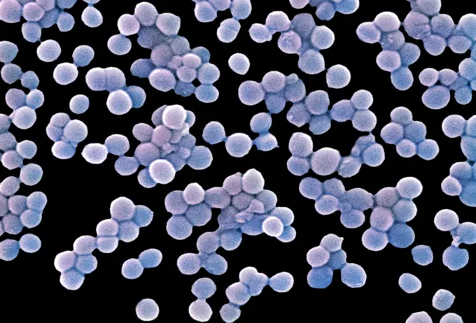
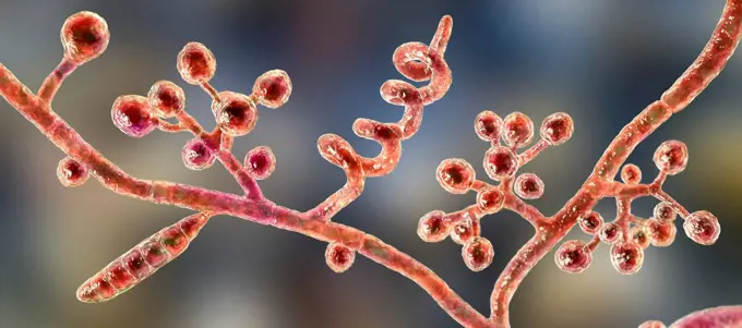
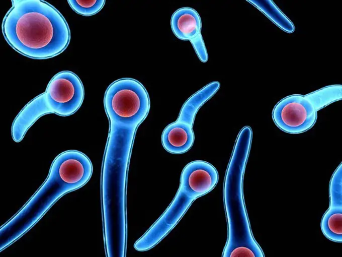
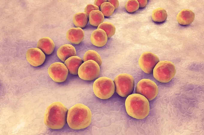
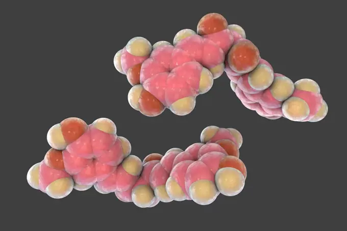
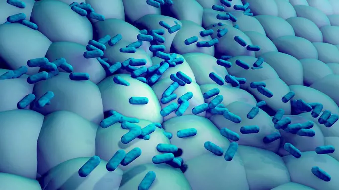
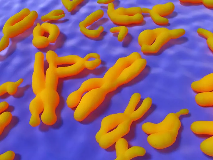
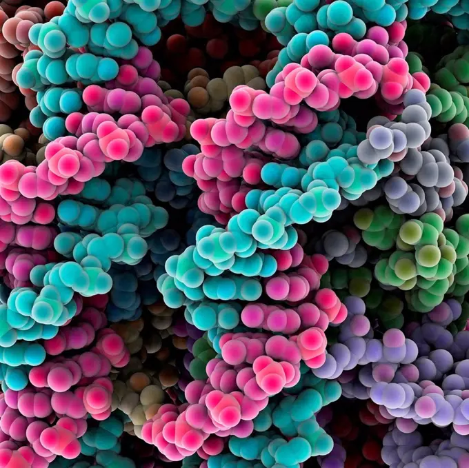
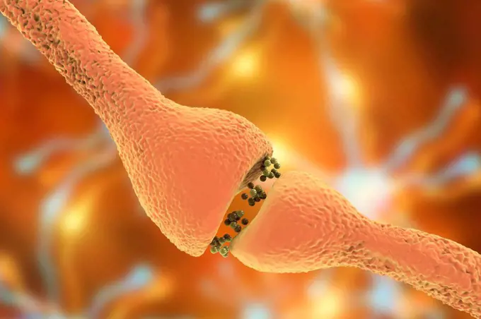
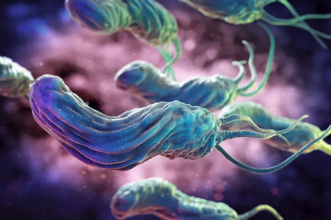
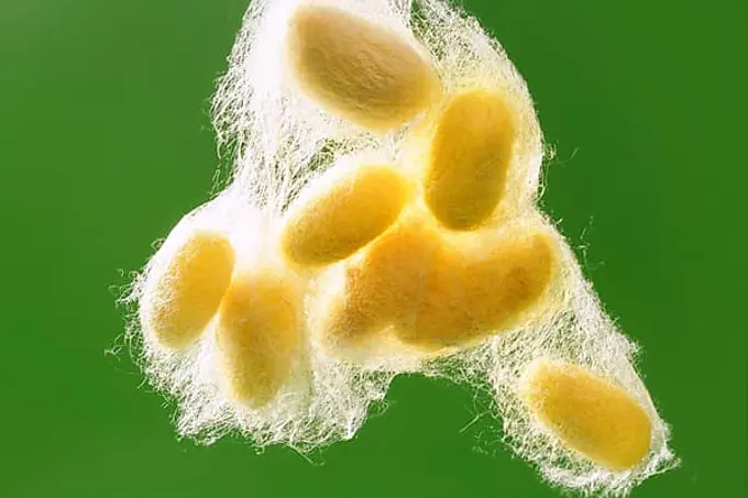
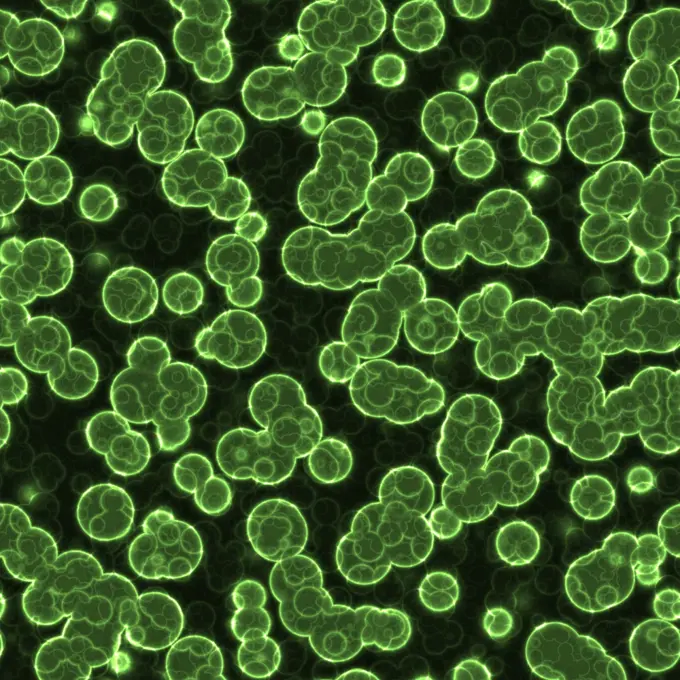
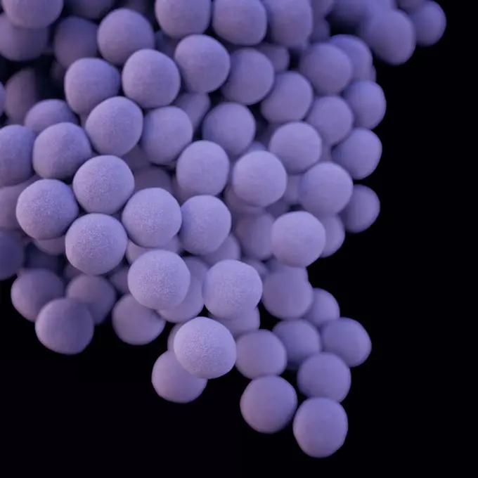
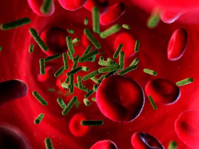
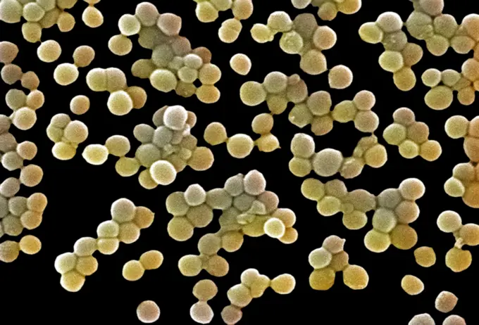
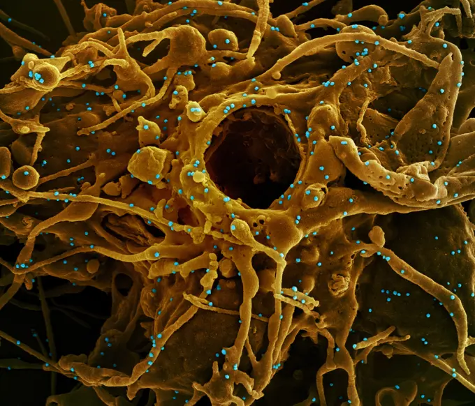
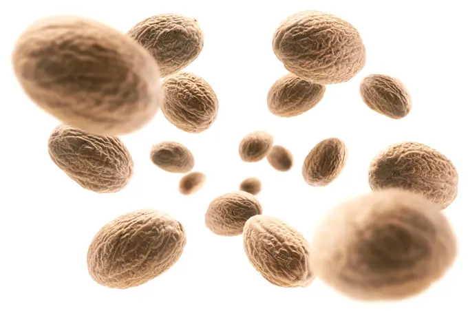
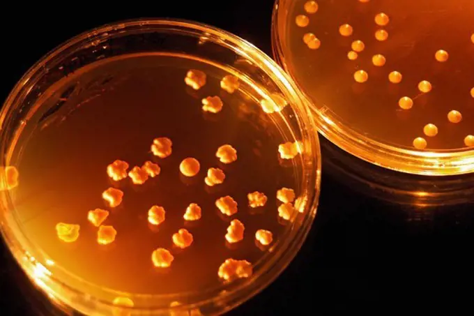
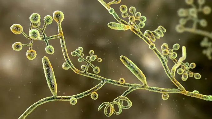
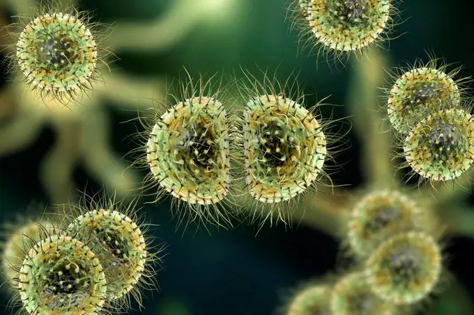
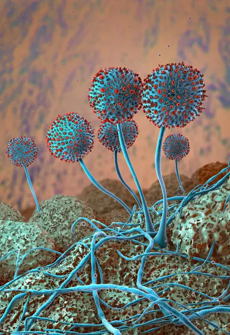
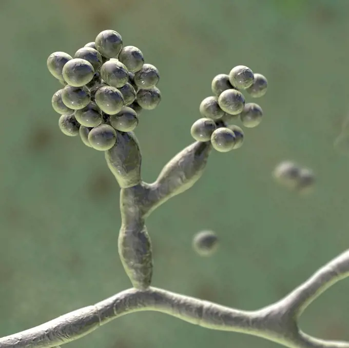
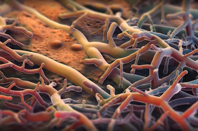
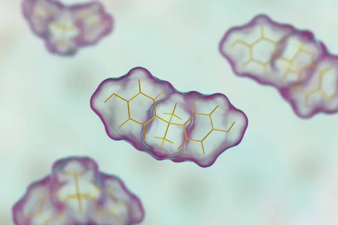
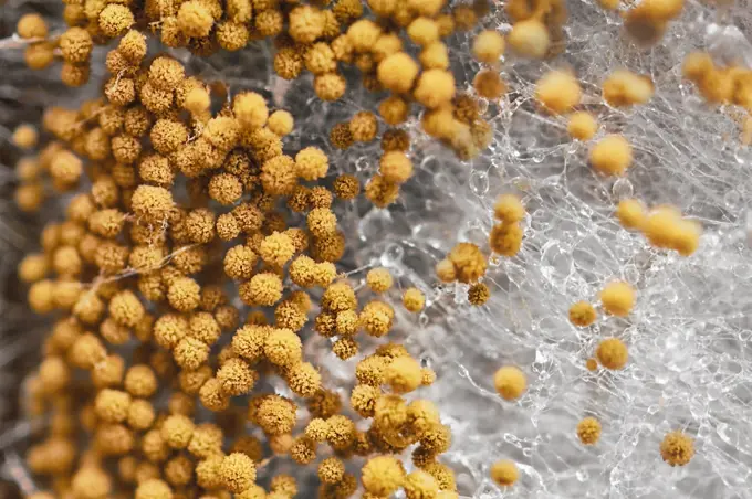
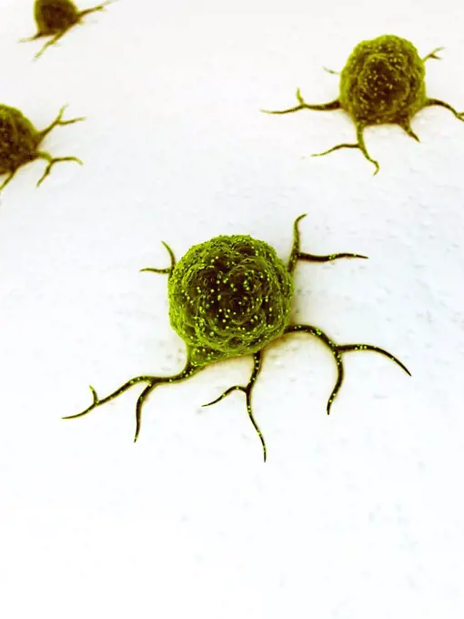
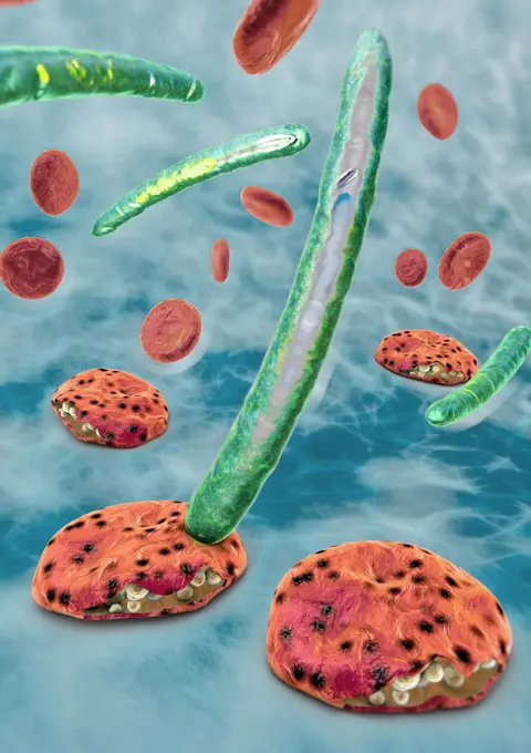
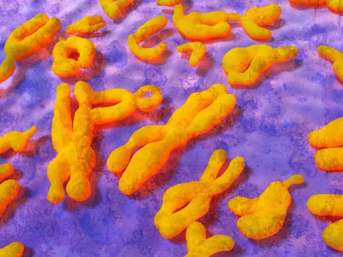
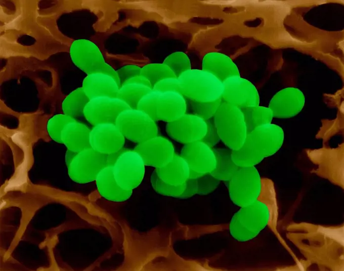
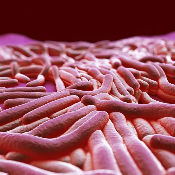
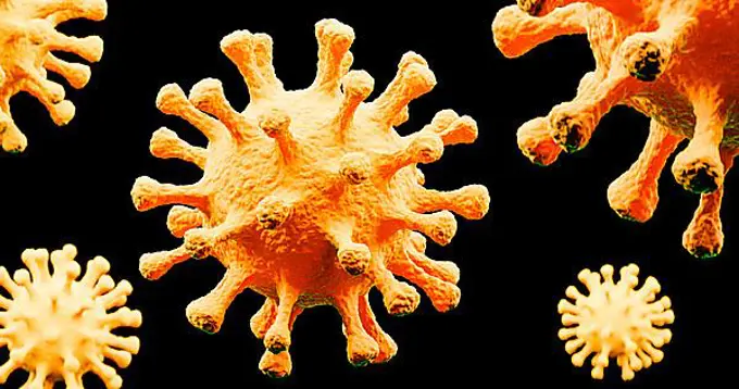
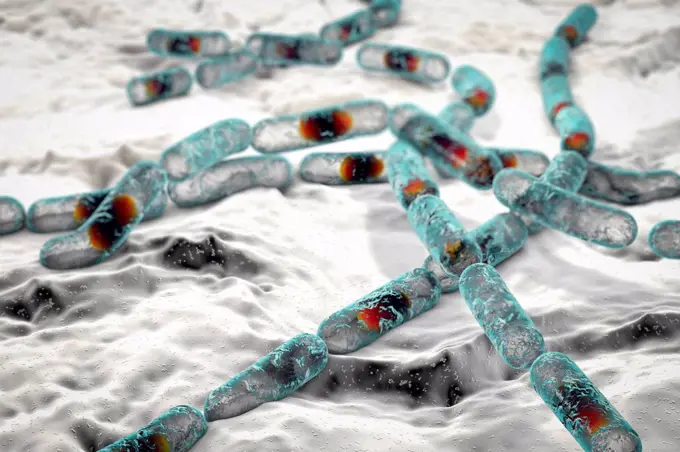
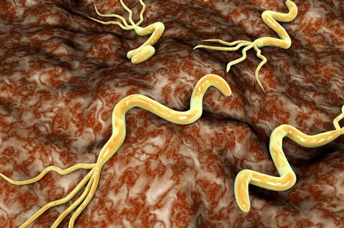
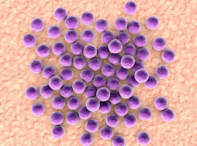
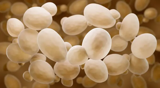
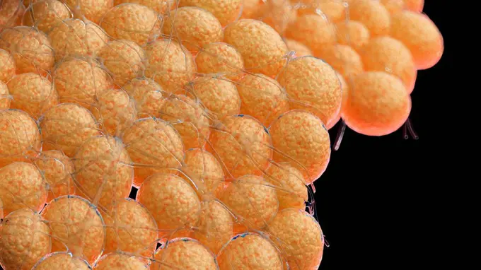
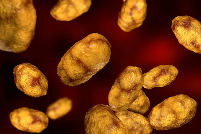
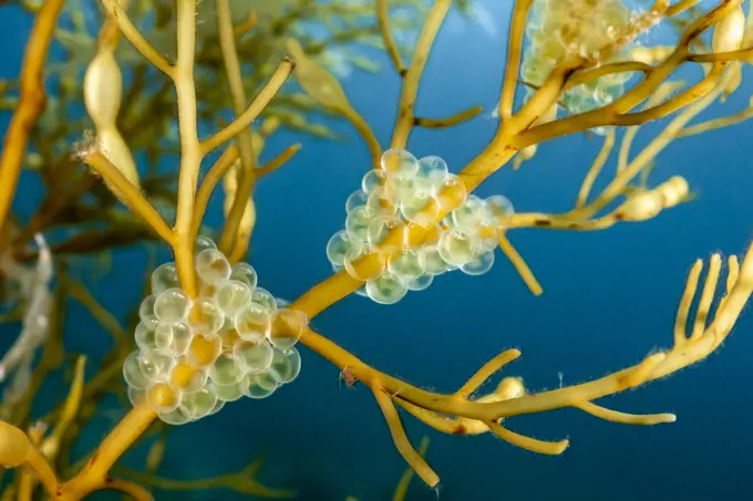
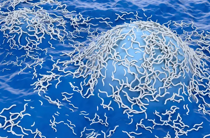
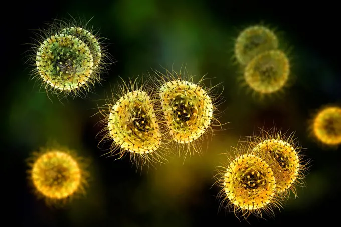
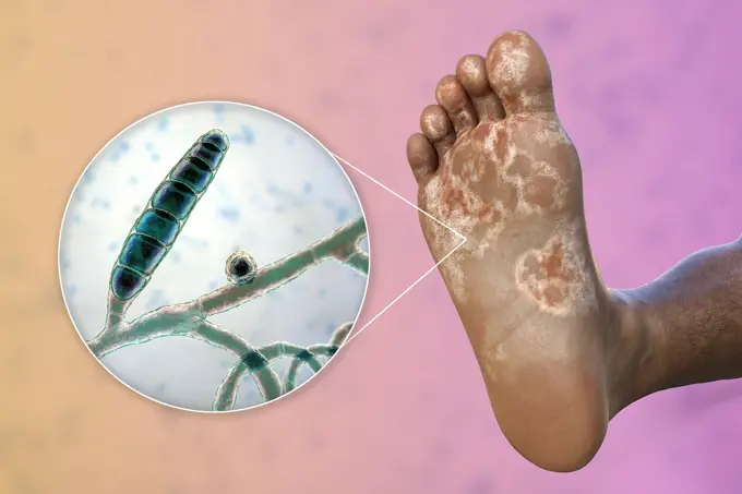
Microbial Cell Illustrations

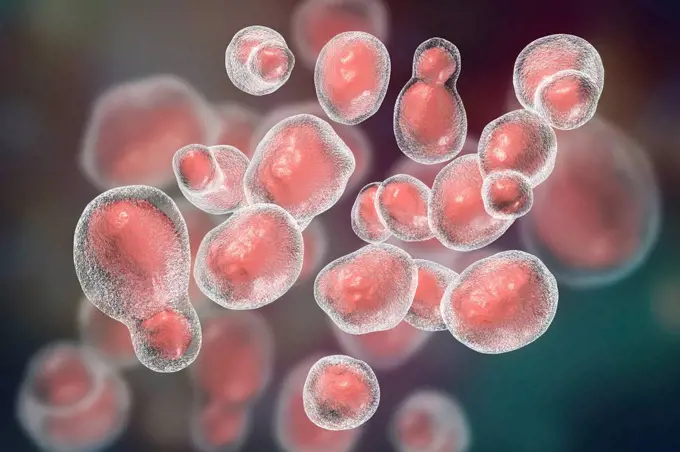
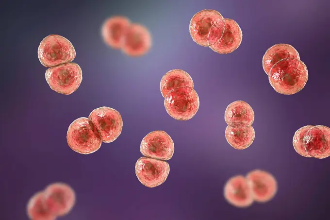
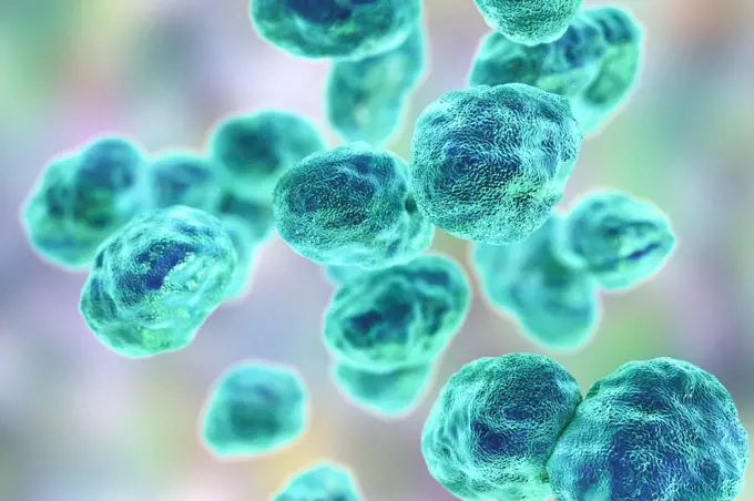
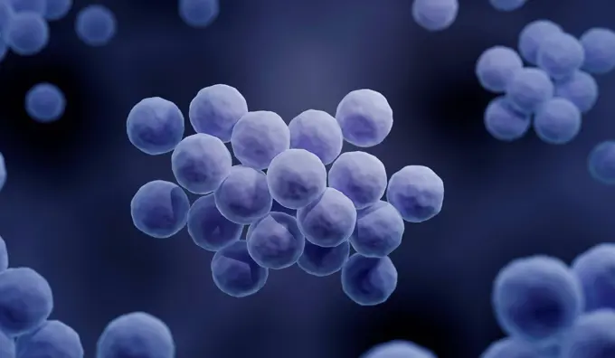
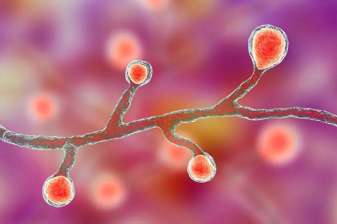
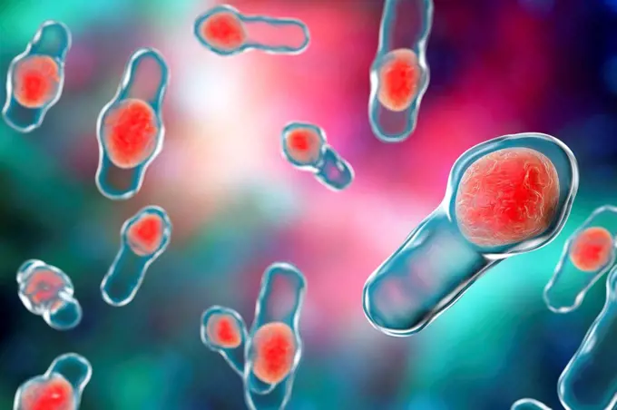
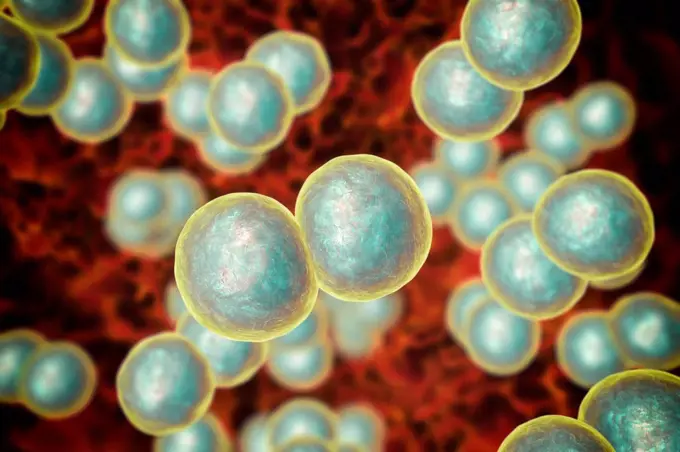
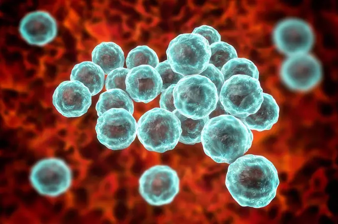
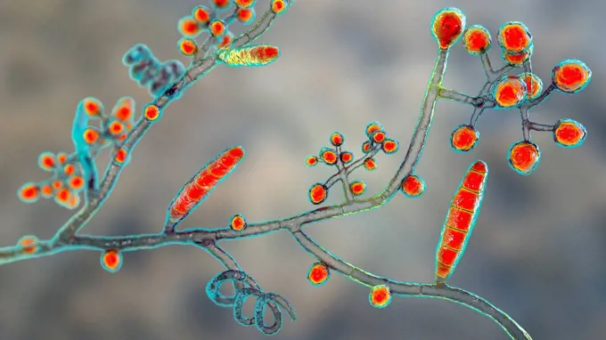
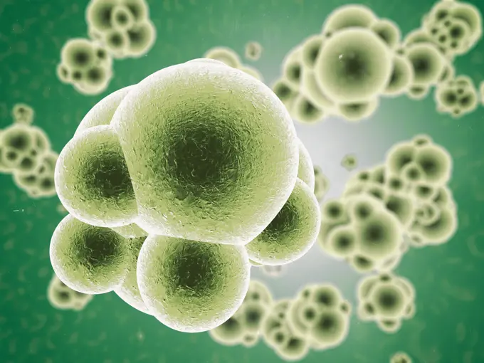
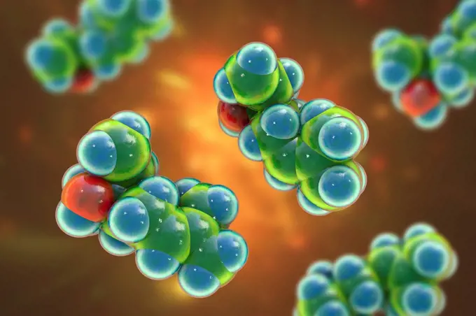
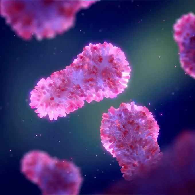
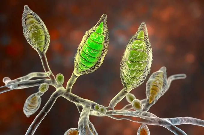
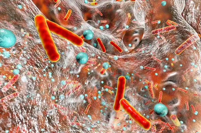
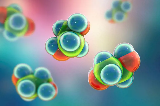
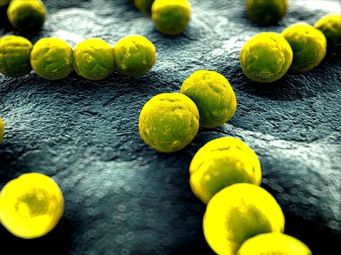
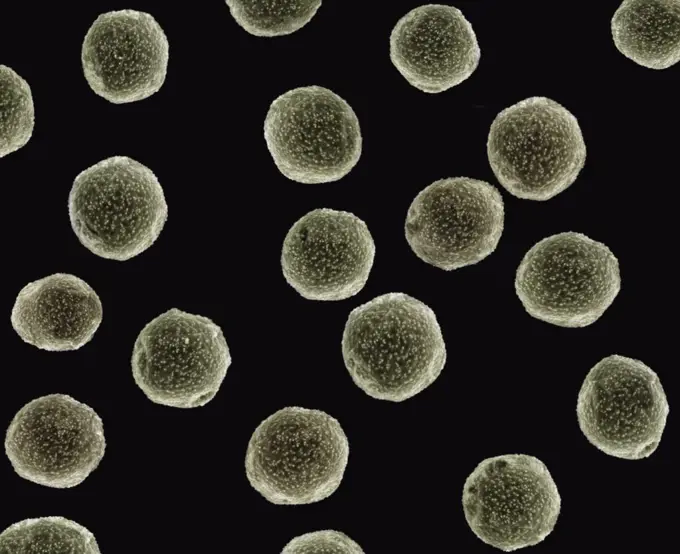
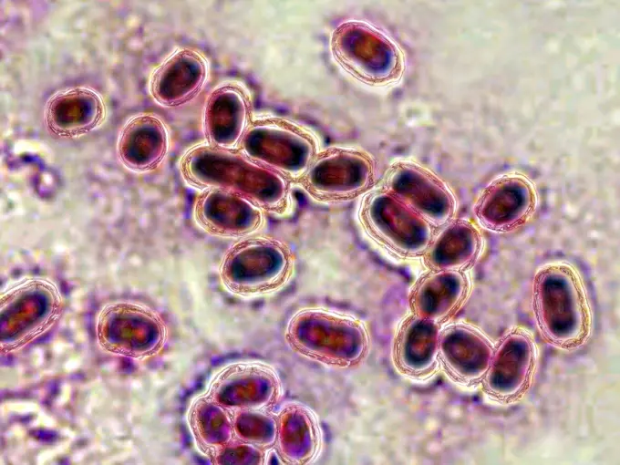
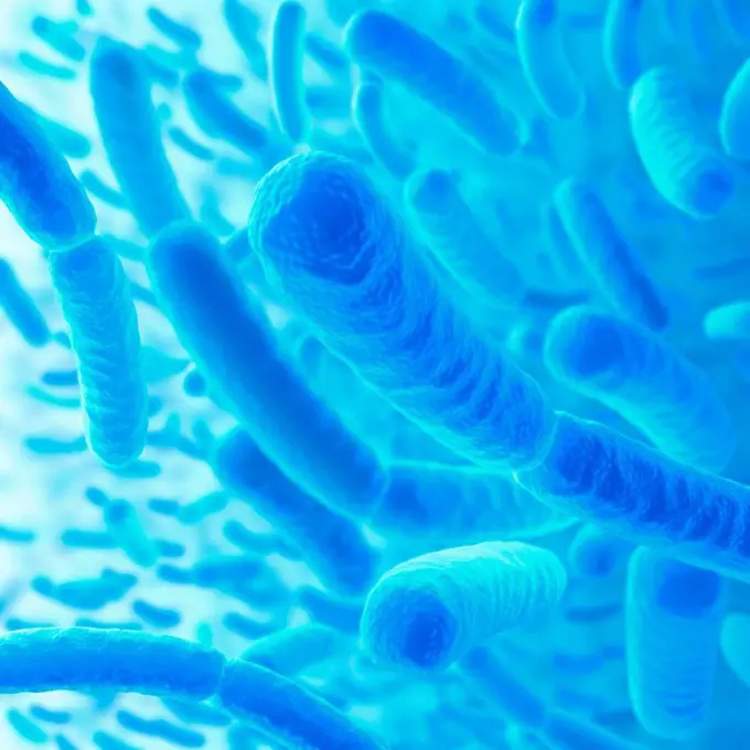
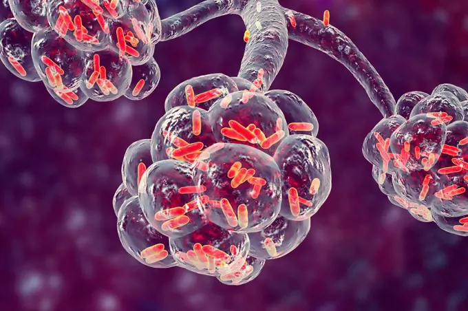
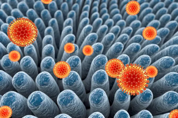
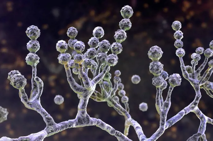
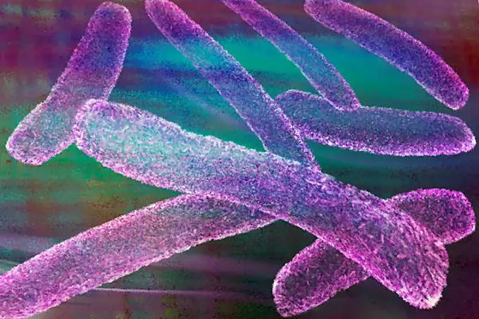
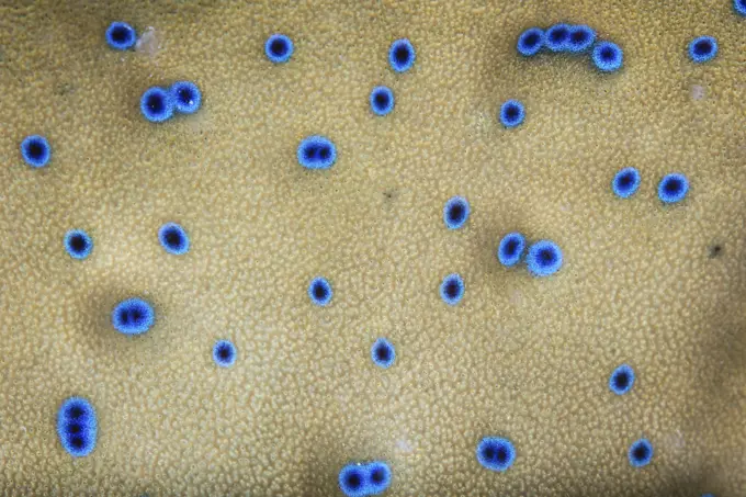
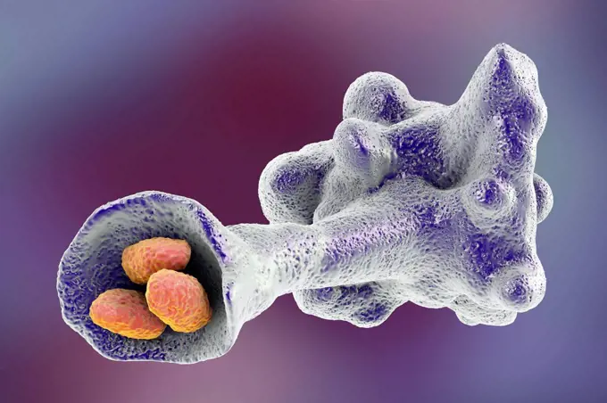
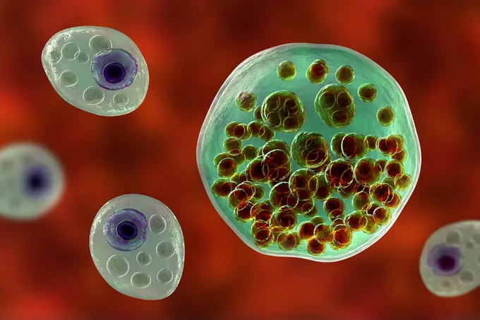
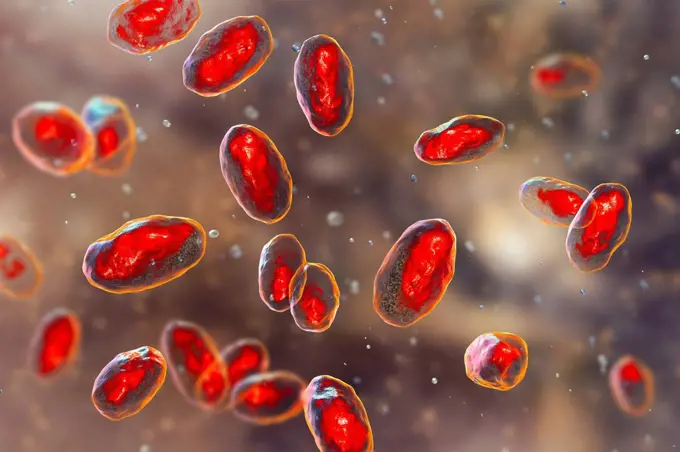
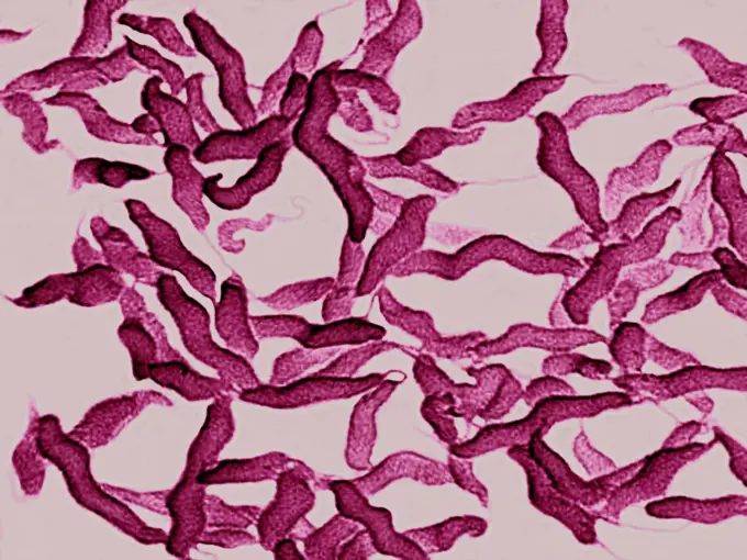
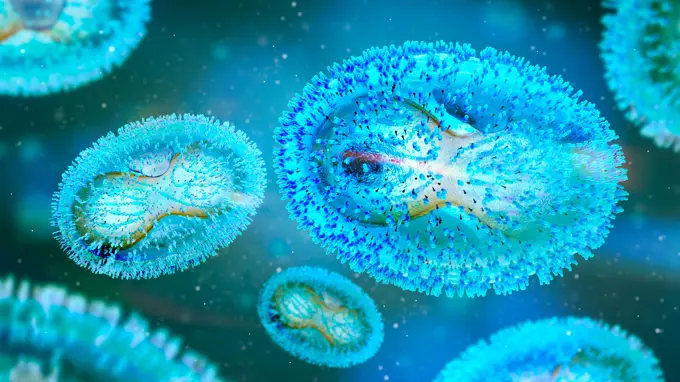
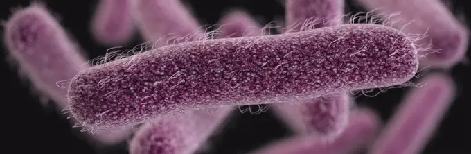
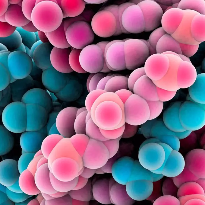
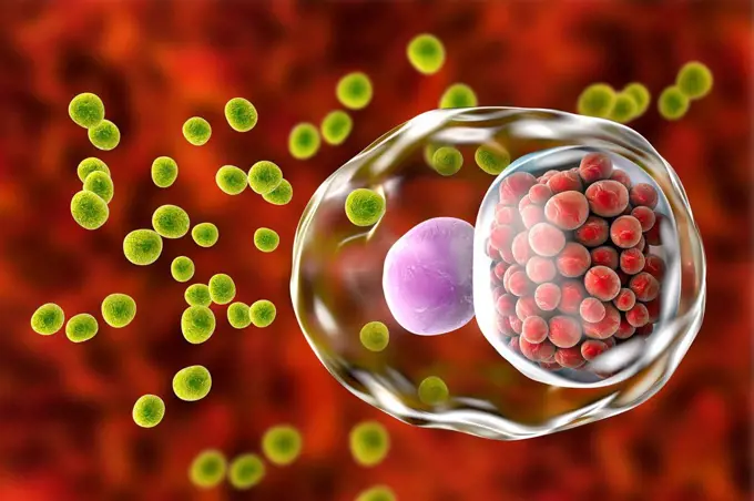
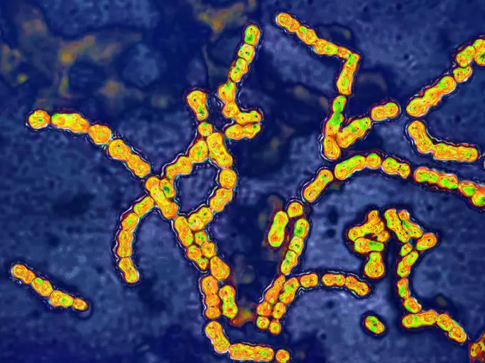
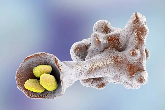
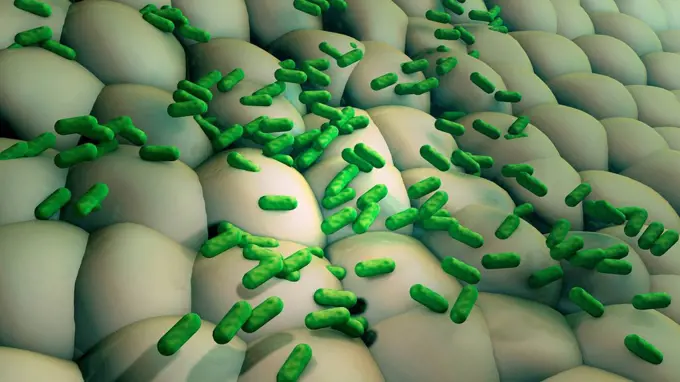
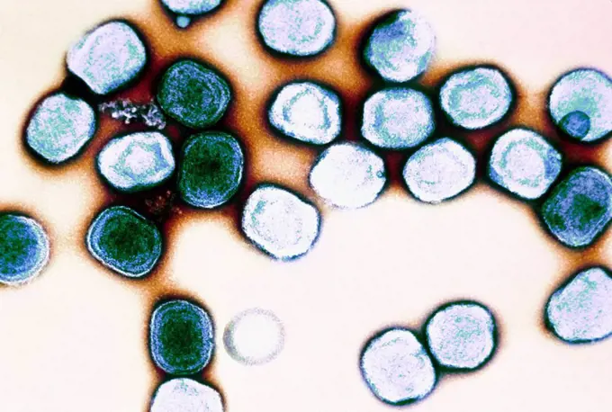
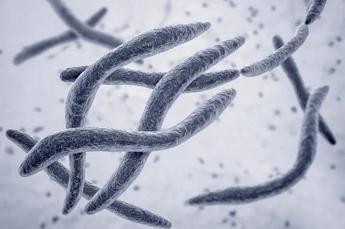
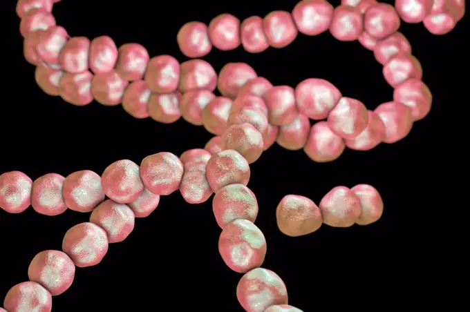
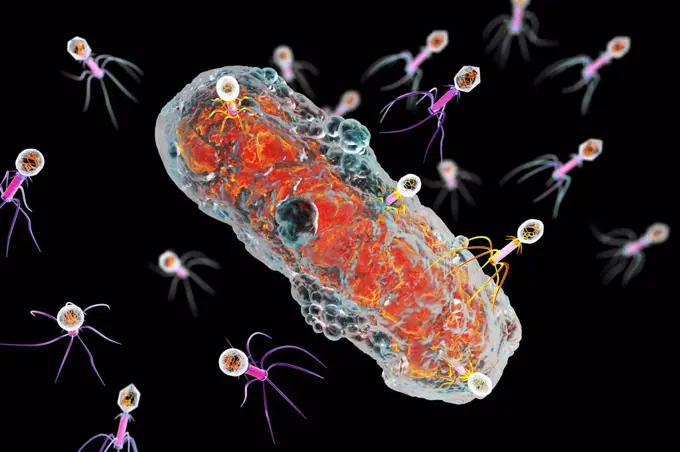
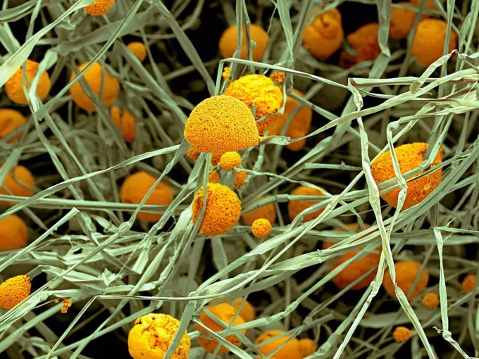
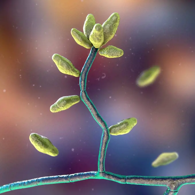
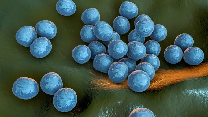
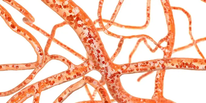
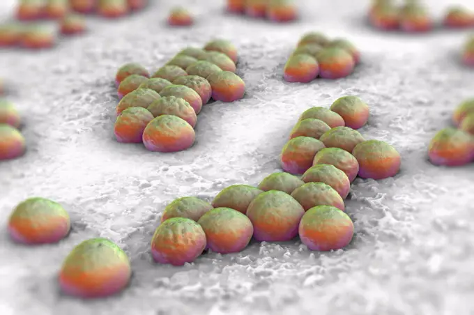
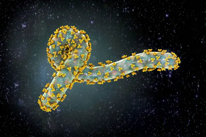
Colorful illustrations of various microorganisms including yeast, bacteria, and fungi. The images highlight intricate cellular structures with vibrant backgrounds.

Peptococcus bacteria, illustration

Assets in this Story

4128R-15405924
Yeast Saccharomyces cerevisiae, illustration
4128R-13940894
Mycoplasma bacteria, computer illustration. Mycoplasmas are the smallest cellular organisms known (diameter 0.3-0.9 microns). They resemble bacteria i...
4128R-14846676
Peptostreptococcus bacteria, illustration
4128R-14587977
Cryptococcus fungus, illustration
4128-30422609
Streptococcus pneumoniae bacteria, illustration
4128R-13763353
Tularaemia bacteria (Francisella tularensis), illustration. F. tularensis is Gram-negative, coccobacillus prokaryote. A zoonotic microorganism that ca...
4128-28968460
MRSA bacteria, illustration
4128R-14587971
Blastomyces fungus, illustration
4128R-14846664
Peptococcus bacteria, illustration
4128R-13282687
Clostridium difficile bacteria, computer illustration. C. difficile is a normal inhabitant of the human intestine, but it can become a pathogen when a...
4128R-15302845
Moraxella catarrhalis bacteria, illustration
4128R-15405900
Chlamydia trachomatis bacteria, illustration
4128-19054739
Microsporum canis fungus, illustration
4128-20044469
Gamma-terpinene molecule, illustration
4128-20044909
Molluscum contagiosum virus, illustration
4128-19489882
Blastomyces fungus, illustration
4128-20044535
Terpinen-4-ol molecule, illustration
4128R-13666937
Cancer cells, illustration.
4128-28767075
Amoxicillin drug molecule, illustration
4128R-13763912
Computer illustration of Haemophilus influenzae, coccobacillus bacteria. These bacteria cause a number of disease including meningitis in children, pn...
1525R-178735
4128-28767188
Conidiobolus coronatus microscopic fungi, illustration.
4128-20038497
Cholesterol molecule, illustration
4128-18799599
Leuconostoc bacteria, illustration
4128-16506558
Fungus Sporothrix schenckii, causative agent of the infection sporotrichosis, computer illustration. Fungal threads of the vegetative mycelium are see...
6188-66486372
Transparente Zellen mit Zellkern auf gelbem Hintergrund - 3d Illustration Transparent cells with nucleus on yellow background - 3d Illustration Copyright: xZoonar.com/ChristophxBurgstedtx 22602995
4128R-15366438
Haemophilus ducreyi bacteria, illustration
4128-19489844
Blastomyces fungus, illustration
4128R-15302841
Moraxella catarrhalis bacteria, illustration
4378-2598
Interior view of a cell showing a mitochondrion.
4128-19489970
Candida tropicalis yeasts, illustration
4378-4619
Blood Cells
4128R-15366378
Pneumococcal pneumonia, illustration
4128R-13711038
Illustration of molecules of antibodies.
4128R-13682067
Spherical bacteria and rod-shaped bacteria inside biofilm with quorum sensing molecules (small red circles), illustration. A biofilm is a colony of ba...
4239-V53647278
4128R-15405878
Candida albicans yeast and hyphae stages, illustration
4128-28767200
Splendore-Hoeppli phenomenon, illustration
4239R-8365
Microscopic view of chlamydia. Chlamydia is a common sexually transmitted disease (STD) caused by a bacterium.
4128R-15302844
Moraxella catarrhalis bacteria, illustration
4128R-13409387
Candida auris fungi, illustration
4128-20041282
Athlete's foot fungus, illustration
4197-63595671
Bacteria, particle and molecule with abstract, render or illustration of virus. Immune system, micro biology and sick for science, microscope and medical research for sepsis or senolytics treatment
4128-20044481
Linalool molecule, illustration
4128-V58569769
4128-19358187
Tupanvirus particles, illustration.
4128R-15290675
Pollen grains of Mimosa flower, illustration
4128-20044527
Terpinen-4-ol molecule, illustration
4128R-15466771
XY chromosomes, illustration
4239R-8021
Microscopic view of streptococcus.
4128R-14587987
Rat bite fever bacteria, illustration
4128R-15366380
Pneumococcal pneumonia, illustration
4128R-13763253
Multi-drug resistant Acinetobacter baumannii bacteria inside biofilm, computer illustration. A. baumannii is a Gram-negative, oxidase negative, aerobi...
4128R-14845712
Naegleria brain-eating amoeba, illustration
4128-28968594
Rhodotorula fungi, illustration
4128R-14057426
Cryptococcus neoformans fungus, illustration
4128-30420519
Lacazia loboi fungi, illustration
4128-20239394
Microsporum canis fungus, illustration
824-63227084
This transmission electron microscope image shows SARS-CoV-2 also known as 2019-nCoV, the virus that causes COVID-19. Virus particles emerge from the surface of a cell cultured in the laboratory. Spikes on the outer edge of virus particles give coronaviruses their name, shaped like a crown. Image captured and colorized at Rocky Mountain Laboratories in Hamilton, Montana. Credit: NIAID
4128-24796440
Polyethylene glycol molecule, illustration
4128R-14060597
Streptococcus pyogenes bacteria, illustration
824-65831019
Colorized scanning electron micrograph of a cell (teal) infected with the Omicron strain of SARS-CoV-2 virus particles (yellow), isolated from a patient sample.
6188-65540477
3d-Illustration von Hunderten von Meningitis-Erregern, den sogenannten Menigokokken 3d illustration of hundreds of meningitis pathogens called menigococcus Copyright: xZoonar.com/ChristophxBurgstedtx 22579383
4128-20040268
Microaneurysms, illustration
824-63223194
Colorized scanning electron micrograph of a white blood cell interacting with an antibiotic resistant strain of Staphylococcus aureus bacteria. Credit: NIAID.
4128R-15405882
Candida albicans yeast and hyphae stages, illustration
4128-19489912
Blastomyces fungus, illustration
4378-3974
A group of human antibodies, the Y-shaped proteins used by the immune system to defend against foreign objects like bacteria and viruses.
4128-28767116
Conidiobolus coronatus microscopic fungi, illustration.
4128-20043170
Monkeypox virus floating, illustration
4128-19054718
Microsporum canis fungus, illustration
4128R-15405520
Trichomonas vaginalis and neutrophils, illustration
4128-19056210
Fungi Paracoccidioides lutzii, illustration
4128-28767119
Conidiobolus coronatus microscopic fungi, illustration.
4128R-14323930
Bacteria in a biofilm, illustration
4128-28575432
Ethylene glycol molecule, illustration
824-63191210
This is a colorized scanning electron micrograph of a white blood cell eating an antibiotic resistant strain of Staphylococcus aureus bacteria, commonly known as MRSA.
1525R-243337
3d rendered scientific illustration of some bacteria
4201-66253
Pellitory (Parietaria sp) SEM close-up view of pollen grains at 1050x magnification, a well-recognized and important inhalant allergen in the Mediterranean area
824-63190249
Pneumococcus (streptococcus pneumoniae). Pneumococcus is the bacterium that causes pneumonia, bronchial pneumonia, purulent pleurisy, bacterial meningitis, ear infections, sinusitis and conjunctivitis. Optical microscopy x 1000.
4128R-14587973
Blastomyces fungus, illustration
1899-65662360
Scanning electron micrograph of Neisseria gonorrhoeae bacteria, which causes gonorrhea.
824-65830673
Scanning electron micrograph of Neisseria gonorrhoeae bacteria, which causes gonorrhea.
4128R-30487
Bacteria, computer artwork.
4128R-15366372
Bacterial pneumonia, illustration
4128R-14057567
Rotaviruses infecting intestine, illustration
4128-19055957
Fertilisation, illustration
4239R-8053
Stylized rabies virus particles, the cause of the viral neuroinvasive disease acute encephalitis.
4128-V58573415
4378-2318
Group of vibrio cholerae bacteria which causes cholera.
6188-66486302
Zytotoxische T-Zellen suchen und zerstören mutierte Krebszellen - 3d-Illustration Cytotoxic T cells search and destroy mutated cancer cells - 3d illustration Copyright: xZoonar.com/ChristophxBurgstedtx 22602869
4128R-13575502
Destruction of Candida fungi, computer illustration. Image can be used to illustrate antifungal treatment concept.
824-63227078
This scanning electron microscope image shows SARS-CoV-2 (round yellow particles) emerging from the surface of a cell cultured in the laboratory. SARS-CoV-2, also known as 2019-nCoV, is the virus that causes COVID-19. Image captured and colorized at Rocky Mountain Laboratories in Hamilton, Montana. Credit: NIAID
4128R-14323920
Bacillus subtilis bacteria, illustration
4128-19054741
Microsporum canis fungus, illustration
4128R-14768496
Gonorrhoeae bacteria, illustration
1525-27220396
3D render of a medical background with Corona virus cells with bloods cells
4128-19056194
Fungi Paracoccidioides lutzii, illustration
4128R-25486
Bacterial infection. Computer artwork of bacteria (green) in the blood stream.
4128-19054730
Scopulariopsis brevicaulis fungus, illustration
1848-49407987
Illustration Bacteria, bacteria are single-celled organisms and the simplest form of life on our planet
4239-69148150
Detail of the mantle of a giant clam.
4128R-13928415
Parasitic Amoeba protozoan engulfing bacteria, computer illustration. The amoeba will engulf and digest the prey in a process called phagocytosis.
4128-24796423
Ethylene glycol molecule, 3D illustration
4128R-13928453
Parasitic amoeba (Entamoeba histolytica) engulfing red blood cells, computer illustration. This single-celled organism causes amoebic dysentery and ul...
4128-V58614830
4128R-12923033
Enterococcus faecalis bacterium
4378-1864
Treponema pallidum, the bacterium responsible for the dangerous sexually transmitted infection syphilis.
4128-111762351
Stachybotrys sp. fruiting structure with spores, computer illustration. Often known as the toxic mould (black mould). Several strains of this fungus (...
4128R-15405927
Yeast Saccharomyces cerevisiae, illustration
4128-V58562075
4128R-26269
MRSA bacteria. Computer artwork of methicillin-resistant Staphylococcus aureus (MRSA) bacteria.
824-63225886
This scanning electron microscope image shows SARS-CoV-2 (round yellow particles) emerging from the surface of a cell cultured in the lab. SARS-CoV-2, also known as 2019-nCoV, is the virus that causes COVID-19. Image captured and colorized at Rocky Mountain Laboratories in Hamilton, Montana.
6188-65540217
3D-Illustration von transparenten und bunten Protozoen oder einzelligen Organismus 3D illustration of transparent and colorful protozoa or unicellular organism Copyright: xZoonar.com/ChristophxBurgstedtx 22577737
4128R-14845675
Chlamydia trachomatis bacteria, illustration
4128R-15302846
Moraxella catarrhalis bacteria, illustration
4128R-15047189
Transparent cells, illustration
1525-57354178
Abstract molecular backrgound, laboratory reasearch and medicine concept, 3D illustration. Abstract molecular backrgound
824-63225885
This scanning electron microscope image shows SARS-CoV-2 (round gold particles) emerging from the surface of a cell cultured in the lab. SARS-CoV-2, also known as 2019-nCoV, is the virus that causes COVID-19. Image captured and colorized at Rocky Mountain Laboratories in Hamilton, Montana.
824-66066009
Colorized scanning electron micrograph of a cell heavily infected with SARS-CoV-2 virus particles (yellow), isolated from a patient sample. The black area in the image is extracellular space between the cells. Image captured at the NIAID Integrated Research Facility (IRF) in Fort Detrick, Maryland. Credit: NIAID/NIH.
4128R-13587567
Plague bacteria (Yersinia pestis), computer illustration. Y. pestis are gram-negative bacteria from Enterobacteriaceae family, they have ovoid shape w...
4128-V58572076
4128R-13242351
Antibodies attacking bacterium, illustration, conceptual image.
4128R-15545931
Atoms, illustration
4269-27468
Candida albicans. Fluorescent antibody stain micrograph of budding yeast cells of Candida albicans. Candida albicans is a single-celled fungus commonly found with man and usually harmless. However, it can sometimes causes infections like oral thrush, vaginal thrush and diaper rash.
4128-18799658
Rhizopus fungus, illustration
4128R-14060581
Mycobacterium avium bacterium, illustration
4128-28767319
Rhinosporidium seeberi parasite, illustration
4128-20045064
Yersinia pseudotuberculosis bacteria, illustration
824-63178972
Campylobacter is a bacterium causing food poisoning which is the cause of: diarrhea, enteritis, fevers and abdominal pain. Optical microscopy
7203-70646315
Natural occurring fluorescence in underwater bubble coral (Euphyllia ancora), captured by using special UV blocking barrier filters. Night dive near Illi Api Island, Selat Boleng Strait, Indonesia
4128-V58574197
6188-65540146
Querschnitt eines Pockenerregers mit Zellmembran, Nukleokapsid, Zellwand und Glykoproteinen - 3d-Illustration Cross section of a smallpox pathogen with cell membrane, nucleocapsid, cell wall and glycoproteins - 3d illustration Copyright: xZoonar.com/ChristophxBurgstedtx 22575853
4239-18641843
3D illustration of a number of rod-shaped, drug-resistant, Shigella bacteria. This artistic recreation is based upon scanning electron microscopic (SEM) imagery. The exterior of the Shigella bacterium is fimbriated, covered by numerous thin, hair-like projections, imparting a furry appearance.
4128R-11476207
Protein molecules, computer artwork.
4128R-14638030
Chlamydia infection, illustration
824-63190276
Group A streptococci pyogenes. It is the cause of skin infections (impetigo), abscesses, bronchopulmonary infections, a bacterial form of red angina which can develop into complications such as acute articular rheumatism. Optical microscopy X 1000.
6188-65540209
3d-Illustration von Meningitis-Erregern namens Menigokokken 3d illustration of meningitis pathogens called menigococcus Copyright: xZoonar.com/ChristophxBurgstedtx 22577690
4128-20045047
Yersinia pseudotuberculosis bacteria, illustration
4128-20044983
Smallpox virus, illustration
4128R-14060532
Toxoplasma gondii parasites, illustration
4128-19490181
Antimicrobial resistance, conceptual illustration
4128R-13410119
Bacteria, illustration
4128-111493676
Biofilm, conceptual illustration.
4128R-13282636
Rod-shaped bacteria inside biofilm, illustration. A biofilm is a colony of bacteria that forms a coating on a surface. Common places for biofilms to d...
4128-28968543
Molecular model of beta-carotene, illustration
1850-49796
Papaver cultivar, Single open flower and several buds on bent and curving stems. Colour has been digitally manipulated.
4128-19358667
Glucagon hormone molecule. Computer model showing the structure of the human hormone glucagon. Glucagon is involved in carbohydrate metabolism and is ...
824-57657667
Colorized transmission electron micrograph of monkeypox virus particles (green) cultivated and purified from cell culture.
4269-7097
Scanning electron micrograph (SEM) of methicillin-resistant Staphylococcus aureus bacteria (MRSA); Magnification 9560x. This strain of Gram-positif bacterium has developed wide-ranging antibiotic resistance such as methicillin, and are hospital pathogens.
4128-20041270
Athlete's foot fungus, illustration
1773R-101715
Illustration of colorful microbes
1525R-175118
4128R-14846739
Veillonella bacteria, illustration
4128R-13928416
Parasitic Amoeba protozoan engulfing bacteria, computer illustration. The amoeba will engulf and digest the prey in a process called phagocytosis.
4128-111495179
Bacteria multiplying on surface, illustration.
4128-V58566370
4128R-4393
Vaccinia virus particles, TEM
4378-1887
Sperm entering an ovum.
4128-19247881
Fusobacterium bacteria, computer illustration. These bacteria are Gram-negative, anaerobic, non-motile rod-shaped prokaryotes (characterized by a long...
4128-20044388
Curcumin molecule, illustration
4128R-11544499
Cells, computer illustration.
4128-111495191
Bacteria multiplying on surface, illustration.
4128R-11477286
Computer artwork of a number of human chromosomes.
4128R-11476239
Protein molecules, computer artwork.
4128R-13763959
Nerve synapse. Computer illustration of a junction, or synapse, between two nerve cells (neurons). As the electrical signal reaches the presynaptic en...
4378-4429
Helicobacter pylori
1848-64959817
Yellow silkworm cocoon over green on silk worm net
1525-25137136
cells. lots of green germ cells under the microscope
824-63191198
This illustration depicts a three-dimensional (3D) computer-generated image of a cluster of sphere-shaped vancomycin-resistant Staphylococcus aureus (VRSA) bacteria. The artistic recreation was based upon scanning electron micrographic imagery. Staphylococcus aureus is a common type of bacteria that is found on the skin. During medical procedures when patients require catheters or ventilators, or undergo surgical procedures, S. aureus can enter the body and cause infections. When S. aureus becomes resistant to vancomycin, there are few treatment options available because vancomycin-resistant S. aureus bacteria identified to date were also resistant to methicillin and other classes of antibiotics. A total of 13 cases of vancomycin-resistant Staphylococcus aureus (VRSA) have been identified in the United States since 2002. VRSA infection continues to be a rare occurrence. A few existing factors seem to predispose case patients to VRSA infection, including: - Prior MRSA and enterococcal i
4128R-29760
Bacterial infection, computer artwork.
4239R-8231
Microscopic view of athlete's foot fungus. Athlete's foot fungus, also known as ringworm of the foot and tinea pedis, is a fungal infection of the skin.
4378-3058
Genetic determining human chromosome.
4128-111493617
Nanomicelles, conceptual illustration.
4269-7096
Scanning electron micrograph (SEM) of methicillin-resistant Staphylococcus aureus bacteria (MRSA); Magnification 9560x. This strain of Gram-positif bacterium has developed wide-ranging antibiotic resistance such as methicillin, and are hospital pathogens.
824-63225867
Colorized scanning electron micrograph of a cell (gold) infected with UK B.1.1.7 variant SARS-CoV-2 virus particles (blue), isolated from a patient sample. Image captured at the NIAID Integrated Research Facility (IRF) in Fort Detrick, Maryland.
4128R-14587963
Aggregatibacter aphrophilus bacteria, illustration
4128-30421538
RAGE receptors in membrane, illustration
6188-64771874
Ripe nutmegs levitate on a white background Ripe nutmegs levitate on a white background. Copyright: xZoonar.com/AlekseyxButenkovx 16251778
4389-2108
SSBRP Petri Dish Specimens
4128-20041284
Athlete's foot fungus, illustration
4128-16222769
Meningitis bacteria, illustration
6188-65540329
Mikroskopische Nahaufnahme von wachsendem Mehltau - 3d Illustration Microscopic closeup of growing mildew - 3d illustration Copyright: xZoonar.com/ChristophxBurgstedtx 22579328
4128-19054665
Madurella fungi, illustration
4378-3253
Close up view of athlete's foot fungus.
4128-28767092
Bisphenol A molecule, illustration
4128R-13587550
Streptococcus bacteria, computer illustration. Streptococcus bacteria are an example of cocci (round bacteria) that form chains. There are over 50 spe...
4128R-13586596
Bacteriophages infecting bacteria, computer illustration. A bacteriophage, or phage, is a virus that infects bacteria. It consists of an icosahedral (...
4128R-12699838
Coloured scanning electron micrograph (SEM) of fungal cells. The round structures are sporangia, which house the fungus' reproductive cells (spores). ...
4128-19358306
Curvularia mould fungus, computer illustration. Curvularia is a group of about 35 species, most of which only infect plants. However, C. geniculata ca...
4378-3320
A telomere is a region of the DNA sequence at the end of a chromosome. Their function is to protect the ends of the chromosome from degradating. Here they are visible as highlights at the tips of the chromosomes.
1525-20622642
macro shot of fuzzy mold in agar-agar
4128R-11544474
Cancer cells, computer illustration.
6188-65540474
3D-Illustration von Blutzellen, Plasmodien, die die Malaria-Krankheit verursachen 3d illustration of blood cells, plasmodium causing malaria illness Copyright: xZoonar.com/ChristophxBurgstedtx 22579378
4128R-11477263
Computer artwork of a number of human chromosomes.
4128-28575417
Staphylococcus lugdunensis bacteria, illustration
1525-22394826
yellow silkworm cocoon over green on silk worm net
4128-20040238
Blood vessels, illustration
4378-2519
Clusters of 'superbug' (MRSA) bacteria.
4128R-15366398
Aspergillus fumigatus fungus, illustration
4128-30420564
Marburg virus, illustration
4128R-13620070
Coloured scanning electron micrograph (SEM) of Enterococcus faecalis (formerly known as Streptococcus faecalis), Gram positive, coccoid prokaryote (di...
4378-16109649
Stylized view of alveolar sacs from within the respiratory system.
4378-5671
Tsetse Fly
1525-56158495
The Santa Convention 3d illustrated
4128-18497162
Nanomaterials for antibiotic-resistant bacteria
4378-4472
Legionella Pneumophila
4128R-14060537
Toxoplasma gondii parasites, illustration
4128-20038806
Sperm cell fertilising an egg, illustration
1525-66336672
Virus particles interacting with antibody molecules
4128R-14637824
Candida albicans, illustration
4128-V58569317
1848-50969307
Image of Flu COVID-19 virus cell under the microscope on the blood.Coronavirus Covid-19 outbreak influenza background.Pandemic medical health risk concept with disease cell as a 3D render
4128R-13587471
Bacillus cereus bacteria, computer illustration. These are Gram-positive rod-shaped spore-producing bacteria, that are often arranged in chains (strep...
4128R-13575565
Illustration of Helicobacter pylori bacteria. It is a curved, or spiral, Gram-negative bacilli with flagella at one end (seen here) that make it highl...
4128R-13048918
Computer illustration of staphylococci bacteria (Staphylococcus aureus).
4128-28968472
Saccharomyces cerevisiae yeast, illustration
4128-19053691
Fat cells, illustration
4128-16222950
Haemophilus influenzae bacteria, illustration
4279-66658
DEU, 2005: Hedgehog Gourd (Cucumis dipsaceus), whole and halfed fruit, studio picture.
4070-28603410
Korean sandlace (Hypoptychus dybowskii) fish eggs freshly spawned and attached to Sargassum (Sargassum horneri) by a sticky secretion, Japan, Pacific Ocean.
4128-48285275
Ependymoma cancer cells, illustration
4128-16222776
Meningitis bacteria, illustration
4128-30420507
Athlete's foot, illustration
6188-68402349
Russische Karte mit Coronavirus unter der Lupe, 3D-Rendering Russian map with coronavirus under magnifier, 3D rendering isolated on white background LicenseRF Copyright: xZoonar.com/alexlmxx 23342444