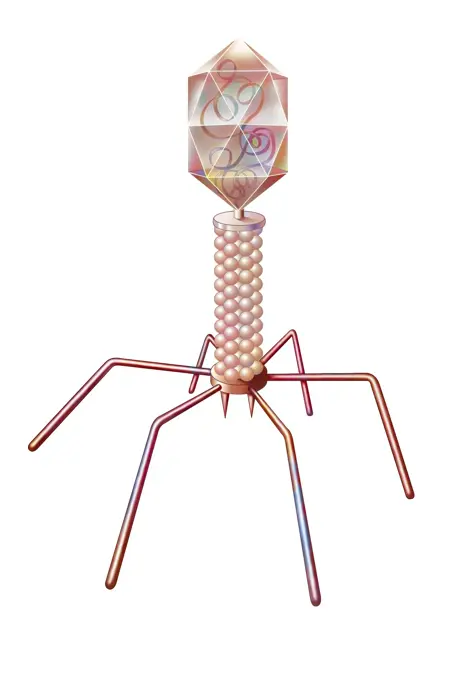
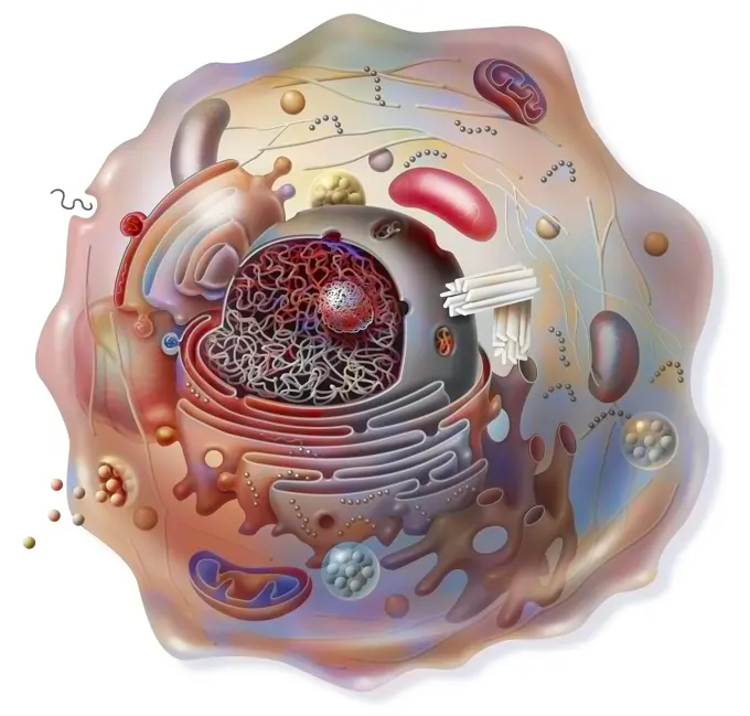
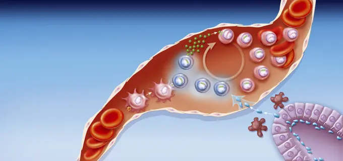
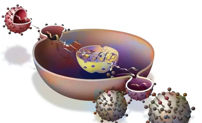
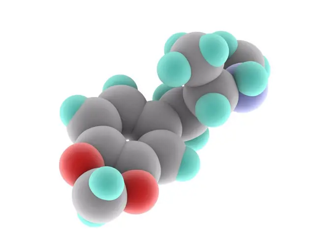
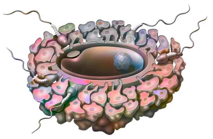
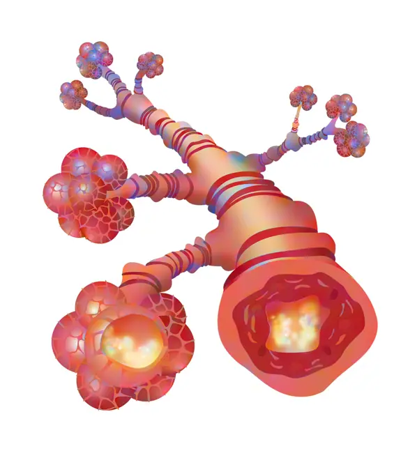
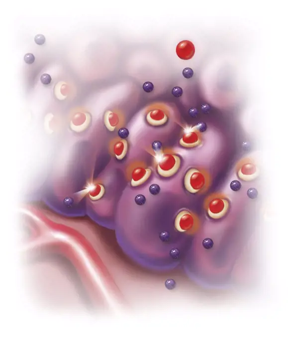
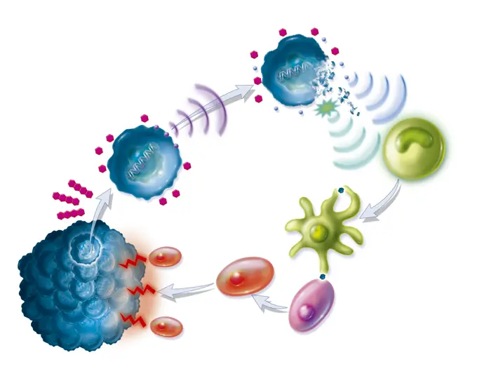
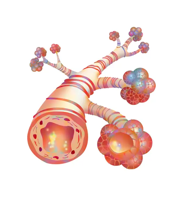
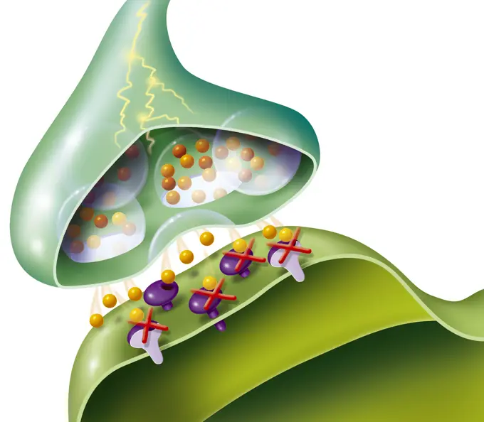
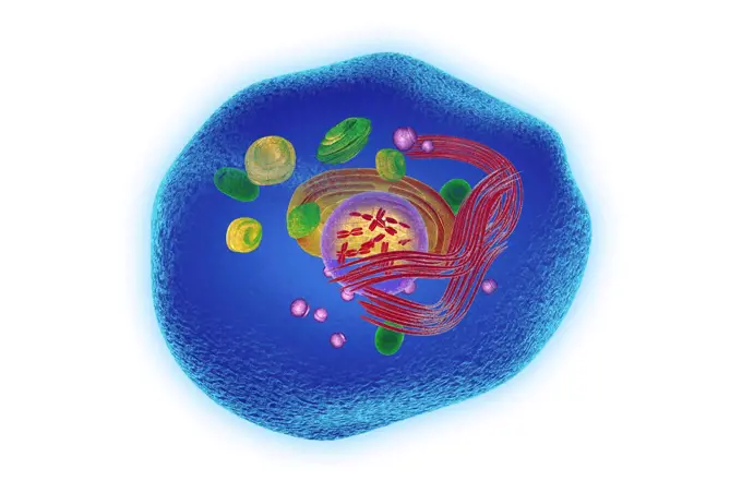
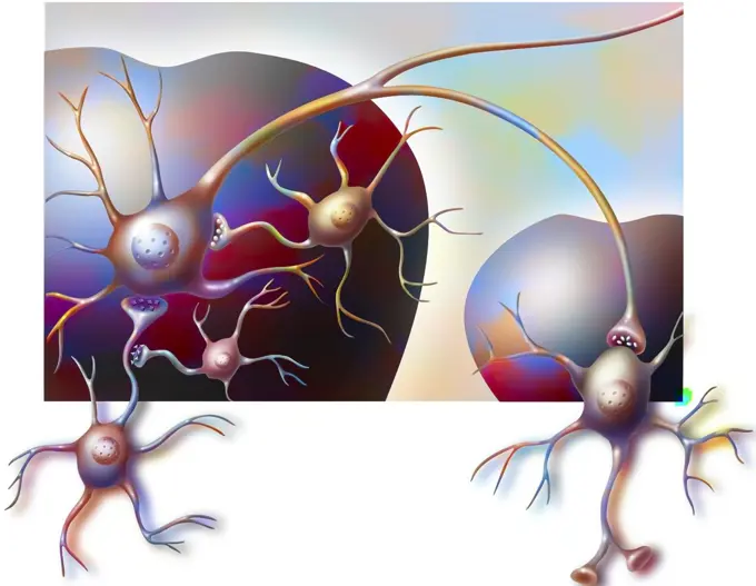
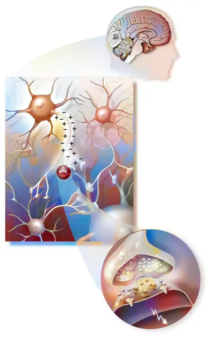
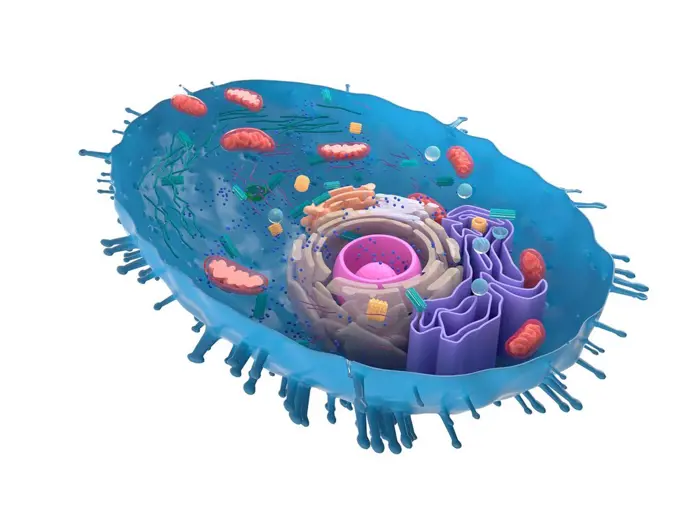
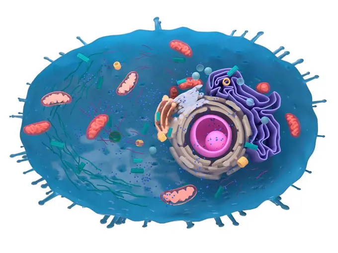
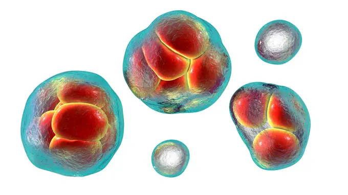
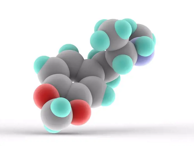
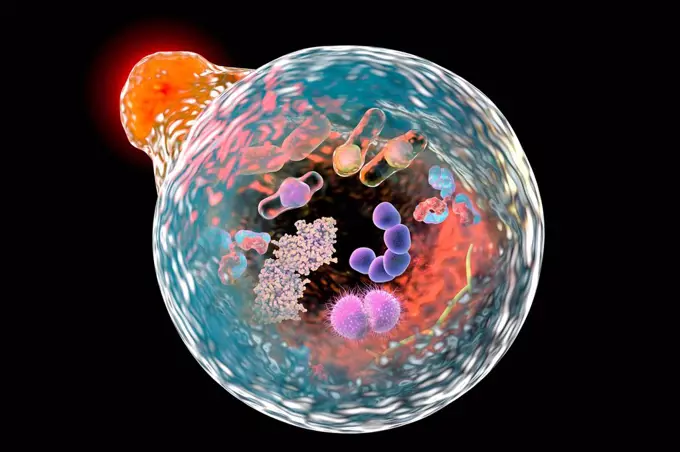
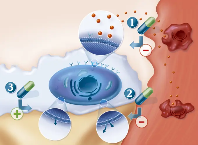
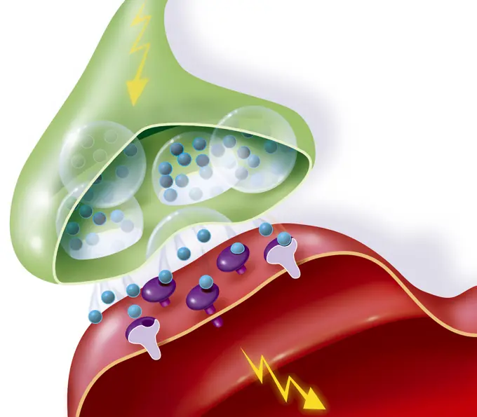
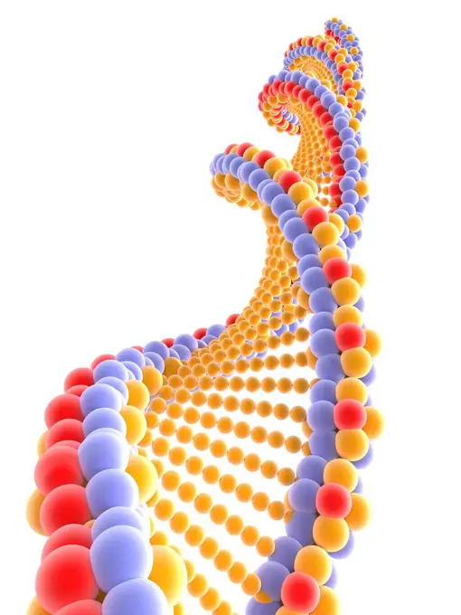
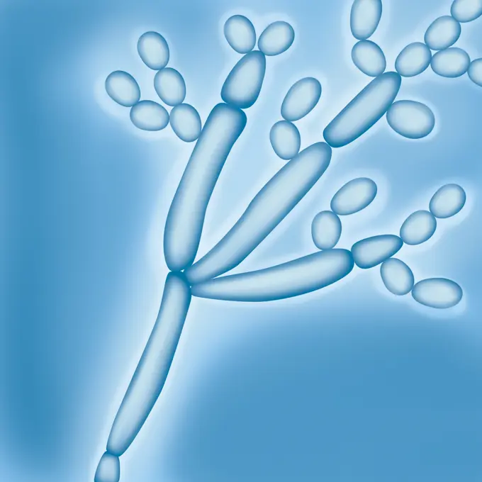
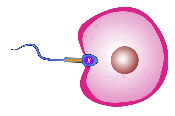
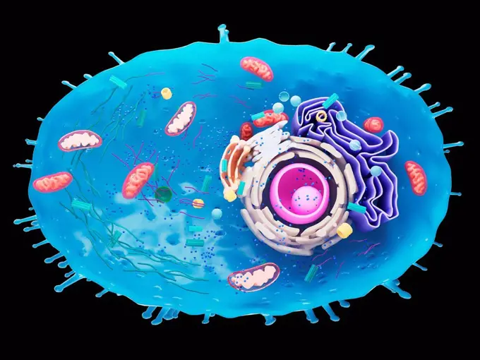
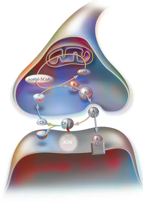
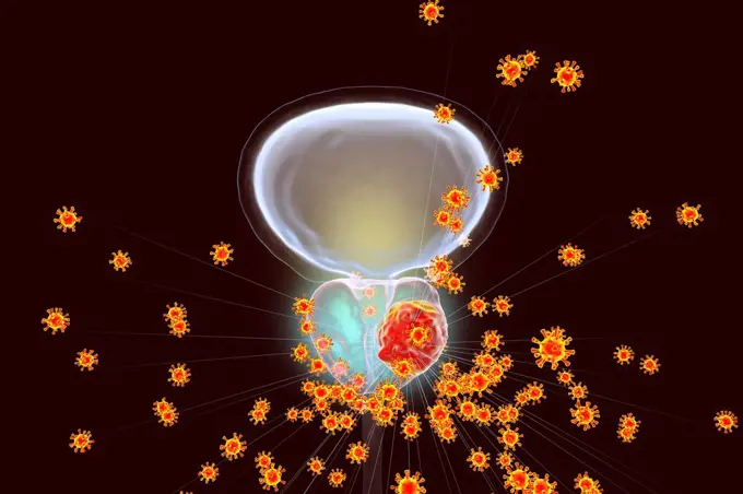
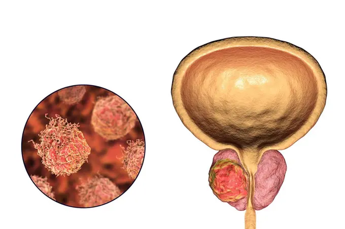
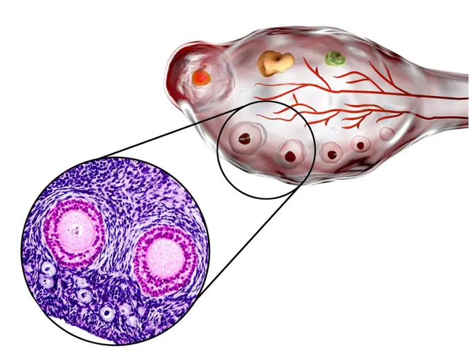
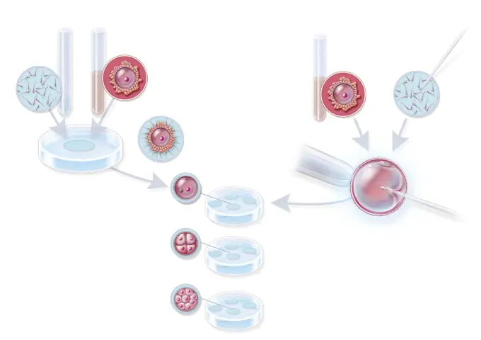
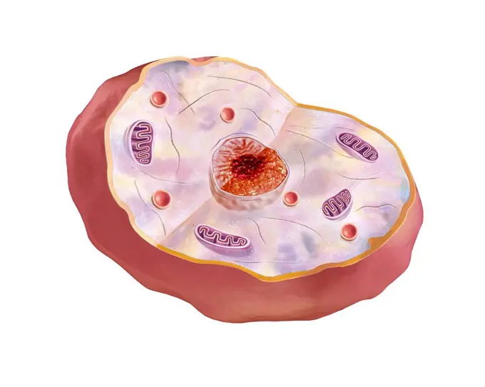
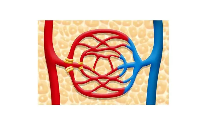
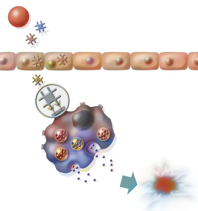
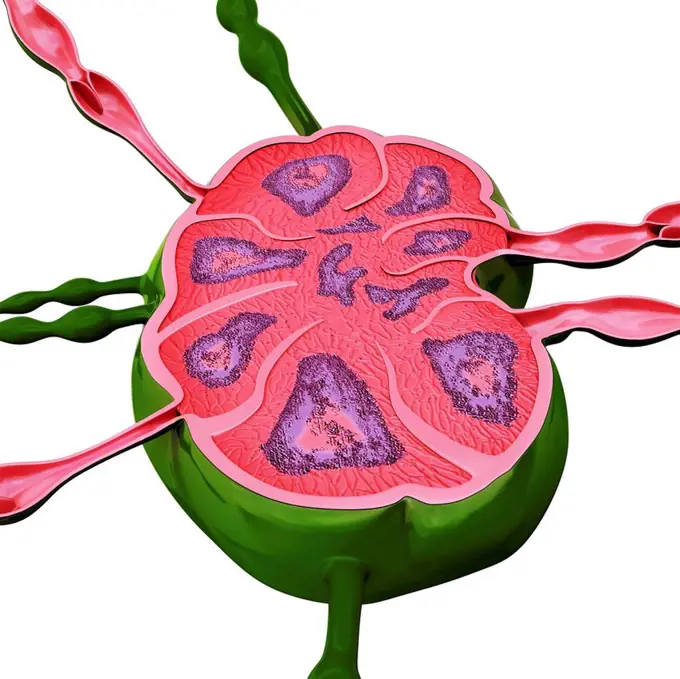
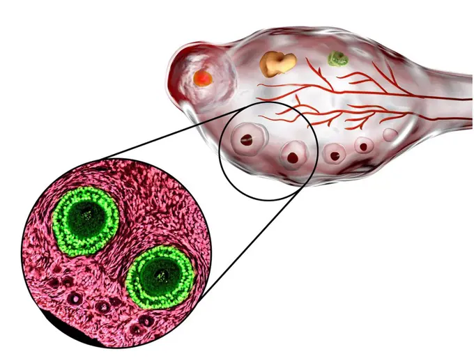
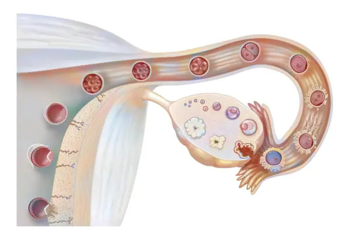
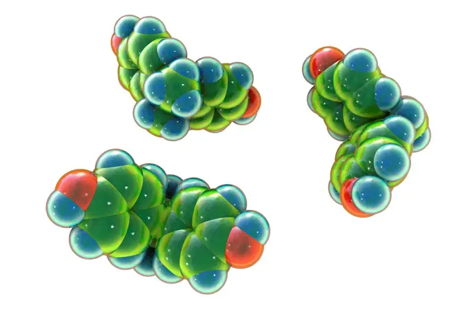
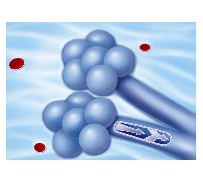
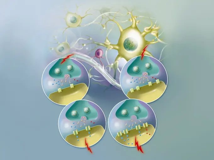
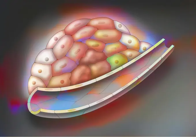
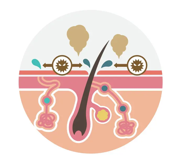
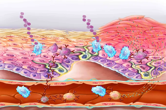
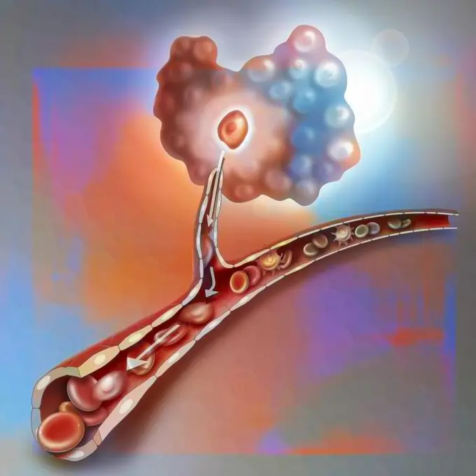
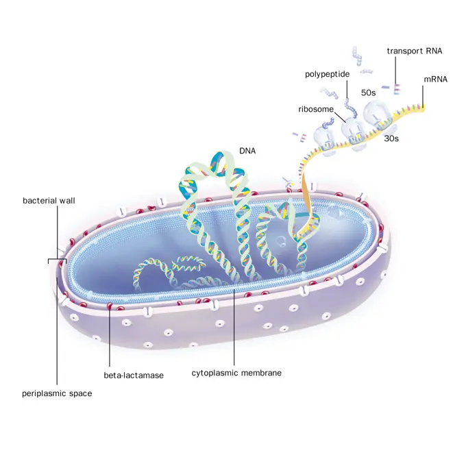
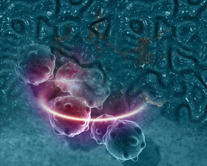
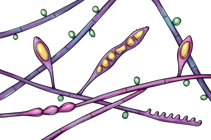
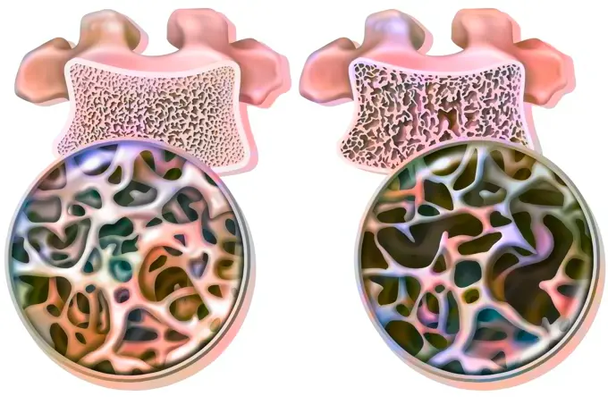
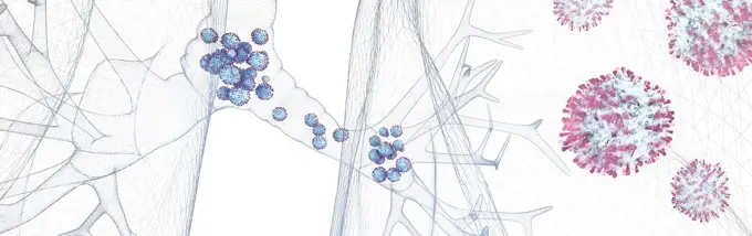
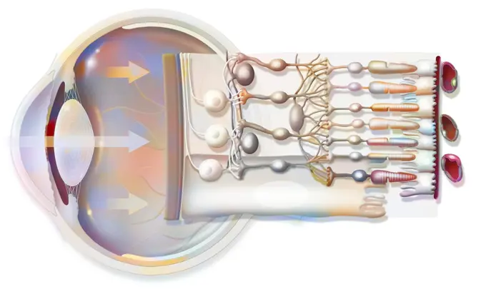
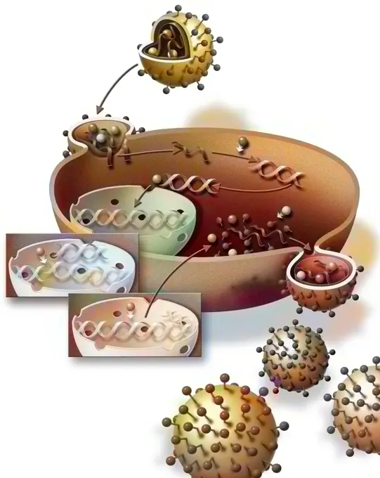
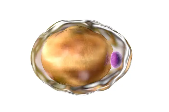
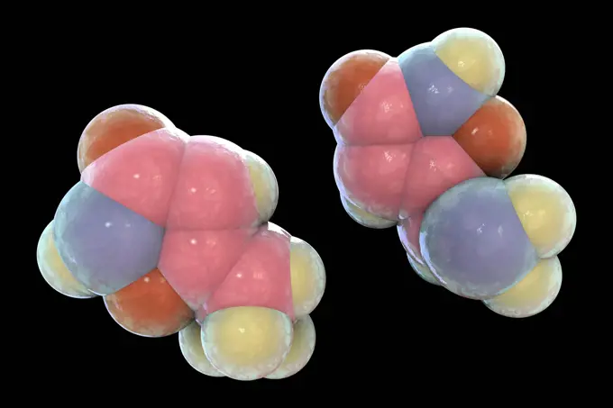
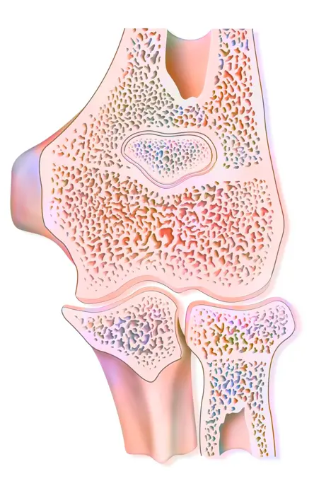
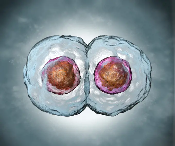
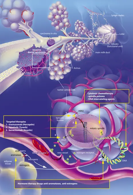
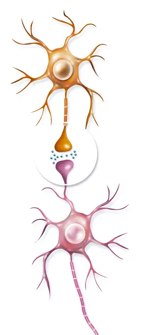
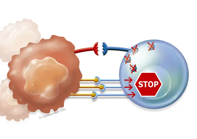
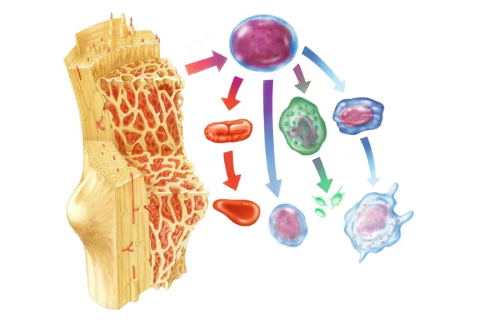
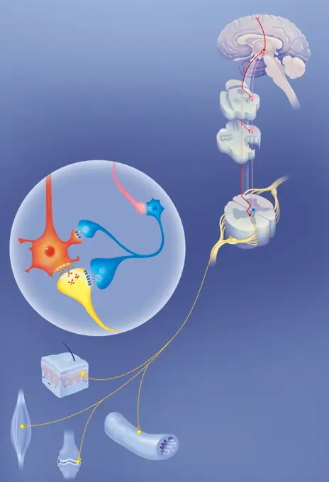
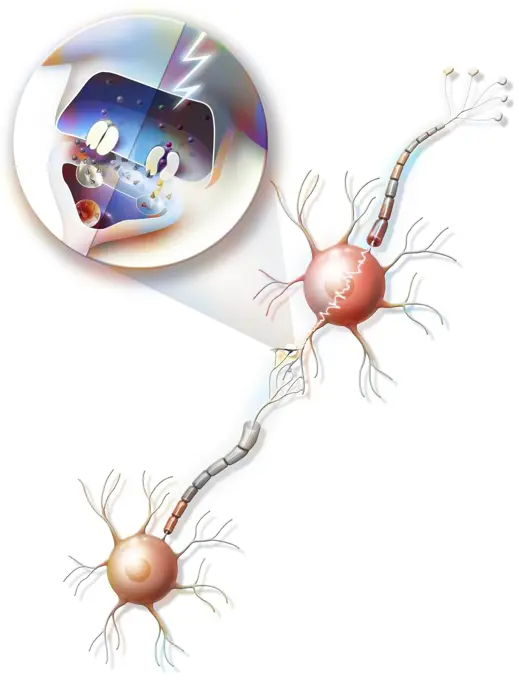
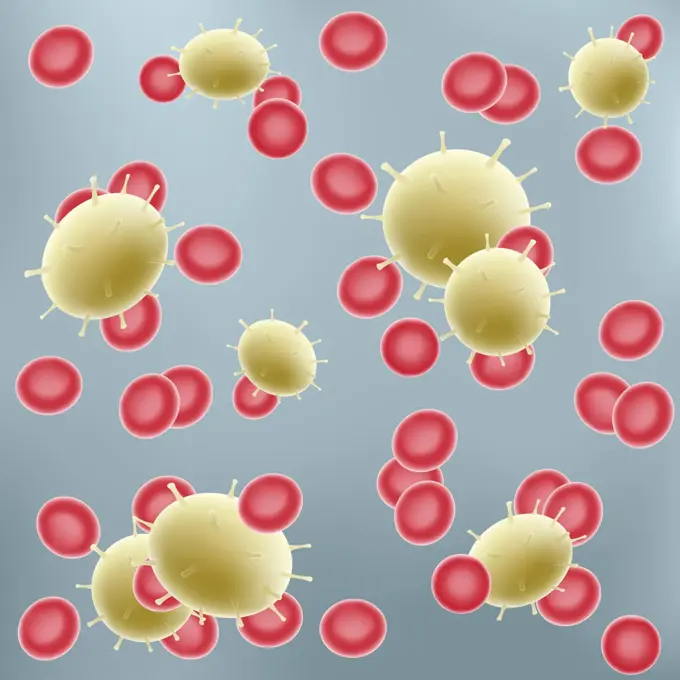
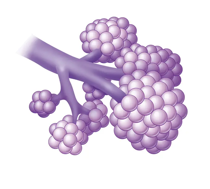
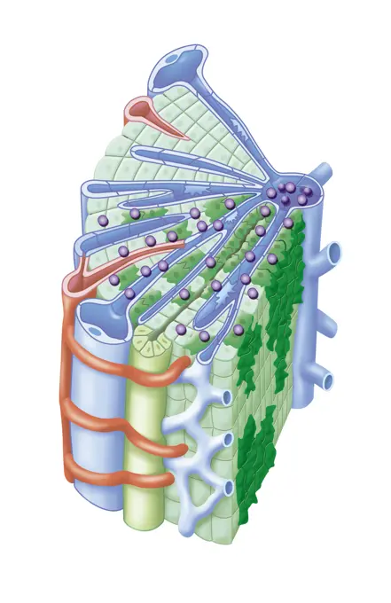
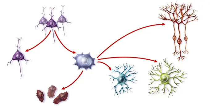
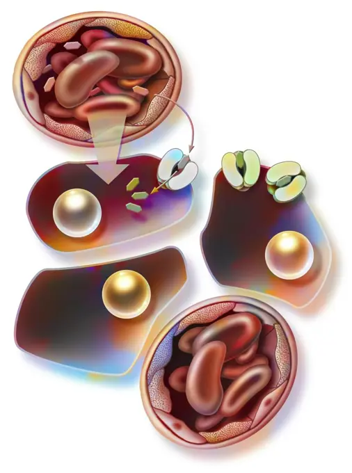
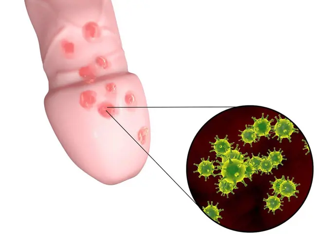
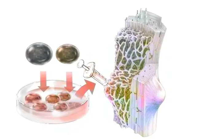
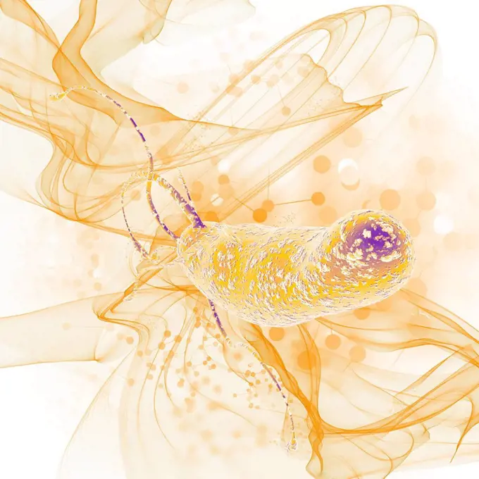
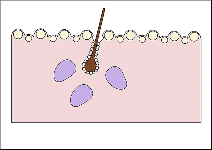
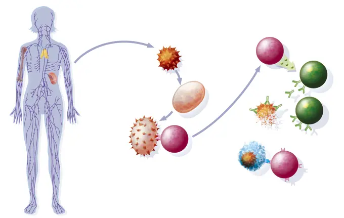
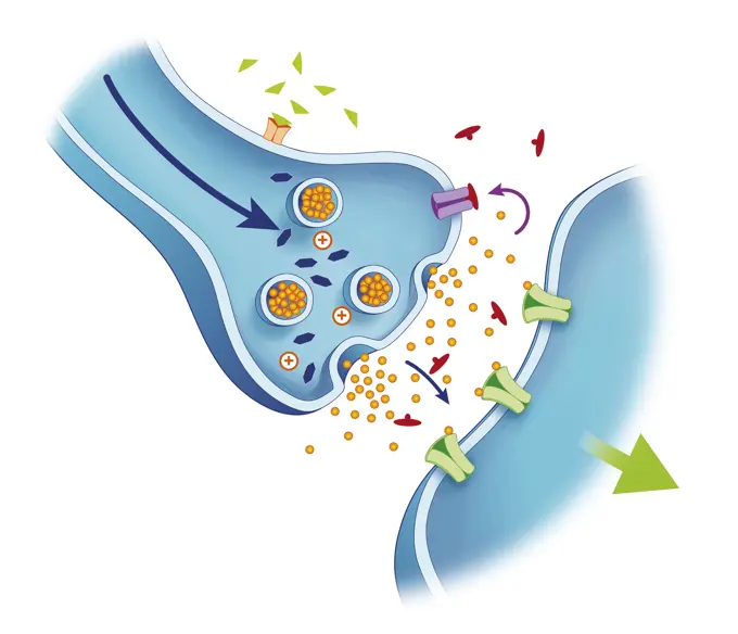
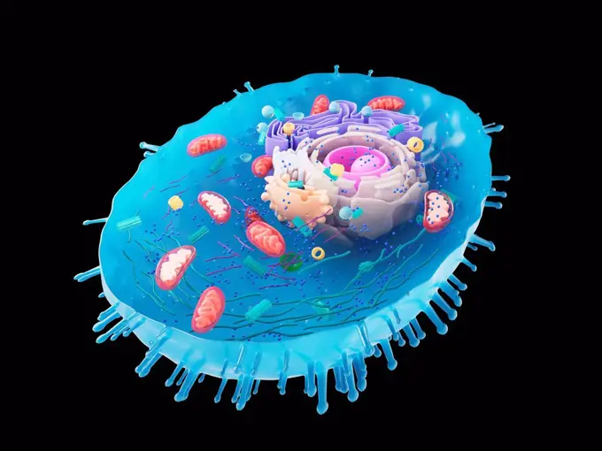
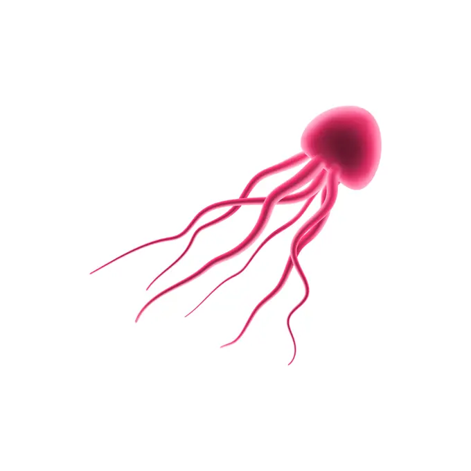
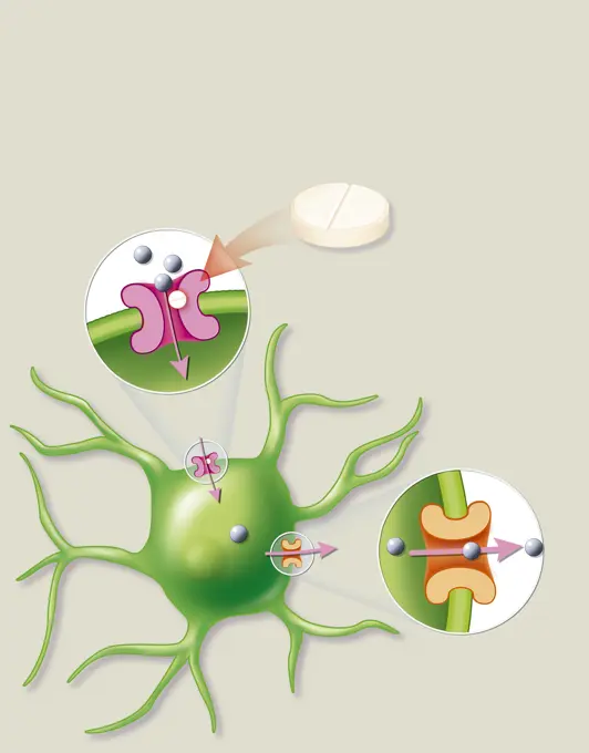
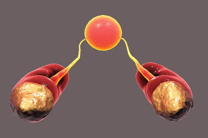
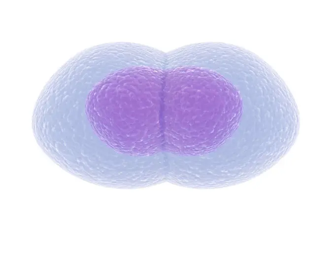
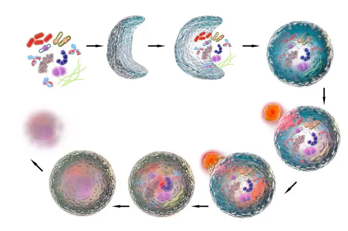
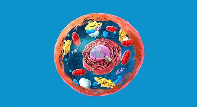
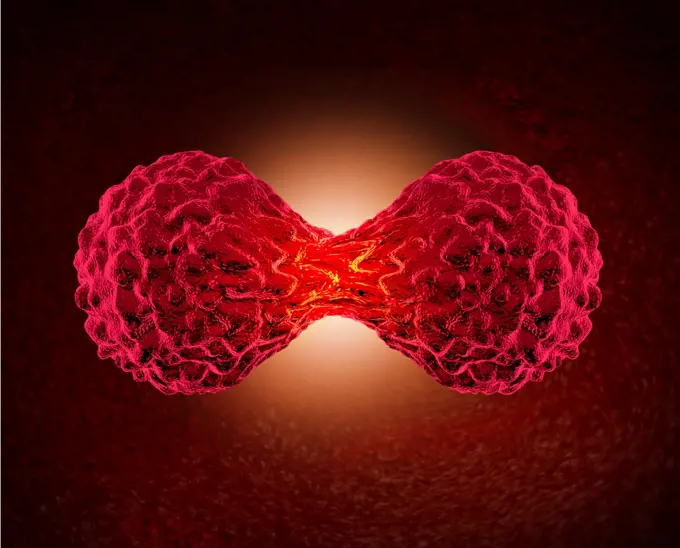
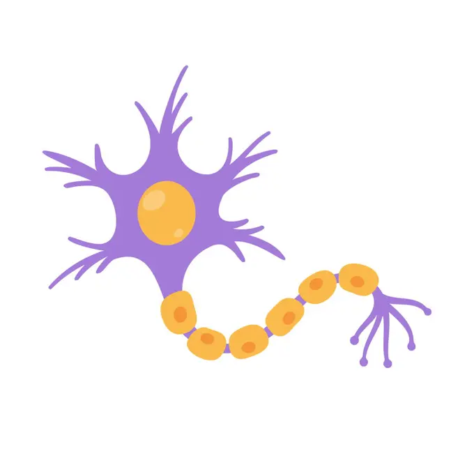
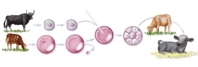
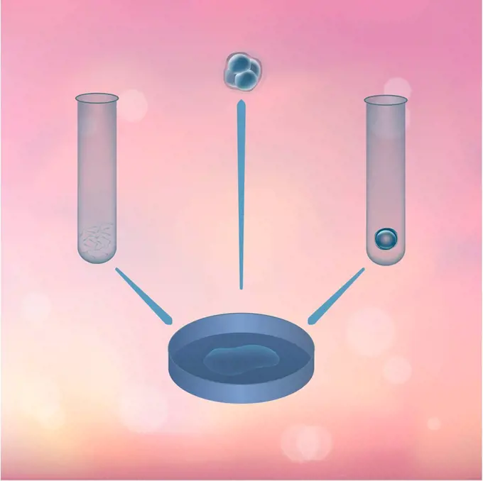
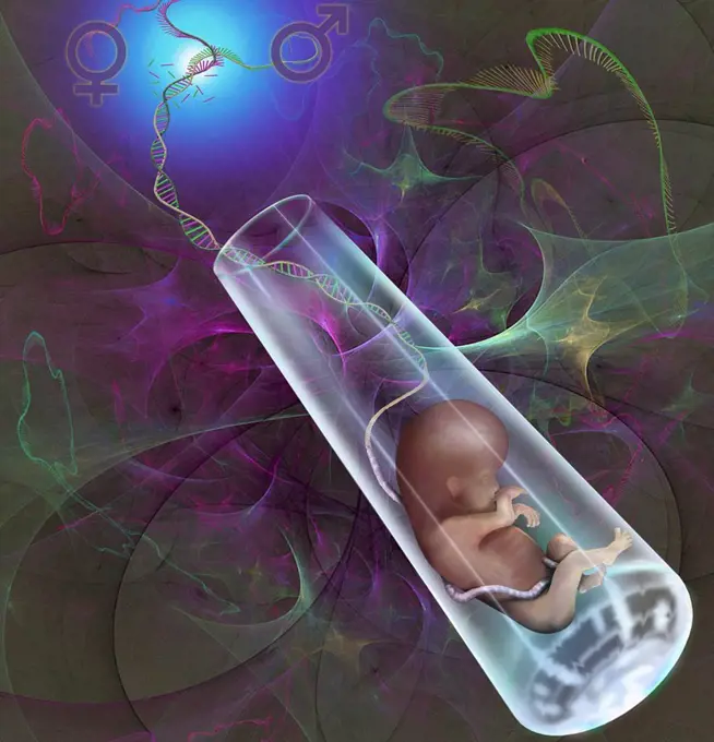
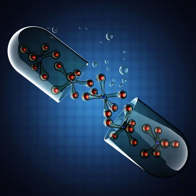
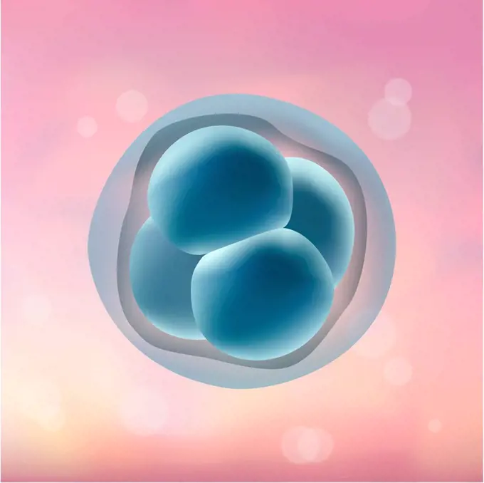
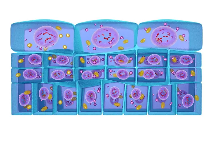
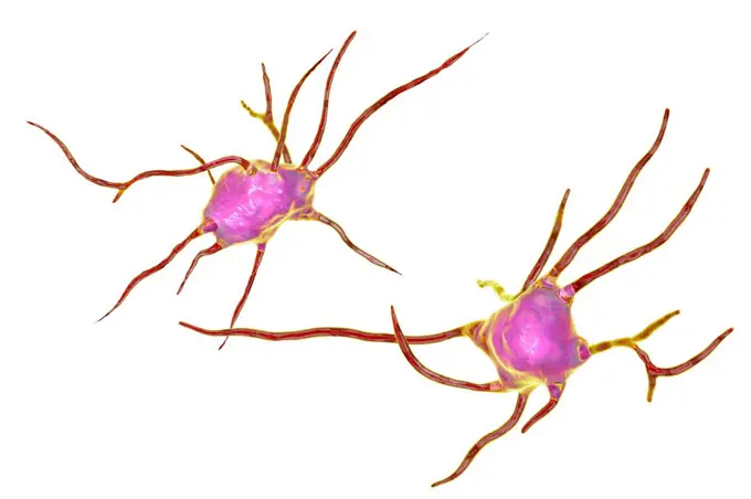
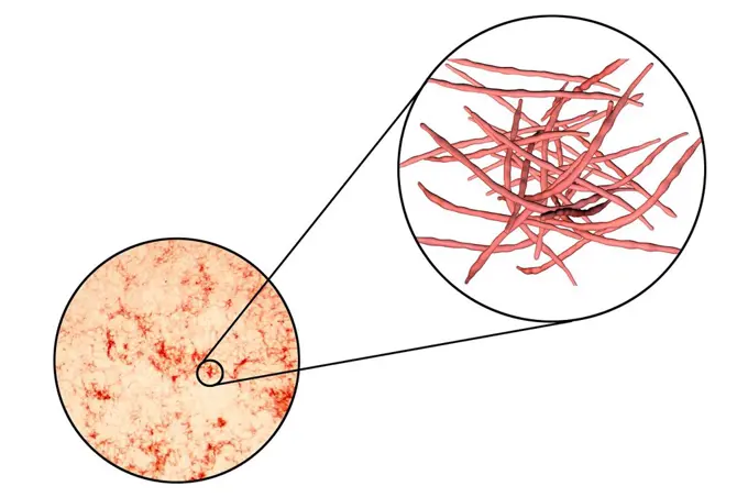
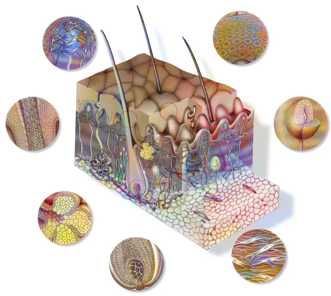
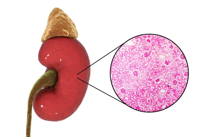
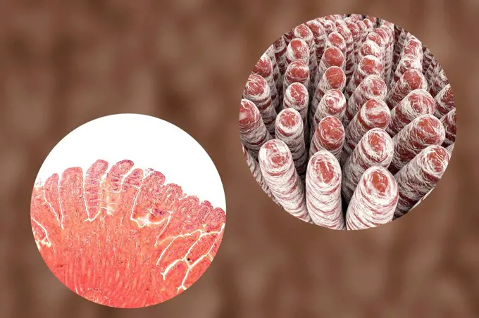
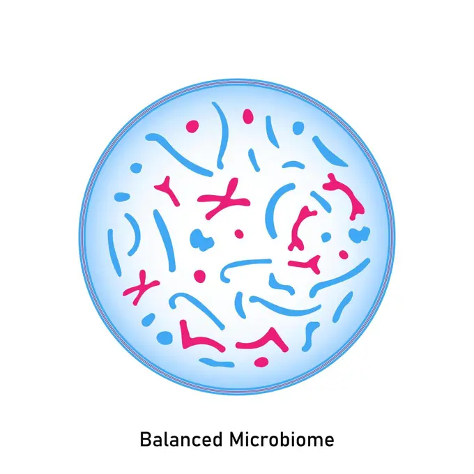
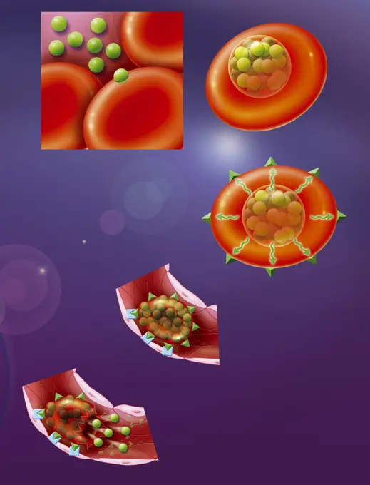
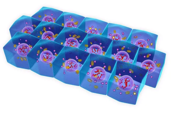
Cellular and Molecular Biology

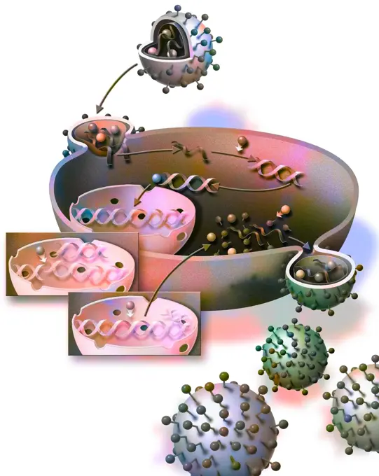
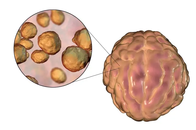
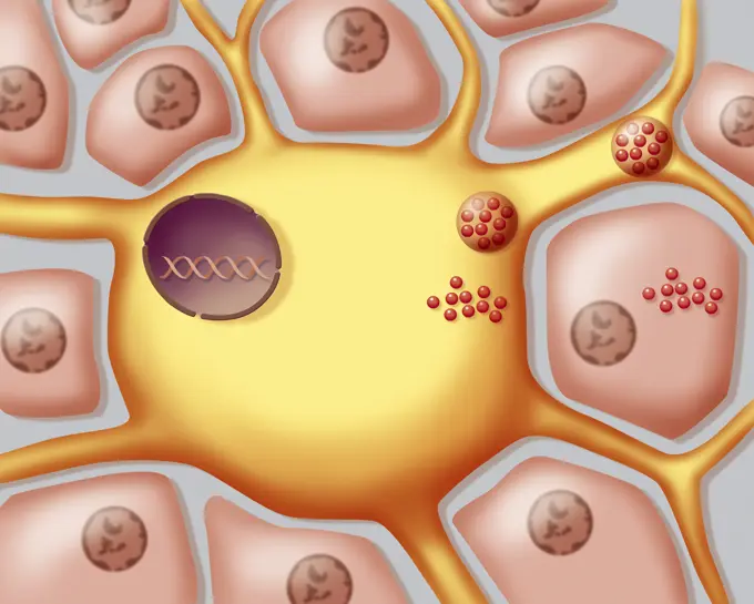
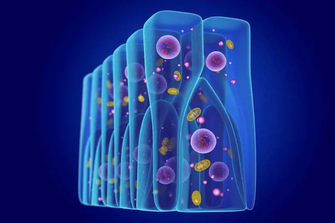
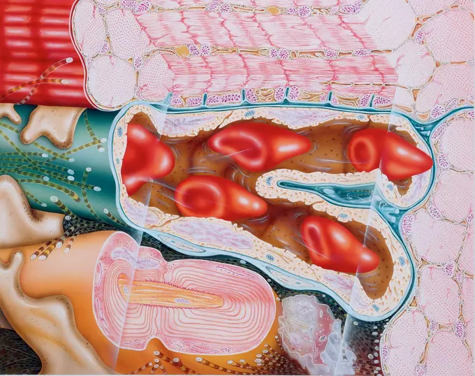
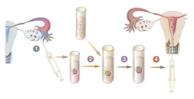
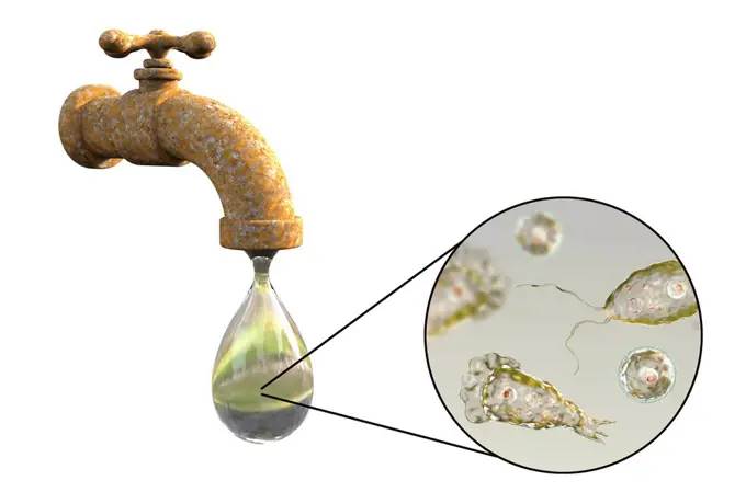
Illustrations depicting cellular structures, organelles, and processes related to HIV infection and molecular models, highlighting biological interactions and therapies.

Substantia nigra in Parkinson's disease, illustration

Assets in this Story

1899-53509134
Cell sectional view with all the main organelles, nucleus, reticulum.
824-63186539
Representation of the self-maintenance of HIV infection. The AIDS virus (in green) which has entered the blood vessel infects and integrates its genome with the inactive T lymphocytes (in pink on the right of the drawing). This triggers the apoptosis of infected T lymphocytes (in brown) which normally constitute the immune barrier preventing the passage of bacteria from the intestine to the bloodstream. The bacteria can therefore reach other regions of the body via the blood network and trigger inflammatory reactions in various organs. In addition, the macrophages (in pink on the left of the drawing), in contact with viruses other than that of AIDS, present the detected antigen of these other viruses to the infected T lymphocytes (in blue). These multiply, triggering the replication of HIV.
1899-53509371
Current therapy most used against AIDS.
4128R-28198
MDMA drug, molecular model. Atoms are represented as spheres and are colour-coded: carbon (grey), hydrogen (blue-green), oxygen (red) and nitrogen (bl...
1899-30608832
Fertilization, penetration of spermatozoa into the oocyte.
6188-66529022
Asthma, drawing Asthma, drawing 016889_257
4239-20481094
Antihistamine in histamine receptors blocking the allergic reaction.
824-63191442
Illustration of the interaction between the immune system and anthracycline chemotherapy. The cancer tumour (blue) is treated with anthracycline chemotherapy (pink). The cancer cell damaged by treatment sequentially emits a combination of specific signals during the various stages of its death. The signals emitted lead to recruitment and maturation of dendritic cells (green) in the immune system. The dendritic cells that have infiltrated the tumour activate naive T lymphocytes (pink) which become specific cytotoxic T lymphocytes (red), which can effectively recognise and attack the cancer tumour.
4128R-15290690
Substantia nigra in Parkinson's disease, illustration
824-63194758
Vaccination (stage 2) : multiplication of the T and B lymphocytes, production of antibodies.
824-63190353
Illustration of the principle of a vaccination. It consists of injecting into the body an infectious agent (virus or bacteria), in an inoffensive form, but that will stimulate the bodys immune response. The immune system has a form of memory, and any previous exposure to the infectious agent will trigger a rapid and more efficient response.
824-63167675
Anatomy of the hen genital system. From left to right : the ovary and its follicles (the mature ones are represented in orange), the infundibulum (funnel-shaped), and the cloaca at the right extermity.
6188-66529021
Bronchus, drawing Bronchus, drawing 016889_255
824-63180730
Representation of the transmission of nerve impulses from a neuron A (dark green) to a neuron B (light green). Vesicles release glutamate (orange balls) which bind to receptors (purple) which open sodium channels and allow Na+ ions to enter the dendritic button of neuron B, thus creating an electric current.
4128R-13844357
Keratinocyte skin cell, illustration. This cell is from the epidermis human skin. It contains the protein keratin (red).
824-63164917
Ventral tegmental area of the brain and nucleus accumbens with their neurons.
824-63165136
Transmission of nerve impulses in the brain from one neuron to another.
4128R-13723643
Illustration of the capillaries and alveoli of the lungs.
1899-53509365
Penetration and duplication of the AIDS virus in the host cell.
4128R-15516384
Illustration of a human cell cross-section
4128R-15516383
Illustration of a human cell cross-section
4128-28767238
Prototheca wickerhamii algae, illustration
4128R-28649
MDMA drug, molecular model. Atoms are represented as spheres and are colour-coded: carbon (grey), hydrogen (blue-green), oxygen (red) and nitrogen (bl...
824-63151715
Bronchus, drawing
4128R-13587439
Autophagy. Computer illustration of a lysosome (orange) fusing with an autophagosome (large sphere). Autophagy (autophagocytosis) is the natural mecha...
4128R-14057408
Cryptococcal meningitis, illustration
824-63163777
Dorsal vertebrae with osteoporosis with partially destroyed cancellous tissue.
824-63181835
Representation of the metabolism allowing the production of melanin in the melanocytes, and transfer of the content of the melanosomes (melanin) into the keratinocytes.
824-63164437
Sagittal section of a breast: breast fat and close-up of a lobule.
824-63199671
Illustration of the action of arthritis treatment on the knee. The treatment acts: 1 on the joint swelling, that it reduces 2 on the deterioration of the cartilage, that it reduces 3 on the consolidation of the cartilage, that it enables
824-63195463
Representation of the transmission of nerve impulses from one neuron (green) to a 2nd neuron (red). The electrical signal is transformed into a chemical signal, vesicles release glutamate (blue) which binds to the receptors (purple) which open the sodium channels and allow Na+ ions to enter the dendritic button, thus creating an electric current.
824-63189061
Illustration of the ovarian cycle in the ovary : the follicle matures (pink) which contains the ovocyte (dark purple), then the egg is released into the Fallopian tube and finally, the formation of corpus luteum (yellow) then disintegration of the egg isnt fertilised.
4128R-11297247
DNA molecule, computer artwork.
824-63180631
Illustration of mold responsible for allergic reactions.
1899-53509166
Influenza viruses and proteins that bind to host cells.
1525-56906583
Sperm cell entry into the ovum egg cell, 3d rendering
4128R-15516386
Illustration of a human cell cross-section
824-63164585
On a cholinergic synapse, the nerve impulse is transmitted between the two neurons thanks to acetylcholine.
4128R-14768525
Viral etiology of prostate cancer, conceptual illustration
4128R-14324155
Prostate cancer, illustration
4128R-13575620
Ovarian follicles, light micrograph (bottom left) and computer illustration (top right). Once sexual maturity is reached up to 20 oocytes start develo...
1899-54028646
Illustration of an IVF (In Vitro Fertilization). On the left, a classic IVF: the spermatozoa and oocytes are placed in an embryo culture environment that favors their survival. Spontaneous fertilization takes place at 37° during incubation. On the right: IVF ICSI: having taken and prepared the sperm and ovules in a favorable embryo culture environment, a spermatozoon is injected into the ovules cytoplasm. After a 48-hour incubation period, the fertilized egg becomes an embryo which can be transferred to the uterus. Several embryos are be obtained, two or three of which will be transferred to the uterus. . (Photo by: BSIP/UIG via Getty Images)
4128R-23614
Animal cell, computer artwork.
4409-17349165
Capillary network.
824-63164928
Female genitals showing the menstrual cycle.
4128R-13844665
Pseudostratified epithelium, illustration.
4413-135029
Microscopic view of Radiolaire
824-63163925
Allergic reaction to pollen: triggering phase of the allergy (second contact).
824-63164841
Transmission of nerve impulses and zoom on a synapse.
824-63224089
All uses except for Canadian college and university publishing - Immunotherapy, cancer treatment. The principle of immunotherapy consists of injecting anti-PD-L1 (or anti-PD-1) immunomodulators into the patient. PD-L1 receptors on the tumor cell (in red) are shown in yellow. While lymphocyte PD-1 receptors (in blue) are shown in light blue. The anti PD-L1 (dark green) and anti PD-1 (light green) immunomodulators bind to the corresponding receptors. Thus they block the connection between these two receptors (yellow and blue). Therefore they block the brake of the immune reaction caused by the PD-L1 PD-1 binding. So the interaction between PD-L1 and PD-1 is prevented. Thus, the T lymphocyte again destroys the cancer cells it recognizes. - Restriction : Canada College and University Publishing Exclusion from 05/03/2021
824-63195039
Illustration of the transmission of information with long-term potentiation. The activation of neurons is more efficient, the number of neurotransmitters in the ion channels is higher. A larger number of sodium ions enter the postsynaptic neuron, increasing the speed and intensity of the electrical signal transmission. This also allows the passage of calcium ions (red), increasing the intensity of the signal.
4128R-11312169
Human lymph node, computer artwork.
4128R-14057543
Ovarian follicles, micrograph and illustration
824-63163616
Female genitalia: ovarian cycle, ovulation, fertilization, embryo segmentation, implantation.
4128-28767088
Bisphenol A molecule, illustration
824-63189389
Illustration of the constitution of part of the prostate. The prostate is a gland in male genitalia. It allows secretion and storing of some of the seminal fluid, one of the constituants of sperm. Acini are illustrated here, linked to ducts constituted of secretory epithelial cells. The blue area surrounding these elements is the fully vascularised stroma.
824-63195033
Illustration of the transmission of information before and with long-term potentiation. The two synapses on the left show how the signal is transmitted from one neuron to another before LTP (long-term potentiation). The electrical signal arrives at the synapse, triggering the opening of vesicles containing neurotransmitters (yellow) released into the intersynaptic space. There are few ion channels (green) at the surface of the post-synaptic neuron, which limits the entrance of sodium ions (blue) in the channels that are open due to neurotransmitters. The signal is therefore weak and transmitted at a slow pace. On the right, two close-ups show what happens during a long-term potentiation nervous signal transmission. The activation of neurons is more efficient, the number of neurotransmitters in the ion channels is higher. A larger number of sodium ions enter the postsynaptic neuron, increasing the speed and intensity of the electrical signal transmission. This also allows the passage of
5507-31584612
Blue evil microbes on a white background
824-63164345
Cancerous tumor, stage 2: Cancer cells have multiplied and transformed.
4128R-13763361
Gram-negative bacterial cell wall, illustration. The horizontal layers include both an external and an internal membrane (blue), both containing trans...
824-63167635
Migraine treatment that inhibits the synapse's release of neurotransmitters via specific receptors.
5507-46944692
Cause of body odor vector flat illustration
1899-53513201
The allergic reaction has two phases. When the body comes into contact with a certain allergen for the very first time, it becomes primed or sensitized to react against the certain substance. Macrophages (in blue) phagocytose the allergen particles, then pass on information about the allergen to the B lymphocytes, which produce IgE antibodies. The antibodies bind to basophils and mast cells. At the next encounter with this allergen, the allergic process is activated -- the allergen binds to the IgE antibodies, triggering the mast cell and basophils to release histamine that orchestrates the allergic reaction (here, skin rash).
1525-24422447
Emphysema as shortness of breath, lung disorder as a COPD illness and Chronic obstructive pulmonary disease medical concept as bronchioles and alveoli are damaged as a 3D illustration,
4128R-14057410
Bacterial meningitis, illustration
4128R-14060497
Liver cirrhosis due to Hepatitis C, illustration
824-63164350
Spread of metastases in the body with the tumor triggering angiogenesis.
824-63219970
Gram negative bacterium in section, DNA, mRNA ribosomes, treatments. The bacterium is cut to show its structure: wall traversed by porins, periplasmic space with the peptidoglycan, and the cytoplasmic membrane. At the heart of the bacterium, the DNA in transcription with a long strand of mRNA, the ribosomes producing polypeptides. Transport RNA.
1525-19757769
cancer cell made in 3d software
4128-20239384
Microsporum audouinii fungus, illustration
1899-53509040
Healthy dorsal vertebrae with osteoporosis. (zooms on the spongy bone).
824-63163769
Healthy dorsal vertebrae with osteoporosis. (zooms on the spongy bone).
4128-28515288
Respiratory syncytial virus particles,
824-63163714
The eye and retina with the vitreous, the internal limiting membrane.
824-63163770
Structure of a healthy dorsal vertebra with spongy and compact tissue.
824-63164795
Penetration and duplication of the AIDS virus in the host cell.
4128R-13372913
Fat cell, computer illustration. An adipocyte form adipose tissue, which stores energy as an insulating layer of fat. White adipose tissue is used as ...
824-63164127
Penetration and replication of a retrovirus (example: AIDS) in a host cell.
4128-24796459
Muscimol molecule, illustration
824-63164293
Healthy elbow joint in sagittal section.
1525-19835687
Mitosis. Stage two
824-63219977
Infiltrating and in situ ductal adenocarcinomas, mitotic spindle zoom, treatments. Bust of a woman with a mammary gland and the axillary lymph nodes related to it. A first zoom starts from this gland to show the anatomical structure of the mammary gland with a main galactophore duct receiving the lobules, themselves made of acinus. Two cancerous tumors are located on the main milk duct: invasive or invasive ductal carcinoma (left) and carcinoma in situ. At the bottom, zoom in on the cancer cells whose nucleus is cut to show the mitotic spindle. A strand of DNA whose structure is modified is extracted from a chromosome. Receptors HER2 (yellow) on the nucleus, VEGF (green) on the neovessel and the estrogen receptor (black) on the DNA strand. Bottom left, adipose tissue site of conversion of androgens to estrogen by aromatases
824-63208834
Illustration of a synapse between two dopamine neurons. Nerve impulse transmission is through dopamine and dopamine receptors. Dopamine is vital for nerve impulse transmission in the control of motor function.
4128R-13242863
Alveoli of the human lung, illustration.
824-63224095
All uses except for Canadian college and university publishing - Cancer, PD-L1 protein, PD-1 receptor, neutralization of the activated immune response against the cancer cell. On contact with a cancer cell, T lymphocytes are activated. They secrete toxic molecules capable of destroying the cancer cell. But the reaction of immune cells against cancer cells is slowed down by the PD-L1 protein produced by the cancer cell. This protein interacts with the PD-1 receptor of the lymphocyte, which inactivates it. So the cancer cell can no longer be destroyed by the T lymphocyte. - Restriction : Canada College and University Publishing Exclusion from 05/03/2021
1899-53513200
Hematopoiesis occurs in three successive cell compartments. Stem cells are pluripotent cells guaranteeing permanent production of blood cells. Precursor cells (or determined cells) are only capable of a limited number of divisions. Maturing cells constitute a transition between the preceding compartments and the bloodstream, cells in the bloodstream have identifiable morphologies.
824-63218265
Site of action of morphine. Increased painful nerve impulses from the cutaneous, muscular, articular or visceral tissues from the nociceptors, in yellow to the spinal cord, then red to the cerebral cortex via the medulla oblongata and the mesencephalon of the brainstem seen in horizontal cuts. Inhibition by descending pathways in pink. zoom on the enkephalin neuron of the spinal cord in blue and the opiate receptors on the yellow neuron
1899-53509294
Transmission of nerve impulses from neuron A to neuron B with zoom on a synapse.
5507-46149225
coronavirus in blood illustration
1788-20762565
Medicine: Cell division: Mitosis and Meiosis, illustration
1899-53510741
This medical illustration represents air sacs in adults. Each cell shown here constitutes a purple ball. They are gathered in clusters to form bags. Each sac is located at the end of a bronchiole that organizes itself like the branches of a tree. It is at the level of the pulmonary alveoli that the gas exchange between air and blood melts during breathing. In adults there are a large number of pulmonary alveoli which form the air sacs. Which gives it a great breathing capacity. Unlike adults, toddlers have a much smaller number of alveoli. They are more scattered and do not form bags. (Photo by: JACOPIN/BSIP/UIG)
824-63191589
Illustration of the Hepatitis C virus that infects the liver. It is transmitted through the bloodstream, reproduces and spreads in the hepatic lobules, causing inflammation, then necrosis of the hepatic lobular tissue.
4128R-19938
Human alveoli, computer artwork.
4128R-14181715
Human cell with chromosomes, illustration
4128R-13372970
Intestinal villi, light micrograph and 3D computer illustration. Villi are finger-like projections that greatly increase the intestinal surface area f...
824-63180562
Illustration of the chain reaction involved in muscle cramp.
4128R-13725447
Illustration of an ovarian cycle and ovulation.
824-63181825
Illustration of stem cell (purple) differentiation into neuronal precursor cells (dark blue), some of which die (brown), others become neurons (orange) or glial cells, astrocytes (turquoise) or oligodendrocytes (green).
824-63164852
Supply of glucose to muscle cells via transporters.
4128-19249286
Atherosclerosis, illustration.
4128R-13409949
Lumbar vertebrae, illustration
4128R-13446581
Genital herpes lesions (blisters) on a penis and close-up view of Herpes simplex virus II, computer illustration. Genital herpes is caused by infectio...
824-63163704
Transplant of hematopoietic stem cells administered to colonize the bone marrow.
4128R-15366669
Helicobacter pylori bacteria, illustration
1848-53216186
Hair growth illustration
824-63189806
Illustration of a womans immune system and the immune reactions that halt progess and eliminate micro-organisms that can invade the organism. A micro-organism (orange cell) present in the organism meets white corpuscles (beige cell) such as macrophages or dendritic cells. The latter can either phagocytose the micro-organisms or destroy them : this is the innate respone; or fragment them and present them to lymphocyte T cells (pink cell). In this way the lymphocyte TCD4 cells, due to the cytokines, will activate the lymphocyte B cells (green cell) which will produce antibodies to destroy the micro-organism. Other lymphocyte TCD8 cells are capable of attacking infected cells by the micro-organism (blue cell in the process of being destroyed) and destroy it.
824-63224065
Synapse of dopamine neurons that causes the effect of most drugs.
1525-20790284
Bloody artery. Blockage of blood vessels. Sick artery with leukocytes. Blood cells in  human vein close-up. Healthy and painful human artery.

4128R-15516385
Illustration of a human cell cross-section
1525-27219011
Pink bacteria with tentacles isolated virus cell. Vector germ causing infections, anti body. Virus antibody isolated vector pink bacteria germ
824-63205888
Illustration of the action principal of a diuretic used in treating autism. The pill lessens the entry of chloride into the neurons by blocking the NKCC1 pump, lowering the concentration of chloride inside the neurons, thus improving neuronal activity and decreasing autistic disorders.
4128R-14939866
Human urinary system, illustration
824-63190012
Illustration of a synapse. When a signal, or action potential, arrives at an axonal terminal, the relevant neurons in the memory release glutamate, the main excitatory neurotransmittor. When the signal arrives in the presynaptic neuron, vesicles containing glutamate merge with the neuron membrane, releasing their contents into the intersynaptic space. In this example, the postsynaptic neuron contains two of the four subtypes of glutamate receptor : NMDA and AMPA. These receptors are channels that respectively let calcium ions and sodium ions through. Generally, the NMDA channel is blocked by a large volume of magesium ion, which prevents the ions from getting through. When glutamate latches onto AMPA receptors, positive ions enter the postsynaptic neuron, stimulating it.
4128R-13682591
Human egg cell division, illustration.
4128R-13587442
Autophagy, computer illustration. Autophagy (autophagocytosis) is the natural mechanism that destroys unnecessary or dysfunctional cellular components...
4128-16005860
Animal cell structure, illustration
1525-20586695
Cancer cell dividing concept as a microscopic cellular cycle of replication or division of malignant cancerous cells in the human body as a health care medical and oncology symbol for dangerous tumor growth.
1525-75738654
human sensory neuron model for biology studies
824-63219062
Cloning of a wild Gaur bull. In order to obtain a Gaur calf, the cloning between the genetic code of a wild Gaur bull and the oocyte of a domestic cow is carried out. The genetic code of the Gaur bull is fused with the oocyte from which the nucleus has been removed. This new cell is stimulated to give an embryo. The embryo is implanted in the uterus of another carrier domestic cow. The result thanks to the genetic identity of the Gaur bull, will give a Gaur calf.
4128R-15290592
In vitro fertilization, illustration
4128-15819144
Test-tube baby. Conceptual illustration of a foetus in a test tube, representing the concept of in vitro fertilization (IVF), where fertilization occu...
6188-58132742
Transparent capsule on blue background Transparent capsule on blue background Copyright: xZoonar.com/OxanaxRishnyakx 5689227
4128R-15290590
4-cell stage embryo, illustration
4128R-13844705
Epithelium transitional, illustration.
824-63163819
Research in cell therapy: obtaining and differentiating embryonic stem cells.
4128R-14846614
Nerve cells, computer illustration
1525-57082490
Mummy in sarcophagus. Body of Pharaoh. Egyptian king. Archaeology and Halloween monster. Golden scepter and symbol of immortality. Flat cartoon illustration. Mummy in sarcophagus. Body of Pharaoh.
4128R-14181666
Actinomycete bacteria, micrograph and illustration
1899-53509339
Cut of skin seen from 3/4 with the layers (epidermis, dermis, hypodermis, muscle) and its structures.
4128R-14939869
Human kidney, illustration and micrograph
4128R-13372975
Intestinal villi, light micrograph and 3D computer illustration. Villi are finger-like projections that greatly increase the intestinal surface area f...
5507-43877285
Normal balanced flora of the skin and mucous membranes
824-63189042
Illustration of how malarial merozoites (Plasmodium) proliferate by using red corpuscles. They penetrate the red corpuscles, ultra rapid transporters, which will allow them to avoid the filtering spleen. The merozoites then produce protein membranes which fix onto the membranes of the red corpuscles. These infected red corpuscles will then fix onto the outside of blood vessels, causing various organs to fail. The sheltered parasites proliferate until they rupture the membrane of the cells after 48 hours, spreading through the blood and parasitising other red corpuscles in order to proliferate again.
4128-19249274
Atherosclerosis, illustration.
4128-30417202
Kissing bug, illustration
4128-20045185
Mitosis, illustration
1525-27646742
Telomere concept isolated and telomeres located on the end caps of a chromosome resulting in aging by damaging DNA or protection resulting in living longer or longevity as a medical microscopic 3D illustration.
824-63164137
Penetration and replication of the H1N1 virus through a host cell.
5507-43617475
Process of muscle's stiffness and pain / circle type illustration
4128R-13844679
Simple columnar epithelium, illustration.
824-63181806
Illustration of a network of presynaptic and postsynaptic neurons attached to each other by the intersynaptic space which allows nervous impulses to flow, from the upper neuron (top) to the lower neuron (bottom).
824-63205846
Illustration of the formation of sperm and their genetic diversity, due to interchromosomal genetic mixing during meiosis. At the top, the process of crossing over between two homologous chromosomes (interchromosomal genetic mixing), which increases the genetic diversity of the gametes.
1788-39597
Medicine: Human anatomy, cell
6188-58062890
Types of human cells Different types of human cells including an egg cell, sperm, red blood cell, osteocyte, neuron, skeletal muscle and columnar epithelial cell. Digital illustration. Copyright: xZoonar.com/AndreaxDantix 17120922
5507-34819360
Fisheries
824-63167609
In the center, cross-section of capillaries. Above, cross-section of a muscle. Below, cross-section of a nerve (a neuraxon)
824-63163619
Diagram of the different stages of in vitro fertilization.
4128R-14845750
Tap water contaminated by amoeba, illustration
824-63163608
Bacteriophage consisting of a protein envelope, containing its nucleic acid, and a tail.