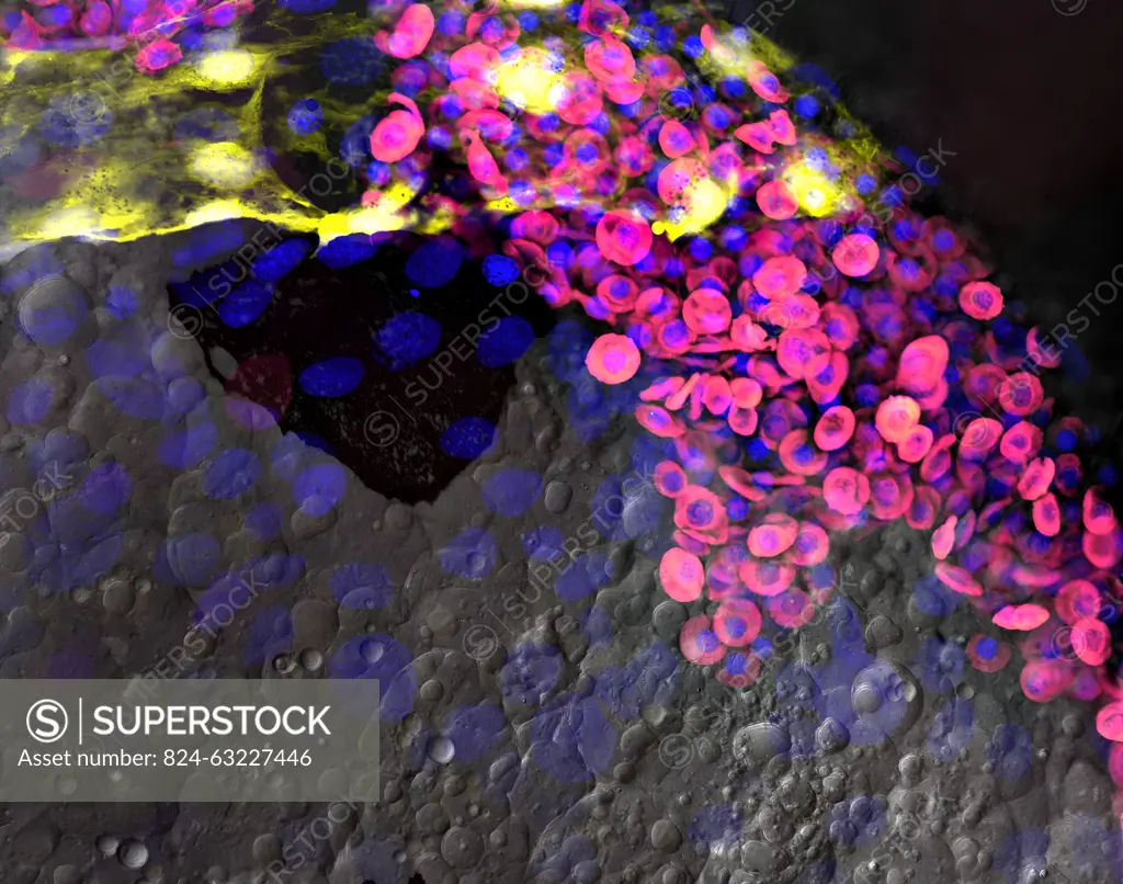
The zebrafish is an excellent model for studying the development of blood cells and blood vessels. This image shows blood cells (magenta) in blood vessels (yellow) on the yolk of a 2-day-old anesthetized transgenic zebrafish embryo that is about the size of a grain of rice. The image was rewarded with a powerful microscope that uses lasers to illuminate the fish.
SuperStock offers millions of photos, videos, and stock assets to creatives around the world. This image of The zebrafish is an excellent model for studying the development of blood cells and blood vessels. This image shows blood cells (magenta) in blood vessels (yellow) on the yolk of a 2-day-old anesthetized transgenic zebrafish embryo that is about the size of a grain of rice. The image was rewarded with a powerful microscope that uses lasers to illuminate the fish. by NIH/IMAGE POINT FR/BSIP is available for licensing today.
Looking for a license?
Click here, and we'll help you find it! Questions? Just ask!
Click here, and we'll help you find it! Questions? Just ask!
DETAILS
Image Number: 824-63227446Rights ManagedCredit Line:NIH/IMAGE POINT FR/BSIP/SuperStockCollection:BSIP Contributor:NIH / IMAGE POINT FR / BSIP Model Release:NoProperty Release:NoResolution:3833×3020