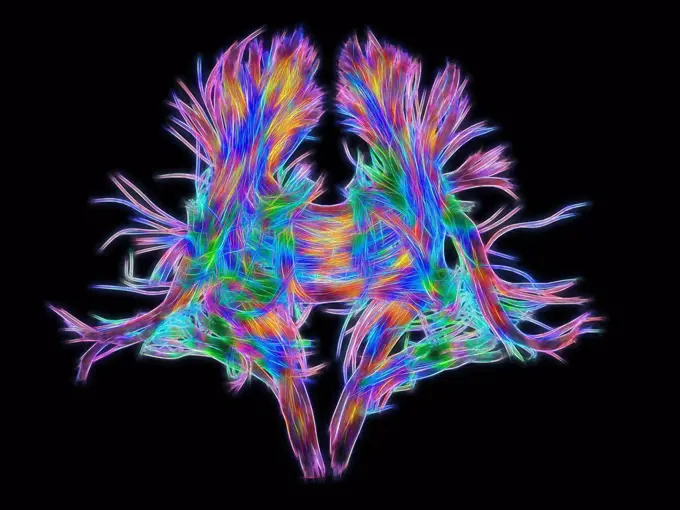
White matter tracts in a mouse brain acquired with diffusion tensioner imaging. The brain is viewed from below and with the front of the brain to the right. The colors represent different fiber directions. White matter integrity can be studied after brain injury using this visualization method.
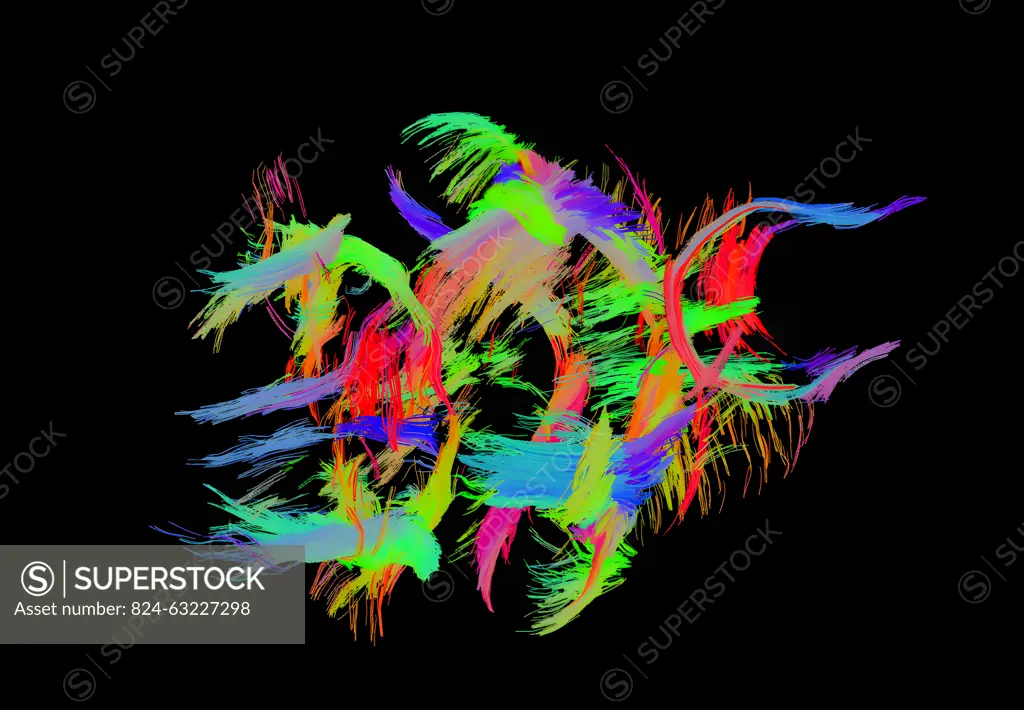
SuperStock offers millions of photos, videos, and stock assets to creatives around the world. This image of White matter tracts in a mouse brain acquired with diffusion tensioner imaging. The brain is viewed from below and with the front of the brain to the right. The colors represent different fiber directions. White matter integrity can be studied after brain injury using this visualization method. by NIH/IMAGE POINT FR/BSIP is available for licensing today.
Visually Similar View more


Looking for a license?
Click here, and we'll help you find it! Questions? Just ask!
Click here, and we'll help you find it! Questions? Just ask!
DETAILS
Image Number: 824-63227298Rights ManagedCredit Line:NIH/IMAGE POINT FR/BSIP/SuperStockCollection:BSIP Story:Molecular Structures and ImagingContributor:NIH / IMAGE POINT FR / BSIP Model Release:NoProperty Release:NoResolution:4768×3308