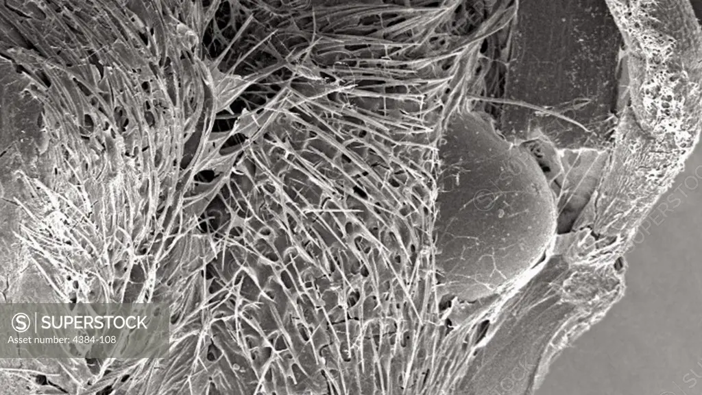
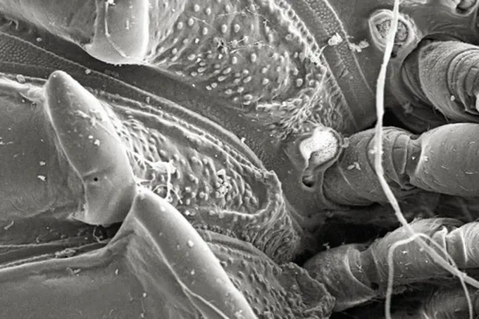
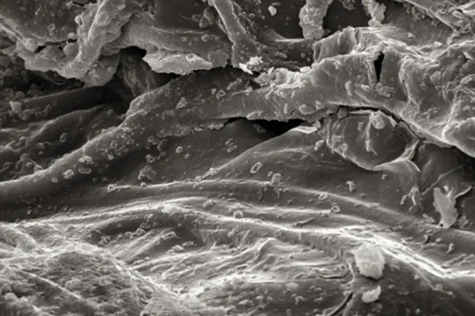
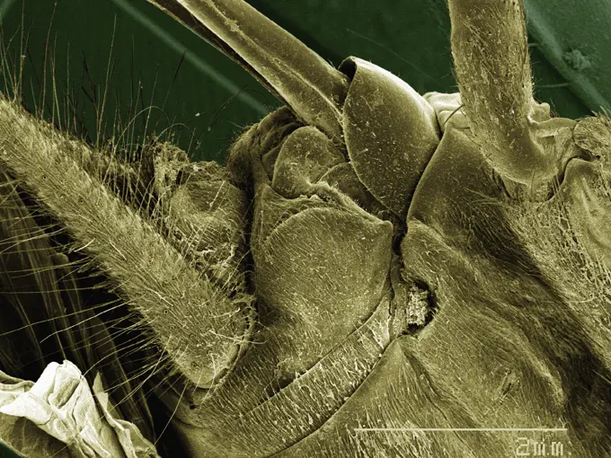
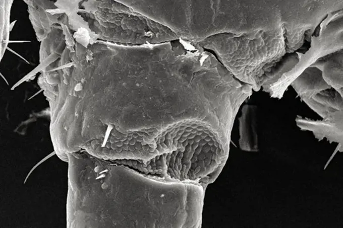
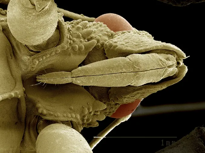
Under a magnification of 40X, this scanning electron micrograph (SEM) revealed some of the ultrastructural architecture found in a brown recluse spiders (Loxesceles reclusa) web that had entrapped an unidentified ant. Though the prey had been enwrapped in a silk cocoon, there were still some of its body parts that were visible such as one of its two compound eyes, and one of its two segmented antennae (background). The antenna is composed of three main regions: scape, pedicle, and flagellum.
SuperStock offers millions of photos, videos, and stock assets to creatives around the world. This image of Under a magnification of 40X, this scanning electron micrograph (SEM) revealed some of the ultrastructural architecture found in a brown recluse spiders (Loxesceles reclusa) web that had entrapped an unidentified ant. Though the prey had been enwrapped in a silk cocoon, there were still some of its body parts that were visible such as one of its two compound eyes, and one of its two segmented antennae (background). The antenna is composed of three main regions: scape, pedicle, and flagellum. by Centers for Disease Control/Centers of Disease Control is available for licensing today.
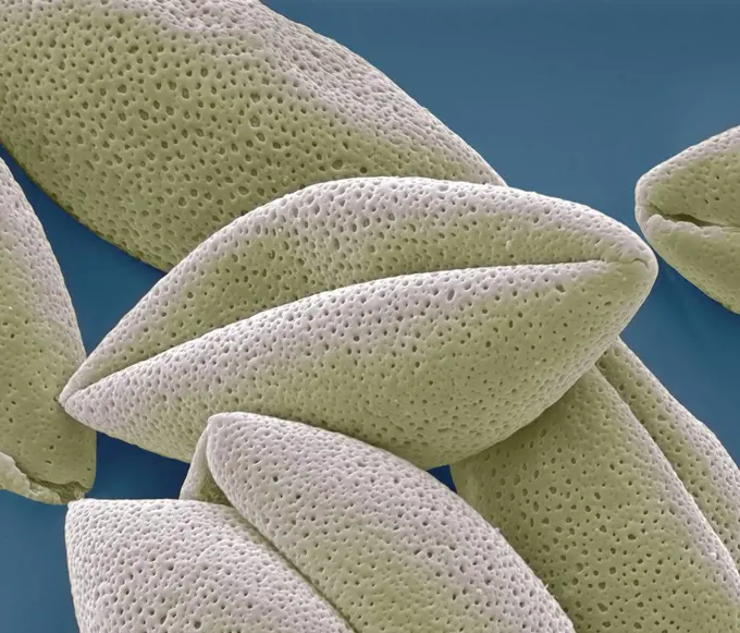
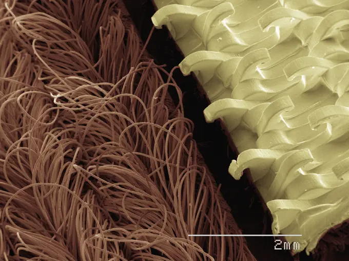
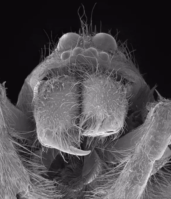
Visually Similar More from Microscopic Wonders of Biology story


Looking for a license?
Click here, and we'll help you find it! Questions? Just ask!
Click here, and we'll help you find it! Questions? Just ask!
DETAILS
Image Number: 4384-108Rights ManagedCredit Line:Centers for Disease Control/Centers of Disease Control/SuperStockCollection:Centers for Disease Control Story:Microscopic Wonders of BiologyModel Release:NoProperty Release:NoResolution:5862×3298