Transmission electron micrograph of HIV-1 virus particles (teal) from infected H9 cells, produced in cell culture. The particles exhibit two stages of replication: the two Arcs are immature particles budding from the plasma membrane of the cell, and the center spherical particle is a mature form in extracellular space.
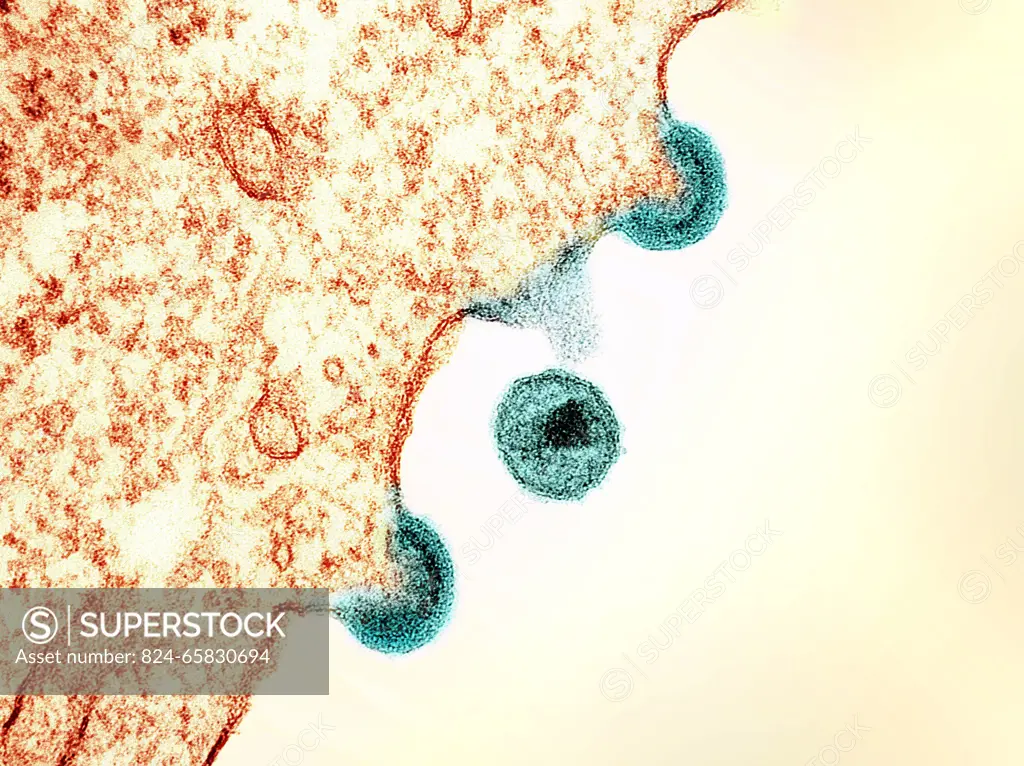
SuperStock offers millions of photos, videos, and stock assets to creatives around the world. This image of Transmission electron micrograph of HIV-1 virus particles (teal) from infected H9 cells, produced in cell culture. The particles exhibit two stages of replication: the two Arcs are immature particles budding from the plasma membrane of the cell, and the center spherical particle is a mature form in extracellular space. by NIH-NIAID/IMAGE POINT FR/BSIP is available for licensing today.
Looking for a license?
Click here, and we'll help you find it! Questions? Just ask!
Click here, and we'll help you find it! Questions? Just ask!
DETAILS
Image Number: 824-65830694Rights ManagedCredit Line:NIH-NIAID/IMAGE POINT FR/BSIP/SuperStockCollection:BSIP Story:Microscopic Organisms ObservedContributor:NIH-NIAID / IMAGE POINT FR / BSIP Model Release:NoProperty Release:NoResolution:4032×3020