This thin-section transmission electron micrograph (TEM) revealed some of the ultrastructural morphology found within the cyst-stage of a Giardia sp. protozoan. The outer cyst wall is composed of filamentous and membranous portions, and is separated from the cytoplasm of the trophozoites contained within by the peritrophic space. This cyst wall is approximately 0.25 microns thick. The protozoan Giardia causes the diarrheal disease called giardiasis. Giardia species exist as free-swimming (by
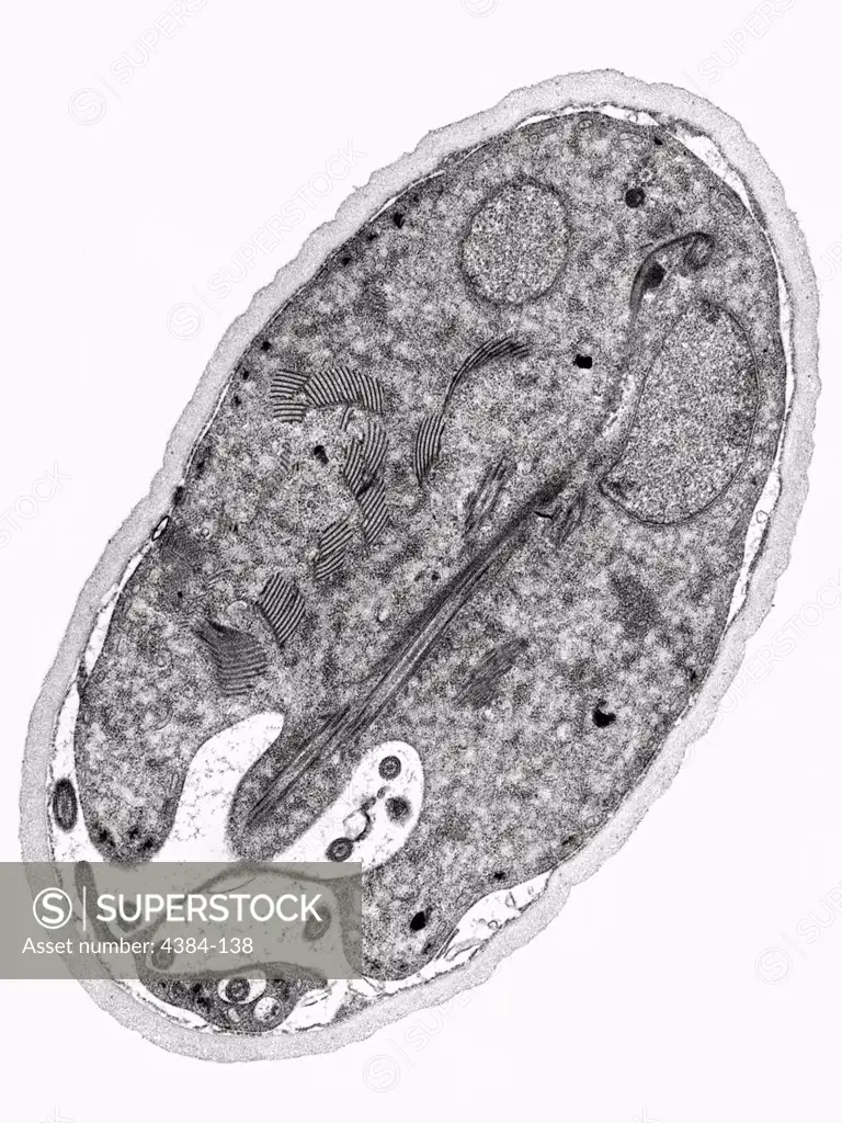
SuperStock offers millions of photos, videos, and stock assets to creatives around the world. This image of This thin-section transmission electron micrograph (TEM) revealed some of the ultrastructural morphology found within the cyst-stage of a Giardia sp. protozoan. The outer cyst wall is composed of filamentous and membranous portions, and is separated from the cytoplasm of the trophozoites contained within by the peritrophic space. This cyst wall is approximately 0.25 microns thick. The protozoan Giardia causes the diarrheal disease called giardiasis. Giardia species exist as free-swimming (by by Centers for Disease Control/Centers of Disease Control is available for licensing today.
Looking for a license?
Click here, and we'll help you find it! Questions? Just ask!
Click here, and we'll help you find it! Questions? Just ask!
DETAILS
Image Number: 4384-138Rights ManagedCredit Line:Centers for Disease Control/Centers of Disease Control/SuperStockCollection:Centers for Disease Control Model Release:NoProperty Release:NoResolution:3802×5068