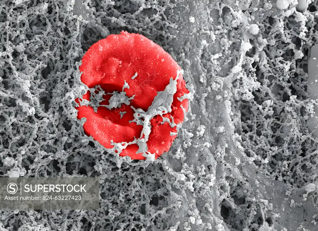
This is a scanning electron microscope image of traumatized muscle tissue taken from a wounded soldier. It shows a red blood cell (false color) entangled in a nanofibrous extracellular matrix. Highly fibrotic regions such as these are thought to precede bone formation during abnormal wound healing, leading to heterotopic ossification, the formation of bone in places outside the skeleton, such as soft tissues.
SuperStock offers millions of photos, videos, and stock assets to creatives around the world. This image of This is a scanning electron microscope image of traumatized muscle tissue taken from a wounded soldier. It shows a red blood cell (false color) entangled in a nanofibrous extracellular matrix. Highly fibrotic regions such as these are thought to precede bone formation during abnormal wound healing, leading to heterotopic ossification, the formation of bone in places outside the skeleton, such as soft tissues. by NIH/IMAGE POINT FR/BSIP is available for licensing today.
Looking for a license?
Click here, and we'll help you find it! Questions? Just ask!
Click here, and we'll help you find it! Questions? Just ask!
DETAILS
Image Number: 824-63227423Rights ManagedCredit Line:NIH/IMAGE POINT FR/BSIP/SuperStockCollection:BSIP Contributor:NIH / IMAGE POINT FR / BSIP Model Release:NoProperty Release:NoResolution:4154×3020