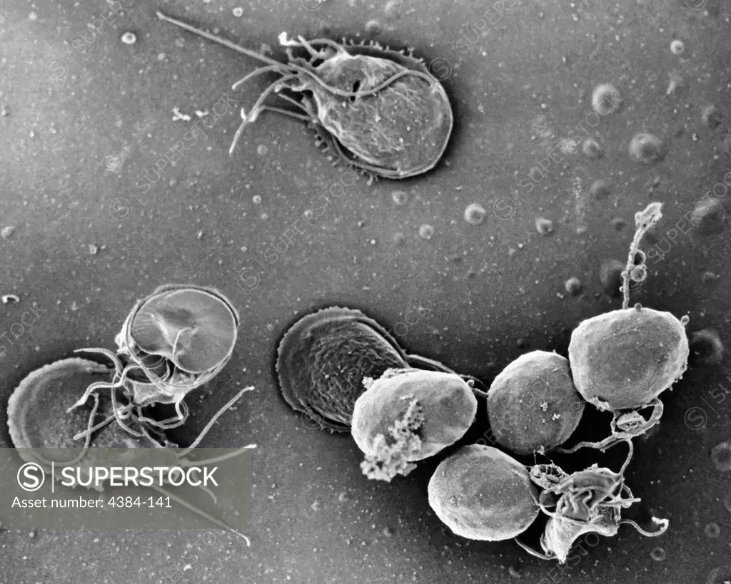
This is a scanning electron micrograph (SEM) of an in vitro Giardia lamblia culture, which had been cultivated in bile-free TYI-S-33 medium for 48 hrs, then incubated 24 hrs with 10 mg/ml bovine bile in order to stimulate cyst formation. This photograph contains both trophozoites, and a cluster of maturing cysts (bottom right). At far left, the two trophozoites-staged organisms are positionally situated opposite to one another, with the farthest left G. lamblia displaying its dorsal, or u
SuperStock offers millions of photos, videos, and stock assets to creatives around the world. This image of This is a scanning electron micrograph (SEM) of an in vitro Giardia lamblia culture, which had been cultivated in bile-free TYI-S-33 medium for 48 hrs, then incubated 24 hrs with 10 mg/ml bovine bile in order to stimulate cyst formation. This photograph contains both trophozoites, and a cluster of maturing cysts (bottom right). At far left, the two trophozoites-staged organisms are positionally situated opposite to one another, with the farthest left G. lamblia displaying its dorsal, or u by Centers for Disease Control/Centers of Disease Control is available for licensing today.
Looking for a license?
Click here, and we'll help you find it! Questions? Just ask!
Click here, and we'll help you find it! Questions? Just ask!
DETAILS
Image Number: 4384-141Rights ManagedCredit Line:Centers for Disease Control/Centers of Disease Control/SuperStockCollection:Centers for Disease Control Model Release:NoProperty Release:NoResolution:4916×3932