Prosthetic hip. 3D computed tomographic (CT) scan reconstruction of a ball-and-socket prosthetic joint used to replace the old hip joint. The upper 'ball' end swivels in the artificial cavity surgically implanted into the pelvis (seen here). The other end extends down into a hole drilled into the femur (thigh bone) (not seen).
This asset has restrictions in United States, but still may be available. Get in touch for more details.
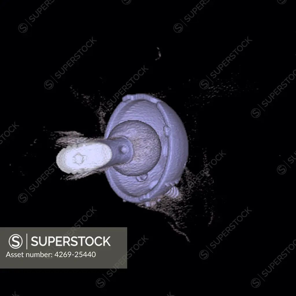
SuperStock offers millions of photos, videos, and stock assets to creatives around the world. This image of Prosthetic hip. 3D computed tomographic (CT) scan reconstruction of a ball-and-socket prosthetic joint used to replace the old hip joint. The upper 'ball' end swivels in the artificial cavity surgically implanted into the pelvis (seen here). The other end extends down into a hole drilled into the femur (thigh bone) (not seen). by Icvi-Ccn-Voisin/Phanie is available for licensing today.
Looking for a license?
Click here, and we'll help you find it! Questions? Just ask!
Click here, and we'll help you find it! Questions? Just ask!
DETAILS
Image Number: 4269-25440Rights ManagedCredit Line:Icvi-Ccn-Voisin/Phanie/SuperStockCollection:Phanie Story:Marine MicroorganismsContributor:Icvi-Ccn-Voisin Model Release:NoProperty Release:NoResolution:4500×4500