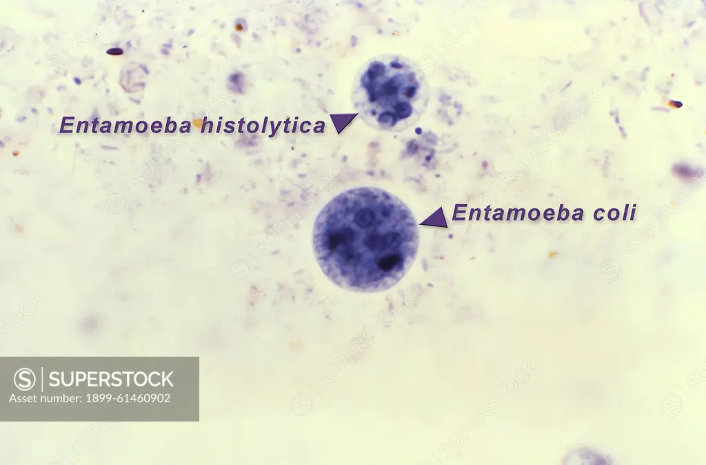
This photomicrograph of an iron-hematoxylin-stained specimen, revealed the presence of two amoebic organisms in their cystic stages of their development. In this focal plane, the smaller Entamoeba histolytica cyst (top), contained four nuclei, and two bluntly-tipped chromatoid bodies, while six nuclei could be detected in the larger Entamoeba coli cyst (bottom), which also contained two chromatoid bodies as well. CDC/ Dr. Mae Melvin 1977
SuperStock offers millions of photos, videos, and stock assets to creatives around the world. This image of This photomicrograph of an iron-hematoxylin-stained specimen, revealed the presence of two amoebic organisms in their cystic stages of their development. In this focal plane, the smaller Entamoeba histolytica cyst (top), contained four nuclei, and two bluntly-tipped chromatoid bodies, while six nuclei could be detected in the larger Entamoeba coli cyst (bottom), which also contained two chromatoid bodies as well. CDC/ Dr. Mae Melvin 1977 by CDC/IMAGE POINT FR/BSIP/Universal Images is available for licensing today.
Looking for a license?
Click here, and we'll help you find it! Questions? Just ask!
Click here, and we'll help you find it! Questions? Just ask!
DETAILS
Image Number: 1899-61460902Rights ManagedCredit Line:CDC/IMAGE POINT FR/BSIP/Universal Images/SuperStockCollection:Universal Images Contributor:CDC / IMAGE POINT FR / BSIP Model Release:NoProperty Release:NoResolution:5100×3358