This photomicrograph of a brain tissue specimen depicts the cytoarchitectural changes associated with a free-living amebic infection, which may have been caused by either a Naegleria fowleri , or an Acanthamoeba sp. The organisms were found in the brain of a Japanese Prisoner of war in the 1950's, before we knew about the free living amebae, and how they attack the brain. CDC/ Dr. Martin D. Hicklin 1964
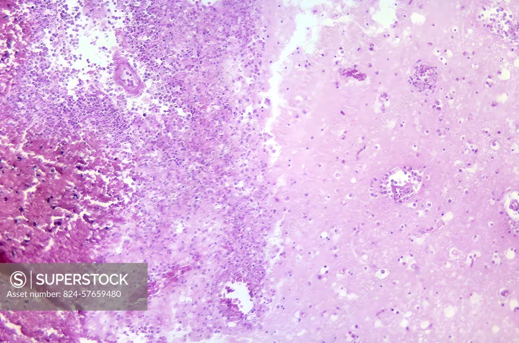
SuperStock offers millions of photos, videos, and stock assets to creatives around the world. This image of This photomicrograph of a brain tissue specimen depicts the cytoarchitectural changes associated with a free-living amebic infection, which may have been caused by either a Naegleria fowleri , or an Acanthamoeba sp. The organisms were found in the brain of a Japanese Prisoner of war in the 1950's, before we knew about the free living amebae, and how they attack the brain. CDC/ Dr. Martin D. Hicklin 1964 by CDC/IMAGE POINT FR/BSIP is available for licensing today.
Looking for a license?
Click here, and we'll help you find it! Questions? Just ask!
Click here, and we'll help you find it! Questions? Just ask!
DETAILS
Image Number: 824-57659480Rights ManagedCredit Line:CDC/IMAGE POINT FR/BSIP/SuperStockCollection:BSIP Contributor:CDC / IMAGE POINT FR / BSIP Model Release:NoProperty Release:NoResolution:5912×3912