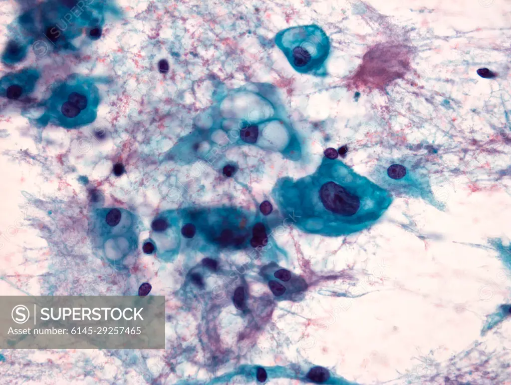
Papanicolaou stained smear of a C2 vertebral chordomal mass, microscopy. Chordomas are cancers formed of cells which resemble those of the notochord (spine) of a developing foetus. Although they can present anywhere within the spine and skull, the majority grow in the sacral region of the spine, corresponding to the lower back. This image shows a Papanicolaou (pap) stained smear obtained from a needle biopsy of a chordoma of the C2 vertebrae, located at the top of the neck just underneath the base of the skull.
SuperStock offers millions of photos, videos, and stock assets to creatives around the world. This image of Papanicolaou stained smear of a C2 vertebral chordomal mass, microscopy. Chordomas are cancers formed of cells which resemble those of the notochord (spine) of a developing foetus. Although they can present anywhere within the spine and skull, the majority grow in the sacral region of the spine, corresponding to the lower back. This image shows a Papanicolaou (pap) stained smear obtained from a needle biopsy of a chordoma of the C2 vertebrae, located at the top of the neck just underneath the base of the skull. by Piemags/PL Photography Limited is available for licensing today.
Looking for a license?
Click here, and we'll help you find it! Questions? Just ask!
Click here, and we'll help you find it! Questions? Just ask!
DETAILS
Image Number: 6145-29257465Royalty FreeCredit Line:Piemags/PL Photography Limited/SuperStockCollection:PL Photography Limited Contributor:Piemags Model Release:NoProperty Release:NoResolution:4080×3072