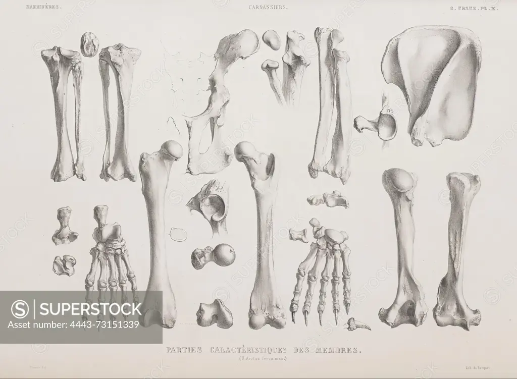
Osteographie ou Description iconographique comparee du squelette et du système dentaire des Mammifères recents et fossiles, Paris, J.B. Baillière et fils, 1839-1864, Henri Marie Ducrotay, A detailed anatomical illustration showcasing the skeletal structure of the human body. The image presents various bones methodically arranged, including upper and lower limbs. Prominently featured are the humerus, radius, and ulna of the upper extremities, alongside the femur, tibia, and fibula of the lower extremities. Additionally, smaller bones such as those in the hands and feet are depicted in a systematic layout, highlighting their connections and relative sizes. Labels accompany each bone, providing clarity on their anatomical names and placements within the skeletal system, emphasizing their significance in human anatomy and physiology.
SuperStock offers millions of photos, videos, and stock assets to creatives around the world. This image of Osteographie ou Description iconographique comparee du squelette et du système dentaire des Mammifères recents et fossiles, Paris, J.B. Baillière et fils, 1839-1864, Henri Marie Ducrotay, A detailed anatomical illustration showcasing the skeletal structure of the human body. The image presents various bones methodically arranged, including upper and lower limbs. Prominently featured are the humerus, radius, and ulna of the upper extremities, alongside the femur, tibia, and fibula of the lower extremities. Additionally, smaller bones such as those in the hands and feet are depicted in a systematic layout, highlighting their connections and relative sizes. Labels accompany each bone, providing clarity on their anatomical names and placements within the skeletal system, emphasizing their significance in human anatomy and physiology. by Artokoloro is available for licensing today.
Looking for a license?
Click here, and we'll help you find it! Questions? Just ask!
Click here, and we'll help you find it! Questions? Just ask!
DETAILS
Image Number: 4443-73151339Rights ManagedCredit Line:Artokoloro/SuperStockCollection:Artokoloro Model Release:NoProperty Release:NoResolution:4521×3320