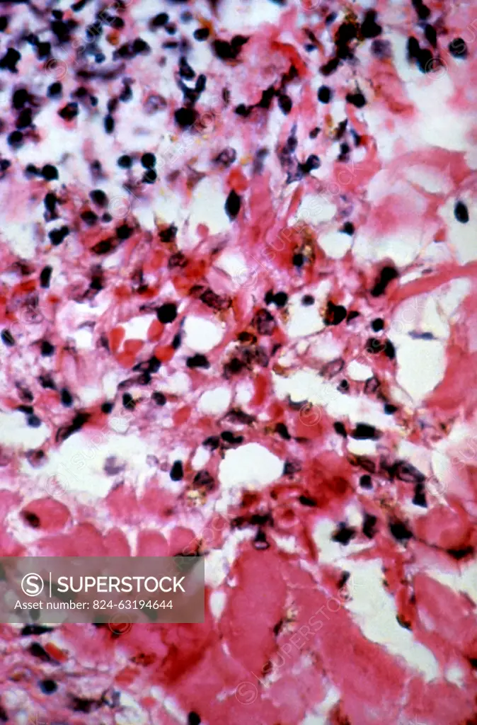
This micrograph depicts the histopathologic changes found in the case of Kaposi's sarcoma lesions. This skin biopsy of what turned out to be a Kaposi's sarcoma lesion at a high magnification depicts two characteristic findings found in such lesions, which includes remnants of extravasated erythrocytes (RBCs), and deposits of hemosiderin.
SuperStock offers millions of photos, videos, and stock assets to creatives around the world. This image of This micrograph depicts the histopathologic changes found in the case of Kaposi's sarcoma lesions. This skin biopsy of what turned out to be a Kaposi's sarcoma lesion at a high magnification depicts two characteristic findings found in such lesions, which includes remnants of extravasated erythrocytes (RBCs), and deposits of hemosiderin. by BSIP is available for licensing today.
Looking for a license?
Click here, and we'll help you find it! Questions? Just ask!
Click here, and we'll help you find it! Questions? Just ask!
DETAILS
Image Number: 824-63194644Rights ManagedCredit Line:BSIP/SuperStockCollection:BSIP Model Release:NoProperty Release:NoResolution:2455×3728