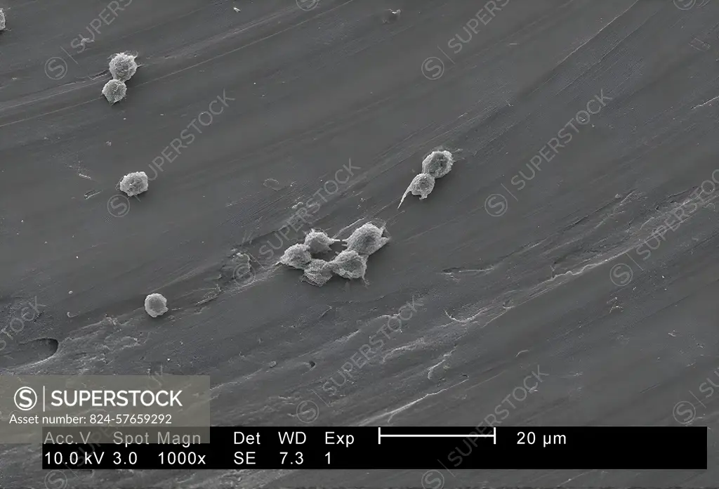
Magnified 1000X, this 2002 scanning electron microscope (SEM) image revealed part of the ultrastructure exhibited by a number of Hartmannella vermiformis amoeba trophozoites. The amoeba assumes this trophozoite stage, which is its vegetative phase, during which it spends time feeding, moving, and reproducing. This free-living protozoan moves in response to chemical cues in its environment by extending pseudopodia or false feet. CDC/Dr Rodney Donlan 2002
SuperStock offers millions of photos, videos, and stock assets to creatives around the world. This image of Magnified 1000X, this 2002 scanning electron microscope (SEM) image revealed part of the ultrastructure exhibited by a number of Hartmannella vermiformis amoeba trophozoites. The amoeba assumes this trophozoite stage, which is its vegetative phase, during which it spends time feeding, moving, and reproducing. This free-living protozoan moves in response to chemical cues in its environment by extending pseudopodia or false feet. CDC/Dr Rodney Donlan 2002 by CDC/IMAGE POINT FR/BSIP is available for licensing today.
Looking for a license?
Click here, and we'll help you find it! Questions? Just ask!
Click here, and we'll help you find it! Questions? Just ask!
DETAILS
Image Number: 824-57659292Rights ManagedCredit Line:CDC/IMAGE POINT FR/BSIP/SuperStockCollection:BSIP Contributor:CDC / IMAGE POINT FR / BSIP Model Release:NoProperty Release:NoResolution:5670×3854