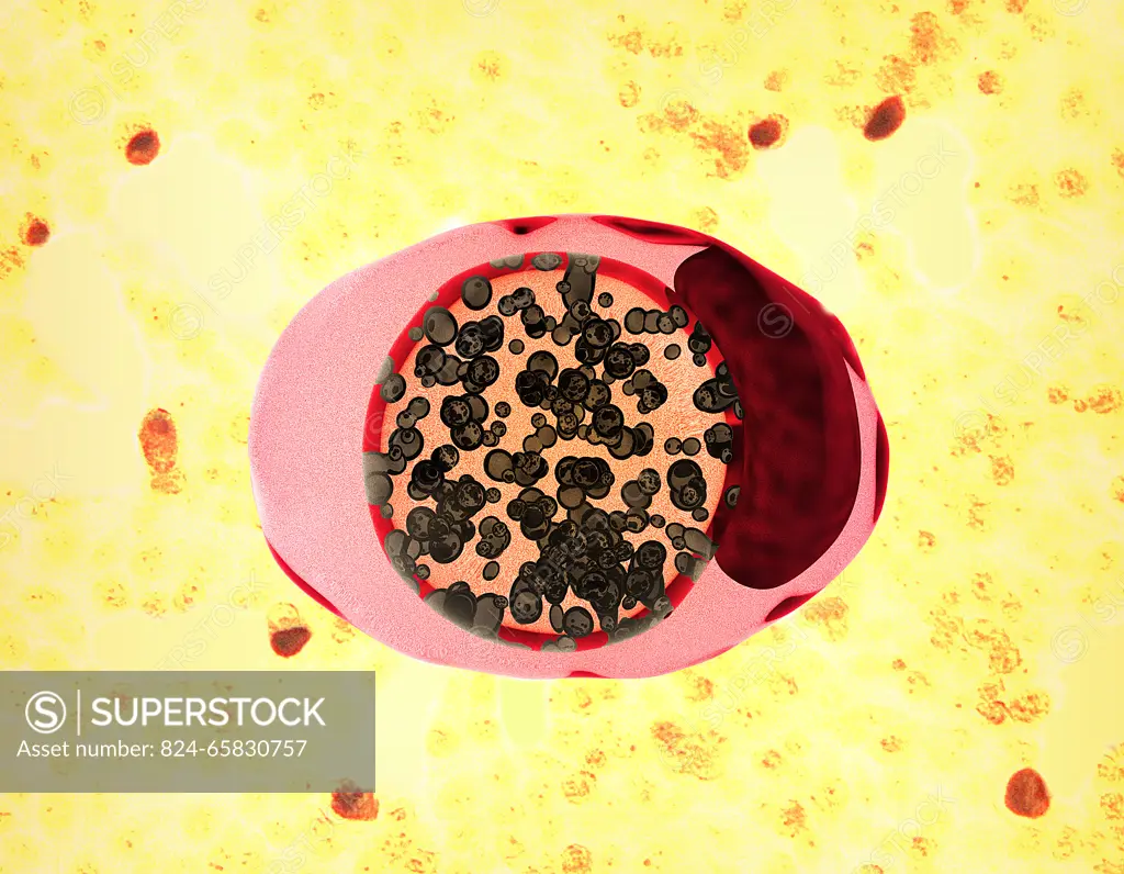
Illustration depicting a chlamydia-infected cell (pink and red oval shape). Chlamydia trachomatis bacteria (represented by small spherical shapes colorized brown) undergo development within a membrane-enclosed inclusion body (center area orange). The dark red area on the right of the illustration denotes the cell nucleus. In the background is a CDC micrograph of cells (yellow) exhibiting chlamydia inclusion bodies (red).
SuperStock offers millions of photos, videos, and stock assets to creatives around the world. This image of Illustration depicting a chlamydia-infected cell (pink and red oval shape). Chlamydia trachomatis bacteria (represented by small spherical shapes colorized brown) undergo development within a membrane-enclosed inclusion body (center area orange). The dark red area on the right of the illustration denotes the cell nucleus. In the background is a CDC micrograph of cells (yellow) exhibiting chlamydia inclusion bodies (red). by NIH-NIAID/IMAGE POINT FR/BSIP is available for licensing today.
Looking for a license?
Click here, and we'll help you find it! Questions? Just ask!
Click here, and we'll help you find it! Questions? Just ask!
DETAILS
Image Number: 824-65830757Rights ManagedCredit Line:NIH-NIAID/IMAGE POINT FR/BSIP/SuperStockCollection:BSIP Contributor:NIH-NIAID / IMAGE POINT FR / BSIP Model Release:NoProperty Release:NoResolution:4123×3206