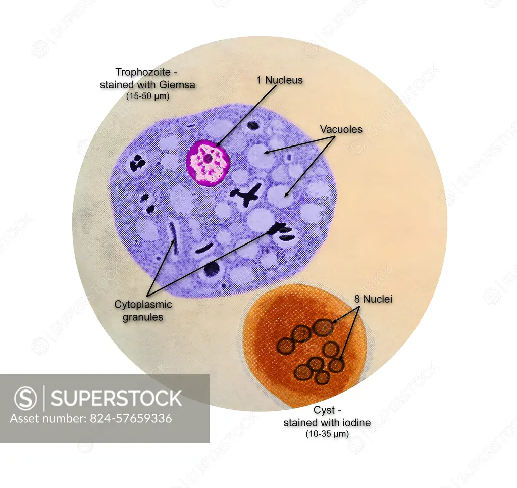
This illustration of a composite photomicrograph reveals the ultrastructural details seen at two stages in the life cycle of the parasite, Entamoeba coli, including its cystic stage at lower right, which was stained with iodine, and its vegetative trophozoite stage stained at the Giemsa in the center left. CDC 1979
SuperStock offers millions of photos, videos, and stock assets to creatives around the world. This image of This illustration of a composite photomicrograph reveals the ultrastructural details seen at two stages in the life cycle of the parasite, Entamoeba coli, including its cystic stage at lower right, which was stained with iodine, and its vegetative trophozoite stage stained at the Giemsa in the center left. CDC 1979 by CDC/IMAGE POINT FR/BSIP is available for licensing today.
Looking for a license?
Click here, and we'll help you find it! Questions? Just ask!
Click here, and we'll help you find it! Questions? Just ask!
DETAILS
Image Number: 824-57659336Rights ManagedCredit Line:CDC/IMAGE POINT FR/BSIP/SuperStockCollection:BSIP Contributor:CDC / IMAGE POINT FR / BSIP Model Release:NoProperty Release:NoResolution:4872×4598