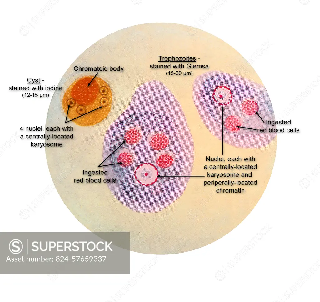
This illustration of a composite photomicrograph reveals the ultrastructural details observed at two stages in the life cycle of the parasite, Entamoeba histolytica, stained with iodine is its cyst stage (lt), , and its Giemsa stain, trophozoite stage (cntr and rt). The cyst contained a chromatoid body and the four characteristic nuclei, each containing a single centrally located karyosome. Each E. histolytica trophozoite contained a single nucleus, within which was a single centrally located karyosome and peripherally located chromatin. Note the red blood cells ingested inside each of the trophozoites. CDC 1954
SuperStock offers millions of photos, videos, and stock assets to creatives around the world. This image of This illustration of a composite photomicrograph reveals the ultrastructural details observed at two stages in the life cycle of the parasite, Entamoeba histolytica, stained with iodine is its cyst stage (lt), , and its Giemsa stain, trophozoite stage (cntr and rt). The cyst contained a chromatoid body and the four characteristic nuclei, each containing a single centrally located karyosome. Each E. histolytica trophozoite contained a single nucleus, within which was a single centrally located karyosome and peripherally located chromatin. Note the red blood cells ingested inside each of the trophozoites. CDC 1954 by CDC/IMAGE POINT FR/BSIP is available for licensing today.
Looking for a license?
Click here, and we'll help you find it! Questions? Just ask!
Click here, and we'll help you find it! Questions? Just ask!
DETAILS
Image Number: 824-57659337Rights ManagedCredit Line:CDC/IMAGE POINT FR/BSIP/SuperStockCollection:BSIP Contributor:CDC / IMAGE POINT FR / BSIP Model Release:NoProperty Release:NoResolution:4872×4598