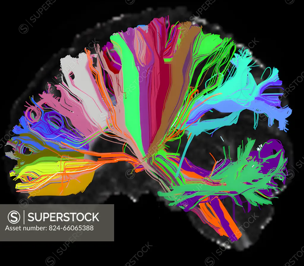
This High Angular Resolution Diffusion Image (HARDI) of the human brain shows long distance connections, or tracts, grouped on the basis of their anatomical neighborhood. Wiring associated with particular brain structures share the same color. In diffusion imaging, the scanner detects movement of water inside neural fibers to reveal their locations. This image is based on first phase HCP data from the MGH/Harvard/UCLA Connectom scanner. Researchers hope to use the same technique to analyze data from a project related to the HCPs second phase that will examine connections in teens with mental illness.
SuperStock offers millions of photos, videos, and stock assets to creatives around the world. This image of This High Angular Resolution Diffusion Image (HARDI) of the human brain shows long distance connections, or tracts, grouped on the basis of their anatomical neighborhood. Wiring associated with particular brain structures share the same color. In diffusion imaging, the scanner detects movement of water inside neural fibers to reveal their locations. This image is based on first phase HCP data from the MGH/Harvard/UCLA Connectom scanner. Researchers hope to use the same technique to analyze data from a project related to the HCPs second phase that will examine connections in teens with mental illness. by NIH/IMAGE POINT FR/BSIP is available for licensing today.
Looking for a license?
Click here, and we'll help you find it! Questions? Just ask!
Click here, and we'll help you find it! Questions? Just ask!
DETAILS
Image Number: 824-66065388Rights ManagedCredit Line:NIH/IMAGE POINT FR/BSIP/SuperStockCollection:BSIP Contributor:NIH / IMAGE POINT FR / BSIP Model Release:NoProperty Release:NoResolution:3888×3400