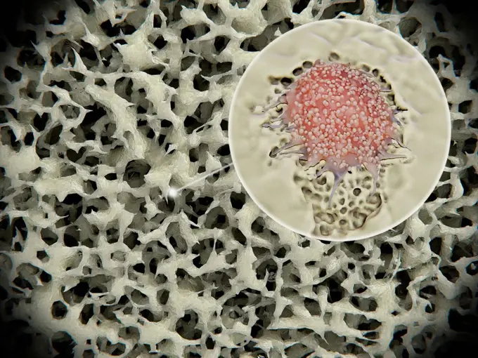
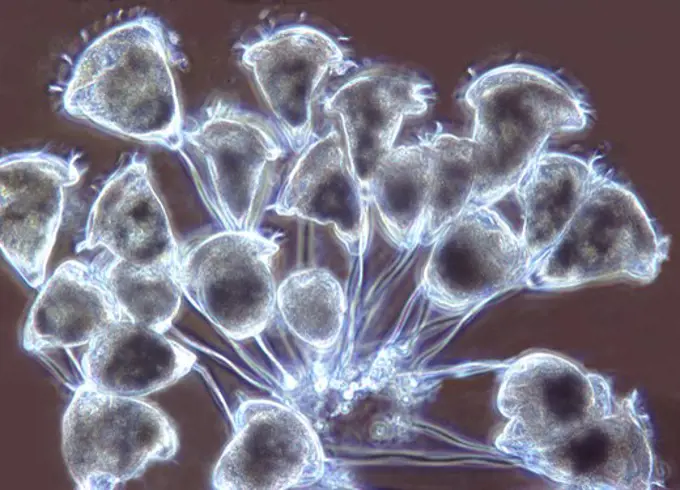
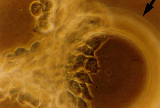
The figure is an electron micrograph showing abnormally shaped and structurally abnormal mitochondria in the liver of a mutant mouse that models methylmalonic acidemia, genetic disease due to a deficiency of the enzyme methylmalonyl CoA mutase.

SuperStock offers millions of photos, videos, and stock assets to creatives around the world. This image of The figure is an electron micrograph showing abnormally shaped and structurally abnormal mitochondria in the liver of a mutant mouse that models methylmalonic acidemia, genetic disease due to a deficiency of the enzyme methylmalonyl CoA mutase. by NIH/IMAGE POINT FR/BSIP is available for licensing today.
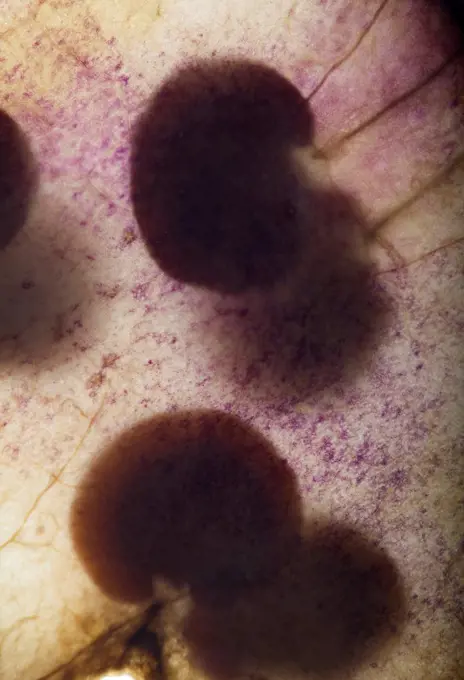
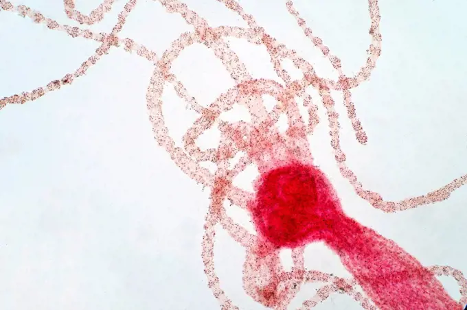
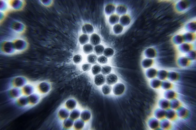
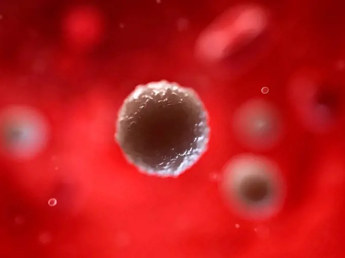
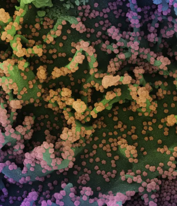
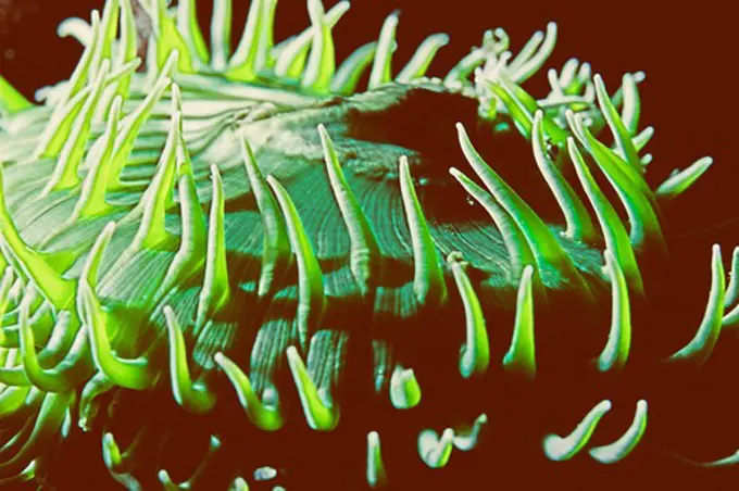
Visually Similar More from Microscopic Viruses and Cells story


Looking for a license?
Click here, and we'll help you find it! Questions? Just ask!
Click here, and we'll help you find it! Questions? Just ask!
DETAILS
Image Number: 824-63227096Rights ManagedCredit Line:NIH/IMAGE POINT FR/BSIP/SuperStockCollection:BSIP Story:Microscopic Viruses and CellsContributor:NIH / IMAGE POINT FR / BSIP Model Release:NoProperty Release:NoResolution:4980×3264