Dislocation of the hip. 3D Computed Tomographic (CT) reconstruction scan of the pelvis showing a posterior hip luxation. The head of the femur (bone of the thigh) has totally slipped out on its insertion point in the pelvis bone.
This asset has restrictions in United States, but still may be available. Get in touch for more details.
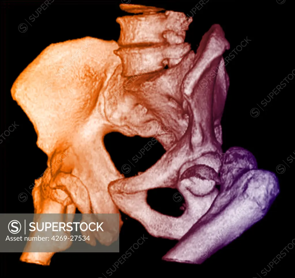
SuperStock offers millions of photos, videos, and stock assets to creatives around the world. This image of Dislocation of the hip. 3D Computed Tomographic (CT) reconstruction scan of the pelvis showing a posterior hip luxation. The head of the femur (bone of the thigh) has totally slipped out on its insertion point in the pelvis bone. by Guilloz-Chu Nancy/Phanie is available for licensing today.
Looking for a license?
Click here, and we'll help you find it! Questions? Just ask!
Click here, and we'll help you find it! Questions? Just ask!
DETAILS
Image Number: 4269-27534Rights ManagedCredit Line:Guilloz-Chu Nancy/Phanie/SuperStockCollection:Phanie Contributor:Guilloz-Chu Nancy Model Release:NoProperty Release:NoResolution:4500×4244