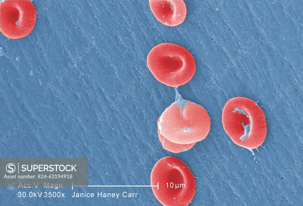
This digitally-colorized scanning electron micrograph (SEM) revealed some of the ultrastructural morphology displayed by red blood cells (RBCs) in a blood specimen of a 6 year old male patient that has sickle cell with hereditary persistence of fetal hemoglobin (S-HPFH) . In these individuals, the presence of the persistent fetal hemoglogin reduces the severity of the consequences of the sickle cell disease, thereby, reducing the degree of cellular deformity, ie sickling, seen in the sickled cells.
SuperStock offers millions of photos, videos, and stock assets to creatives around the world. This image of This digitally-colorized scanning electron micrograph (SEM) revealed some of the ultrastructural morphology displayed by red blood cells (RBCs) in a blood specimen of a 6 year old male patient that has sickle cell with hereditary persistence of fetal hemoglobin (S-HPFH) . In these individuals, the presence of the persistent fetal hemoglogin reduces the severity of the consequences of the sickle cell disease, thereby, reducing the degree of cellular deformity, ie sickling, seen in the sickled cells. by CDC/IMAGE POINT FR/BSIP is available for licensing today.
Looking for a license?
Click here, and we'll help you find it! Questions? Just ask!
Click here, and we'll help you find it! Questions? Just ask!
DETAILS
Image Number: 824-63194918Rights ManagedCredit Line:CDC/IMAGE POINT FR/BSIP/SuperStockCollection:BSIP Contributor:CDC / IMAGE POINT FR / BSIP Model Release:NoProperty Release:NoResolution:3665×2491