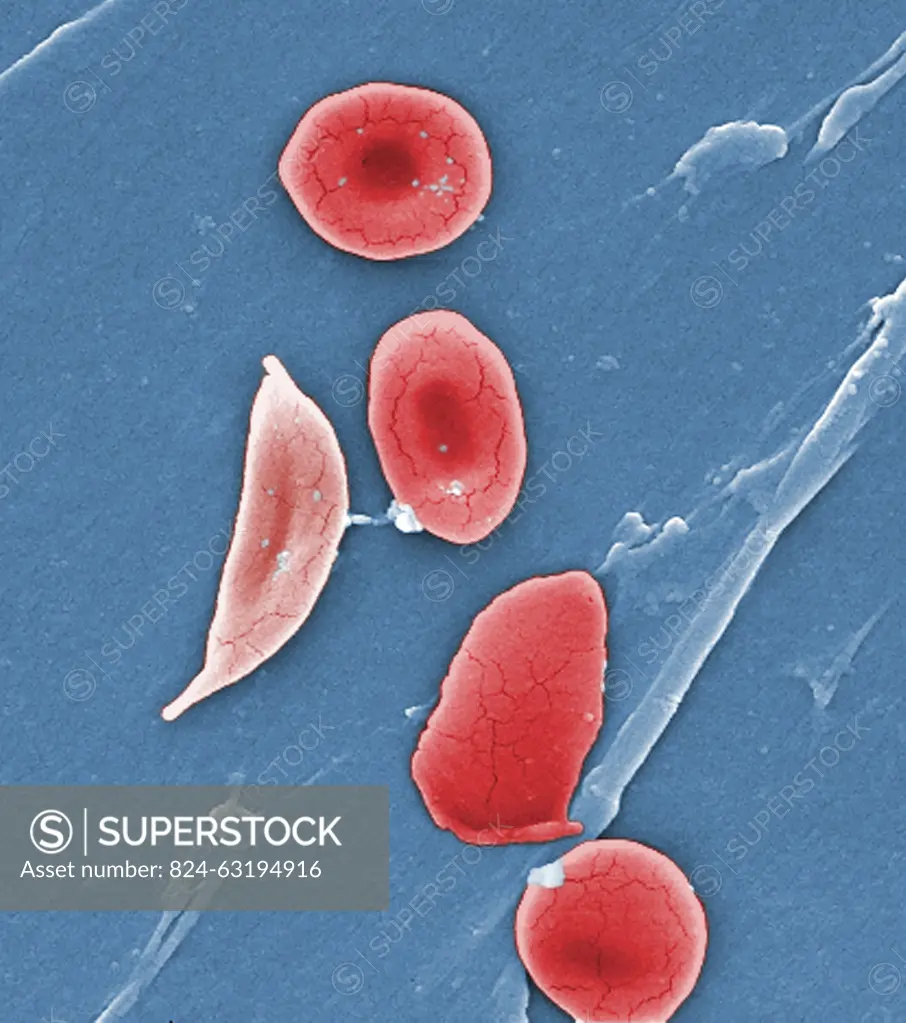
This digitally-colorized scanning electron micrograph (SEM) revealed some of the comparative ultrastructural morphology between normal red blood cells (RBCs), and a sickle cell RBC (left) found in a blood specimen of an 18 year old female patient with sickle cell anemia , (HbSS), People who have this form of sickle cell disease inherit two sickle cell genes (S), one from each parent. This is commonly called sickle cell anemia, and is usually the most severe form of the disease.
SuperStock offers millions of photos, videos, and stock assets to creatives around the world. This image of This digitally-colorized scanning electron micrograph (SEM) revealed some of the comparative ultrastructural morphology between normal red blood cells (RBCs), and a sickle cell RBC (left) found in a blood specimen of an 18 year old female patient with sickle cell anemia , (HbSS), People who have this form of sickle cell disease inherit two sickle cell genes (S), one from each parent. This is commonly called sickle cell anemia, and is usually the most severe form of the disease. by CDC/IMAGE POINT FR/BSIP is available for licensing today.
Looking for a license?
Click here, and we'll help you find it! Questions? Just ask!
Click here, and we'll help you find it! Questions? Just ask!
DETAILS
Image Number: 824-63194916Rights ManagedCredit Line:CDC/IMAGE POINT FR/BSIP/SuperStockCollection:BSIP Contributor:CDC / IMAGE POINT FR / BSIP Model Release:NoProperty Release:NoResolution:2841×3210