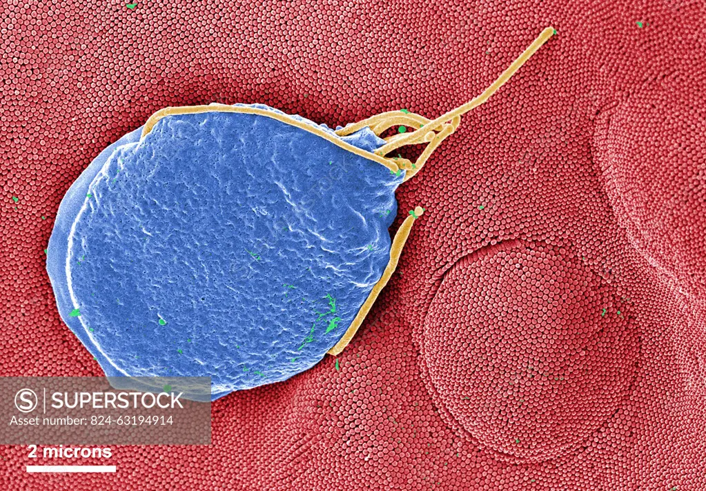
This digitally-colorized scanning electron micrograph (SEM) depicted a Giardia muris protozoan adhering itself to the microvillous border of an intestinal epithelial cell. Each small circular profile under the protozoan represents the rounded tip of a single microvillous, and it is estimated that 2000 to 3000 microvilli cover the surface of a single intestinal epithelial cell. The two circular lesions on the right side of the photograph are impressions made by the ventral adhesive disk of other G. muris organisms. This disk acts like a suction cup, facilitating the organism's attachment to the intestinal surface. The protozoan Giardia causes the diarrheal disease called giardiasis.
SuperStock offers millions of photos, videos, and stock assets to creatives around the world. This image of This digitally-colorized scanning electron micrograph (SEM) depicted a Giardia muris protozoan adhering itself to the microvillous border of an intestinal epithelial cell. Each small circular profile under the protozoan represents the rounded tip of a single microvillous, and it is estimated that 2000 to 3000 microvilli cover the surface of a single intestinal epithelial cell. The two circular lesions on the right side of the photograph are impressions made by the ventral adhesive disk of other G. muris organisms. This disk acts like a suction cup, facilitating the organism's attachment to the intestinal surface. The protozoan Giardia causes the diarrheal disease called giardiasis. by CDC/IMAGE POINT FR/BSIP is available for licensing today.
Looking for a license?
Click here, and we'll help you find it! Questions? Just ask!
Click here, and we'll help you find it! Questions? Just ask!
DETAILS
Image Number: 824-63194914Rights ManagedCredit Line:CDC/IMAGE POINT FR/BSIP/SuperStockCollection:BSIP Contributor:CDC / IMAGE POINT FR / BSIP Model Release:NoProperty Release:NoResolution:3622×2520