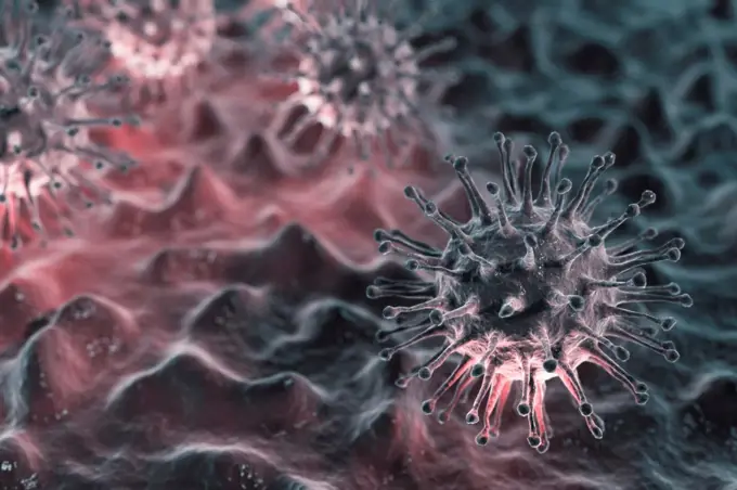
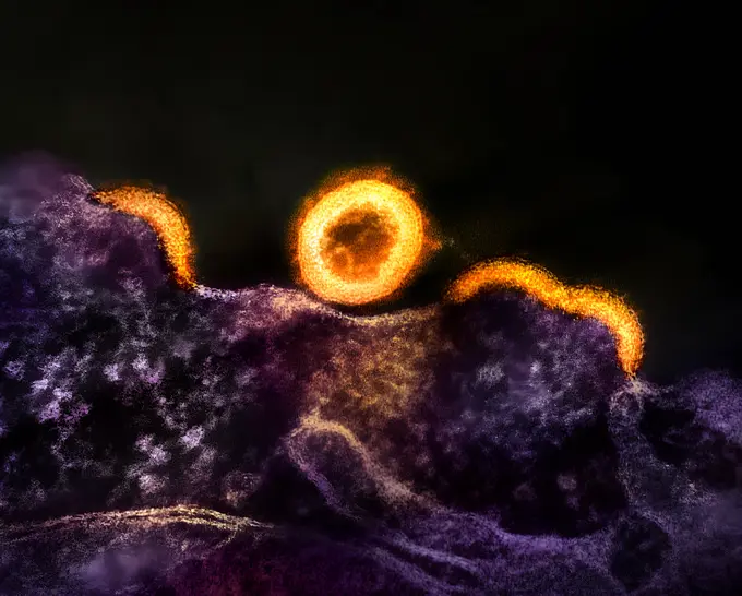
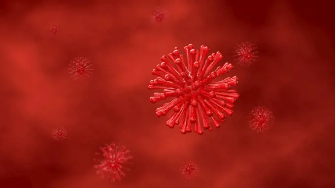
Colorized transmission electron micrograph of two HIV-1 virus particles (yellow) budding from the plasma membrane of an infected H9 T cell (blue).
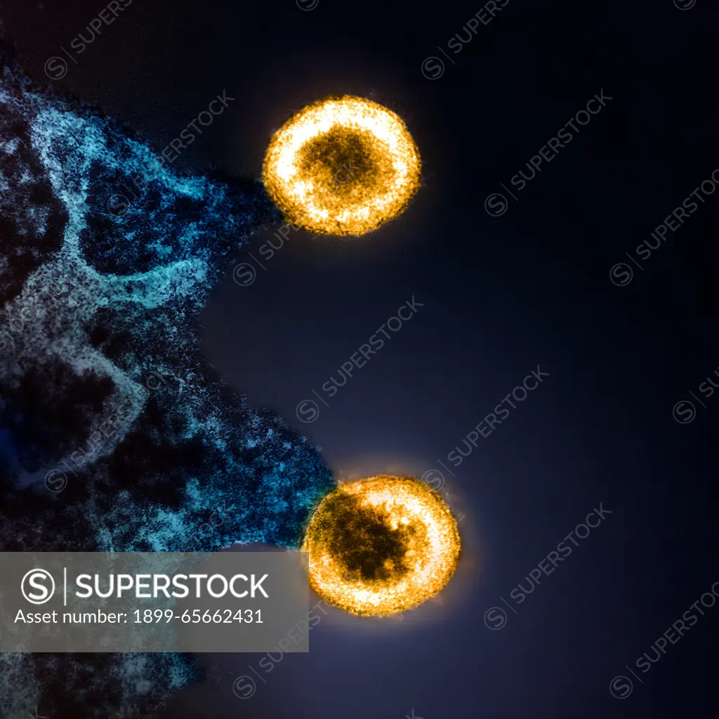
SuperStock offers millions of photos, videos, and stock assets to creatives around the world. This image of Colorized transmission electron micrograph of two HIV-1 virus particles (yellow) budding from the plasma membrane of an infected H9 T cell (blue). by NIH-NIAID/IMAGE POINT FR/BSIP/Universal Images is available for licensing today.
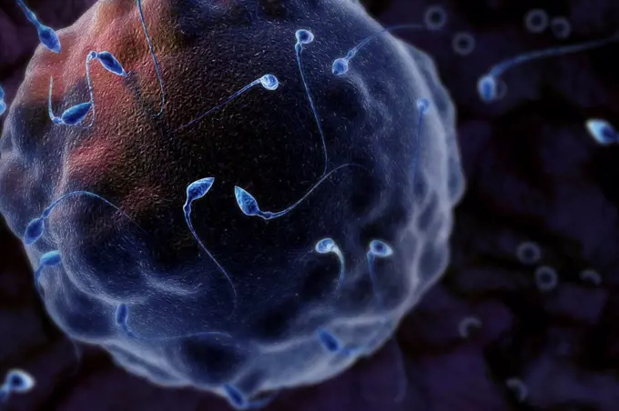
Visually Similar More from Cellular and Viral Illustration story


Looking for a license?
Click here, and we'll help you find it! Questions? Just ask!
Click here, and we'll help you find it! Questions? Just ask!
DETAILS
Image Number: 1899-65662431Rights ManagedCredit Line:NIH-NIAID/IMAGE POINT FR/BSIP/Universal Images/SuperStockCollection:Universal Images Story:Cellular and Viral IllustrationContributor:NIH-NIAID / IMAGE POINT FR / BSIP Model Release:NoProperty Release:NoResolution:4712×4712