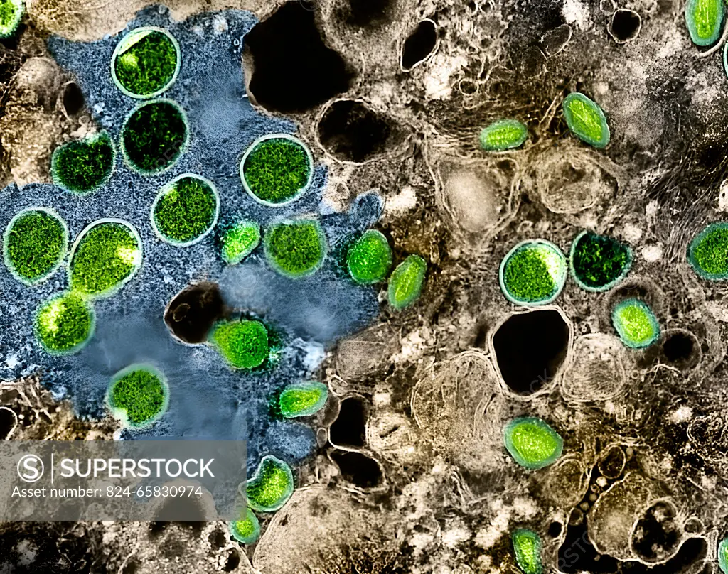
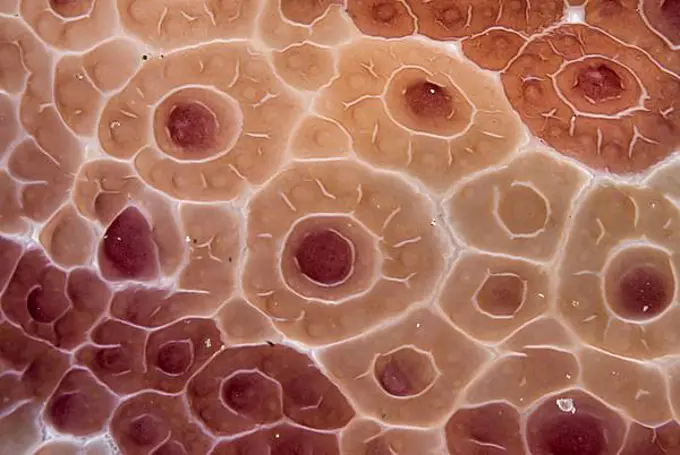
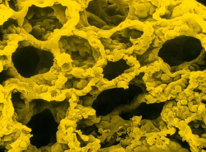
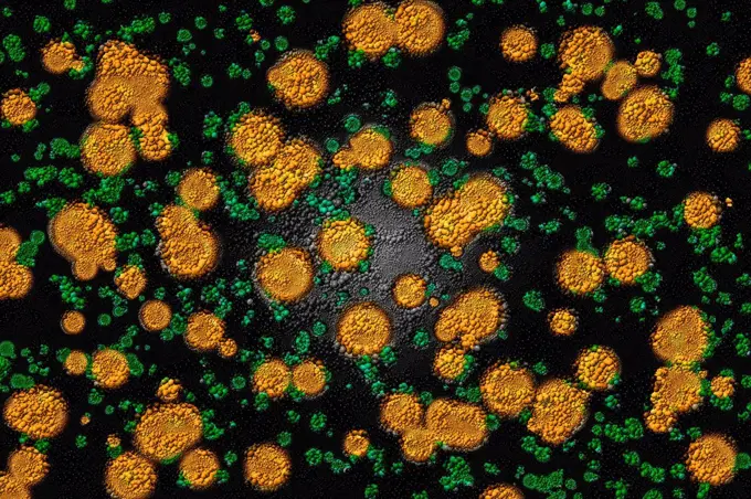
Colorized transmission electron micrograph of mpox virus particles (green) found within infected VERO E6 cells (brown). The virus particles are in various stages of maturity, which accounts for differences in shape. This image also features virus factories (blue), which are inclusions within infected cells where virus replication, maturation, and assembly occurs.
SuperStock offers millions of photos, videos, and stock assets to creatives around the world. This image of Colorized transmission electron micrograph of mpox virus particles (green) found within infected VERO E6 cells (brown). The virus particles are in various stages of maturity, which accounts for differences in shape. This image also features virus factories (blue), which are inclusions within infected cells where virus replication, maturation, and assembly occurs. by NIH-NIAID/IMAGE POINT FR/BSIP is available for licensing today.
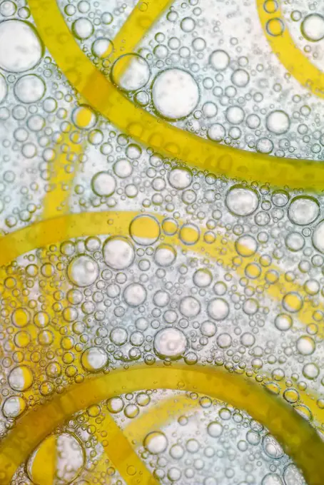
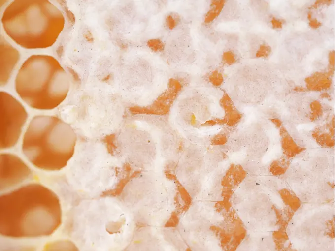
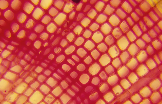
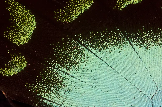
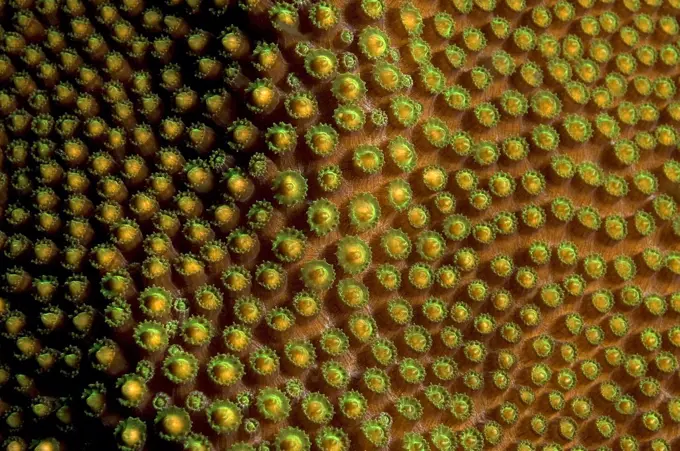
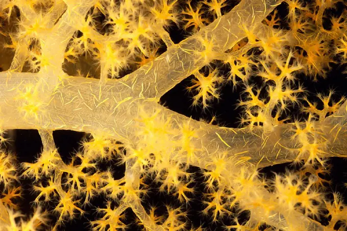
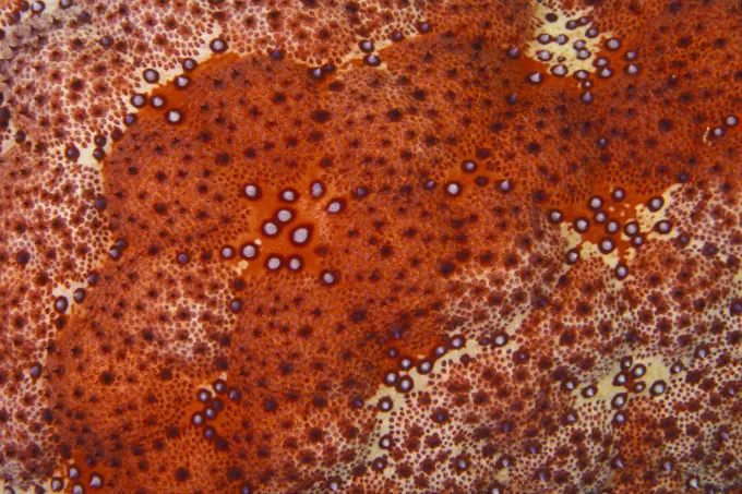
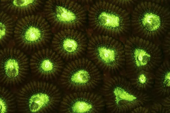
Visually Similar More from Fluorescent Coral Patterns story


Looking for a license?
Click here, and we'll help you find it! Questions? Just ask!
Click here, and we'll help you find it! Questions? Just ask!
DETAILS
Image Number: 824-65830974Rights ManagedCredit Line:NIH-NIAID/IMAGE POINT FR/BSIP/SuperStockCollection:BSIP Story:Fluorescent Coral PatternsContributor:NIH-NIAID / IMAGE POINT FR / BSIP Model Release:NoProperty Release:NoResolution:3825×3020